

The DOI matches its Crossref record; 78 of the 78 references it cites were found in a registry or an index.
Sister Dehalobacter Genomes Reveal Specialization in Organohalide Respiration and Recent Strain Differentiation Likely Driven by Chlorinated Substrates
Abstract
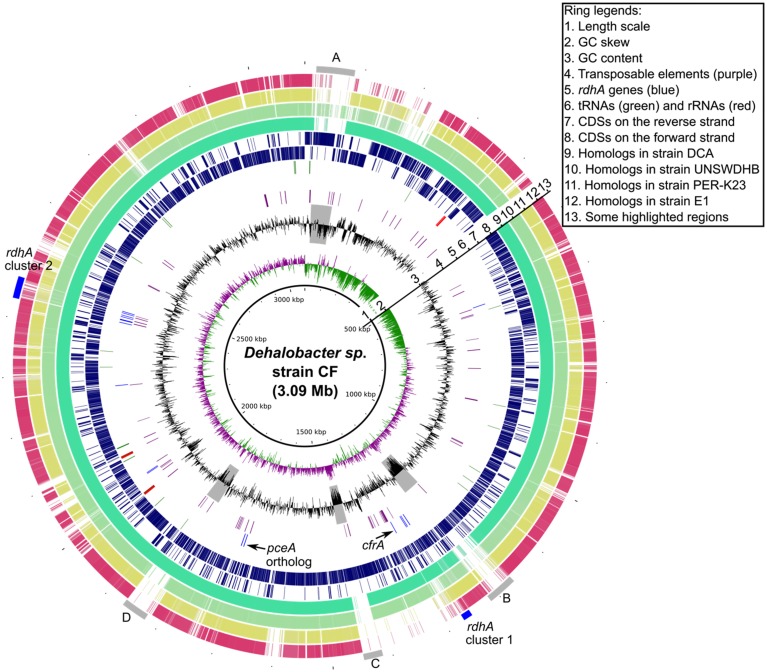
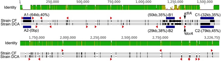
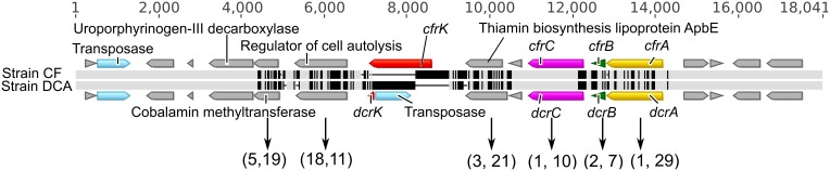
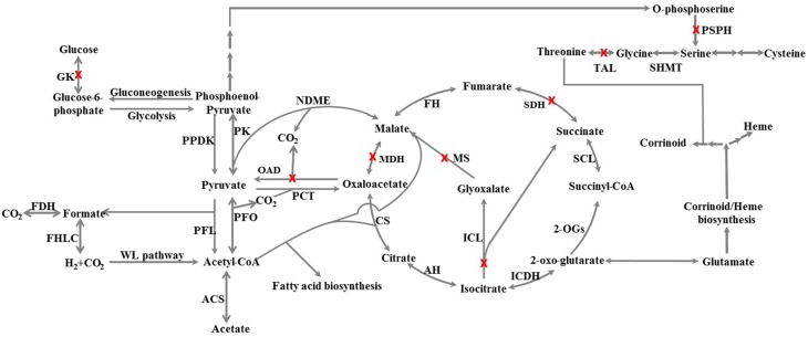
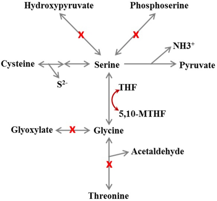
The genomes of two closely related Dehalobacter strains (strain CF and strain DCA) were assembled from the metagenome of an anaerobic enrichment culture that reductively dechlorinates chloroform (CF), 1,1,1-trichloroethane (1,1,1-TCA) and 1,1-dichloroethane (1,1-DCA). The 3.1 Mbp genomes of strain CF (that dechlorinates CF and 1,1,1-TCA) and strain DCA (that dechlorinates 1,1-DCA) each contain 17 putative reductive dehalogenase homologous (rdh) genes. These two genomes were systematically compared to three other available organohalide-respiring Dehalobacter genomes (Dehalobacter restrictus strain PER-K23, Dehalobacter sp. strain E1 and Dehalobacter sp. strain UNSWDHB), and to the genomes of Dehalococcoides mccartyi strain 195 and Desulfitobacterium hafniense strain Y51. This analysis compared 42 different metabolic and physiological categories. The genomes of strains CF and DCA share 90% overall average nucleotide identity and >99.8% identity over a 2.9 Mbp alignment that excludes large insertions, indicating that these genomes differentiated from a close common ancestor. This differentiation was likely driven by selection pressures around two orthologous reductive dehalogenase genes, cfrA and dcrA, that code for the enzymes that reduce CF or 1,1,1-TCA and 1,1-DCA. The many reductive dehalogenase genes found in the five Dehalobacter genomes cluster into two small conserved regions and were often associated with Crp/Fnr transcriptional regulators. Specialization is on-going on a strain-specific basis, as some strains but not others have lost essential genes in the Wood-Ljungdahl (strain E1) and corrinoid biosynthesis pathways (strains E1 and PER-K23). The gene encoding phosphoserine phosphatase, which catalyzes the last step of serine biosynthesis, is missing from all five Dehalobacter genomes, yet D. restrictus can grow without serine, suggesting an alternative or unrecognized biosynthesis route exists. In contrast to D. mccartyi, a complete heme biosynthesis pathway is present in the five Dehalobacter genomes. This pathway corresponds to a newly described alternative heme biosynthesis route first identified in Archaea. This analysis of organohalide-respiring Firmicutes and Chloroflexi reveals profound evolutionary differences despite very similar niche-specific metabolism and function.
Overview
The claims and statistics of this paper haven’t been checked yet.
Run the check — free, no account needed. It reads the open-access text and checks the sources it cites.
Integrity signals
Registry
Cites
Statistics
Figures
Verification
Claims the paper makes
The claims and statistics of this paper haven’t been checked yet.
Run the check — free, no account needed. It reads the open-access text and checks the sources it cites.
Claims other papers make about this one
Each is checked against this paper’s own text: does it say what the citing paper says it does?
The source backs part of the claim, not all of it — or backs it for a different population, condition or comparison.
In the citing paper
“This process of organochlorine and organobromine respiration is now considered to be reasonably common, evolutionarily ancient, and further adapted in response to the many chlorinated and brominated natural products produced in the environment (Mayer‐Blackwell et al., 2016; Tang et al., 2016).”
From Nothing lasts forever: understanding microbial biodegradation of polyfluorinated compounds and perfluorinated alkyl substances (2022) · cites this paper as Tang et al., 2016
Experimental — accuracy not yet measured
Self-check
Percentages, sums and reported differences re-computed from the paper’s own numbers.
The self-check needs the paper’s full text.
Citations
Cites
MUSCLE: multiple sequence alignment with high accuracy and high throughput
Edgar, R. C. (2004) · Nucleic Acids Research · doi:10.1093/nar/gkh340 · Cited by 44,662
The Pfam Protein Families Database
Bateman, A. (2002) · Nucleic Acids Research · doi:10.1093/nar/30.1.276 · Cited by 14,000
The RAST Server: Rapid Annotations using Subsystems Technology
Aziz, R. K., Bartels, D., Best, A. A., DeJongh, M., Disz, T., Edwards, R. A., Formsma, K., Gerdes, S., Glass, E. M., Kubal, M., et al. (2008) · BMC Genomics · doi:10.1186/1471-2164-9-75 · Cited by 11,729
progressiveMauve: Multiple Genome Alignment with Gene Gain, Loss and Rearrangement
Darling, A. E., Mau, B., & Perna, N. T. (2010) · PLoS ONE · doi:10.1371/journal.pone.0011147 · Cited by 3,848
BLAST Ring Image Generator (BRIG): simple prokaryote genome comparisons
Alikhan, N.-F., Petty, N. K., Ben Zakour, N. L., & Beatson, S. A. (2011) · BMC Genomics · doi:10.1186/1471-2164-12-402 · Cited by 3,303
Identifying bacterial genes and endosymbiont DNA with Glimmer
Delcher, A. L., Bratke, K. A., Powers, E. C., & Salzberg, S. L. (2007) · Bioinformatics · doi:10.1093/bioinformatics/btm009 · Cited by 3,157
The Pfam protein families database
Finn, R. D., Mistry, J., Tate, J., Coggill, P., Heger, A., Pollington, J. E., Gavin, O. L., Gunasekaran, P., Ceric, G., Forslund, K., et al. (2010) · Nucleic Acids Research · doi:10.1093/nar/gkp985 · Cited by 2,694
Making sense of it all: bacterial chemotaxis
Wadhams, G. H., & Armitage, J. P. (2004) · Nature Reviews Molecular Cell Biology · doi:10.1038/nrm1524 · Cited by 1,489
IMG ER: a system for microbial genome annotation expert review and curation
Markowitz, V. M., Mavromatis, K., Ivanova, N. N., Chen, I.-M. A., Chu, K., & Kyrpides, N. C. (2009) · Bioinformatics · doi:10.1093/bioinformatics/btp393 · Cited by 817
DNA uptake during bacterial transformation
Chen, I., & Dubnau, D. (2004) · Nature Reviews Microbiology · doi:10.1038/nrmicro844 · Cited by 769
Transposable Elements: An Abundant and Natural Source of Regulatory Sequences for Host Genes
Rebollo, R., Romanish, M. T., & Mager, D. L. (2012) · Annual Review of Genetics · doi:10.1146/annurev-genet-110711-155621 · Cited by 592
MOLYBDENUM-COFACTOR–CONTAINING ENZYMES: Structure and Mechanism
Kisker, C., Schindelin, H., & Rees, D. C. (1997) · Annual Review of Biochemistry · doi:10.1146/annurev.biochem.66.1.233 · Cited by 520
Major, D. W., McMaster, M. L., Cox, E. E., Edwards, E. A., Dworatzek, S. M., Hendrickson, E. R., Starr, M. G., Payne, J. A., & Buonamici, L. W. (2002) · Environmental Science & Technology · doi:10.1021/es0255711 · Cited by 506
Self-citationKörner, H., Sofia, H. J., & Zumft, W. G. (2003) · FEMS Microbiology Reviews · doi:10.1016/s0168-6445(03)00066-4 · Cited by 473
Genome Sequence of the PCE-Dechlorinating Bacterium Dehalococcoides ethenogenes
Seshadri, R., Adrian, L., Fouts, D. E., Eisen, J. A., Phillippy, A. M., Methe, B. A., Ward, N. L., Nelson, W. C., Deboy, R. T., Khouri, H. M., et al. (2005) · Science · doi:10.1126/science.1102226 · Cited by 435
Holliger, C., Hahn, D., Harmsen, H. J. M., Ludwig, W., Schumacher, W., Tindall, B. J., Vazquez, F., Weiss, N., & Zehnder, A. J. B. (1998) · Archives of Microbiology · doi:10.1007/s002030050577 · Cited by 395
Holliger, C., Schraa, G., Stams, A. J. M., & Zehnder, A. J. B. (1993) · Applied and Environmental Microbiology · doi:10.1128/aem.59.9.2991-2997.1993 · Cited by 389
Structure and function of enzymes in heme biosynthesis
Layer, G., Reichelt, J., Jahn, D., & Heinz, D. W. (2010) · Protein Science · doi:10.1002/pro.405 · Cited by 375
Lendvay, J. M., Löffler, F. E., Dollhopf, M. E., Aiello, M. R., Daniels, G., Fathepure, B. Z., Gebhard, M., Heine, R., Helton, R. R., Shi, J., et al. (2003) · Environmental Science & Technology · doi:10.1021/es025985u · Cited by 327
Self-citationDehalogenation: Microbial Processes and Environmental Applications
(2003) · doi:10.1007/b101852 · Cited by 324
Hug, L. A., Maphosa, F., Leys, D., Löffler, F. E., Smidt, H., Edwards, E. A., & Adrian, L. (2013) · Philosophical Transactions of the Royal Society B: Biological Sciences · doi:10.1098/rstb.2012.0322 · Cited by 294
Self-citationWarburton, P. E., Giordano, J., Cheung, F., Gelfand, Y., & Benson, G. (2004) · Genome Research · doi:10.1101/gr.2542904 · Cited by 287
Harnessing microbial activities for environmental cleanup
Löffler, F. E., & Edwards, E. A. (2006) · Current Opinion in Biotechnology · doi:10.1016/j.copbio.2006.05.001 · Cited by 268
Self-citationThe MiST2 database: a comprehensive genomics resource on microbial signal transduction
Ulrich, L. E., & Zhulin, I. B. (2010) · Nucleic Acids Research · doi:10.1093/nar/gkp940 · Cited by 262
Villemur, R., Lanthier, M., Beaudet, R., & Lepine, F. L. (2006) · FEMS Microbiology Reviews · doi:10.1111/j.1574-6976.2006.00029.x · Cited by 233
Sulavik, M. C., Gambino, L., & Miller, P. (1995) · Molecular Medicine · doi:10.1007/bf03401581 · Cited by 192
Genesis, effects and fates of repeats in prokaryotic genomes
Treangen, T. J., Abraham, A., Touchon, M., & Rocha, E. P. C. (2009) · FEMS Microbiology Reviews · doi:10.1111/j.1574-6976.2009.00169.x · Cited by 184
Kemp, L. R., Dunstan, M. S., Fisher, K., Warwicker, J., & Leys, D. (2013) · Philosophical Transactions of the Royal Society B Biological Sciences · doi:10.1098/rstb.2012.0323 · Cited by 182
Exploiting the ecogenomics toolbox for environmental diagnostics of organohalide-respiring bacteria
Maphosa, F., de Vos, W. M., & Smidt, H. (2010) · Trends in biotechnology · doi:10.1016/j.tibtech.2010.03.005 · Cited by 169
Localized Plasticity in the Streamlined Genomes of Vinyl Chloride Respiring Dehalococcoides
McMurdie, P. J., Behrens, S., Müller, J. A., Göke, J., Ritalahti, K. M., (257632), R. W., Goltsman, E., Lapidus, A. L., Holmes, S., Löffler, F. E., et al. (2016) · Figshare · doi:10.1371/journal.pgen.1000714 · Cited by 164
Self-citationDoesburg, W., Eekert, M. H. A. V., Middeldorp, P. J. M., Balk, M., Schraa, G., & Stams, A. J. M. (2005) · FEMS Microbiology Ecology · doi:10.1016/j.femsec.2005.03.003 · Cited by 160
Nonaka, H., Keresztes, G., Shinoda, Y., Ikenaga, Y., Abe, M., Naito, K., Inatomi, K.-I., Furukawa, K., Inui, M., & Yukawa, H. (2006) · Journal of Bacteriology · doi:10.1128/jb.188.6.2262-2274.2006 · Cited by 156
Weber, I. T., Steitz, T. A., Bubis, J. J., et al. (1987) · Biochemistry · doi:10.1021/bi00376a003 · Cited by 153
Growth of Dehalobacter and Dehalococcoides spp. during Degradation of Chlorinated Ethanes
Grostern, A., & Edwards, E. A. (2006) · Applied and Environmental Microbiology · doi:10.1128/aem.72.1.428-436.2006 · Cited by 153
Self-citationSulavik, M. C., Dazer, M. E., & Miller, P. F. (1997) · Journal of Bacteriology · doi:10.1128/jb.179.6.1857-1866.1997 · Cited by 131
Microbial Dehalorespiration with 1,1,1-Trichloroethane
Sun, B., Griffin, B. M., Ayala-del-Rı́o, H. L., Hashsham, S. A., & Tiedje, J. M. (2002) · Science · doi:10.1126/science.1074675 · Cited by 129
Wild, A., Hermann, R., & Leisinger, T. (1997) · Biodegradation · doi:10.1007/bf00115297 · Cited by 123
Molecular hijacking of siroheme for the synthesis of heme and d 1 heme
Bali, S., Lawrence, A. D., Lobo, S. A., Saraiva, L. M., Golding, B. T., Palmer, D. J., Howard, M. J., Ferguson, S. J., & Warren, M. J. (2011) · Proceedings of the National Academy of Sciences · doi:10.1073/pnas.1108228108 · Cited by 120
Complete chloroform dechlorination by organochlorine respiration and fermentation
Lee, M. J., Low, A., Zemb, O., Koenig, J. C., Michaelsen, A., & Manefield, M. (2011) · Environmental Microbiology · doi:10.1111/j.1462-2920.2011.02656.x · Cited by 119
Grostern, A., & Edwards, E. A. (2006) · Applied and Environmental Microbiology · doi:10.1128/aem.01269-06 · Cited by 112
Self-citationSchumacher, W., & Holliger, C. (1996) · Journal of Bacteriology · doi:10.1128/jb.178.8.2328-2333.1996 · Cited by 106
Terrestrial natural sources of trichloromethane (chloroform, CHCl3) – An overview
Laturnus, F., Haselmann, K. F., Borch, T., & Grøn, C. (2002) · Biogeochemistry · doi:10.1023/a:1019887505651 · Cited by 105
Chloroform respiration to dichloromethane by a Dehalobacter population
Grostern, A., Duhamel, M., Dworatzek, S. M., & Edwards, E. A. (2010) · Environmental Microbiology · doi:10.1111/j.1462-2920.2009.02150.x · Cited by 102
Self-citationSmidt, H., van Leest, M., John van der, O. O. S. T., & de Vos, W. M. (2000) · Journal of Bacteriology · doi:10.1128/jb.182.20.5683-5691.2000 · Cited by 101
Effects of chromosomal inversion on cell fitness in Escherichia coli K-12.
Hill, C. W., & Gray, J. A. (1988) · Genetics · doi:10.1093/genetics/119.4.771 · Cited by 101
Kim, S., Harzman, C., Davis, J. K., Hutcheson, R. U., Broderick, J. B., Marsh, T. L., & Tiedje, J. M. (2012) · BMC Microbiology · doi:10.1186/1471-2180-12-21 · Cited by 99
Justicia-Leon, S. D., Ritalahti, K. M., Mack, E. E., & Löffler, F. E. (2011) · Applied and Environmental Microbiology · doi:10.1128/aem.07325-11 · Cited by 88
Self-citationTang, S., & Edwards, E. A. (2013) · Philosophical Transactions of the Royal Society B Biological Sciences · doi:10.1098/rstb.2012.0318 · Cited by 85
Self-citationDing, C., Zhao, S., & He, J. (2014) · Environmental Microbiology · doi:10.1111/1462-2920.12387 · Cited by 81
Rupakula, A., Kruse, T., Boeren, S. A., Holliger, C., Smidt, H., & Maillard, J. (2013) · Philosophical Transactions of the Royal Society B Biological Sciences · doi:10.1098/rstb.2012.0325 · Cited by 78
Repseek, a tool to retrieve approximate repeats from large DNA sequences
Achaz, G., Boyer, F., Rocha, E. P. C., Viari, A., & Coissac, É. (2006) · Bioinformatics · doi:10.1093/bioinformatics/btl519 · Cited by 78
Anantharaman, V., & Aravind, L. (2002) · FEMS Microbiology Letters · doi:10.1111/j.1574-6968.2002.tb11028.x · Cited by 77
Nelson, J. L., Fung, J. M., Cadillo‐Quiroz, H., Cheng, X., & Zinder, S. H. (2011) · Environmental Science & Technology · doi:10.1021/es200480k · Cited by 77
Skovran, E., & Downs, D. M. (2002) · Journal of Bacteriology · doi:10.1128/jb.185.1.98-106.2003 · Cited by 75
Maillard, J., Regeard, C., & Holliger, C. (2004) · Environmental Microbiology · doi:10.1111/j.1462-2920.2004.00671.x · Cited by 71
Maphosa, F., van Passel, M. W. J., de Vos, W. M., & Smidt, H. (2012) · Environmental Microbiology Reports · doi:10.1111/j.1758-2229.2012.00376.x · Cited by 71
Dehalogenation of Chlorobenzenes, Dichlorotoluenes, and Tetrachloroethene by Three Dehalobacter spp.
Nelson, J. L., Jiang, J., & Zinder, S. H. (2014) · Environmental Science & Technology · doi:10.1021/es4044769 · Cited by 66
The Alternative Route to Heme in the Methanogenic Archaeon Methanosarcina barkeri
Kühner, M., Haufschildt, K., Neumann, A., Storbeck, S., Streif, J., & Layer, G. (2014) · Archaea · doi:10.1155/2014/327637 · Cited by 64
The quest for microbial reductive dechlorination of C 2 to C 4 chloroalkanes is warranted
Wildeman, S. M. A. D., & Verstraete, W. (2003) · Applied Microbiology and Biotechnology · doi:10.1007/s00253-002-1174-6 · Cited by 60
Siddaramappa, S., Challacombe, J. F., Delano, S. F., Green, L. D., Daligault, H. E., Bruce, D. C., Detter, C., Tapia, R., Han, C. S., Goodwin, L., et al. (2012) · Standards in Genomic Sciences · doi:10.4056/sigs.2806097 · Cited by 60
Characterizing the Metabolism of Dehalococcoides with a Constraint-Based Model
Islam, M. A., Edwards, E. A., & Mahadevan, R. (2010) · PLoS Computational Biology · doi:10.1371/journal.pcbi.1000887 · Cited by 59
Self-citationTang, S., Gong, Y., & Edwards, E. A. (2012) · PLoS ONE · doi:10.1371/journal.pone.0052038 · Cited by 58
Self-citationSchlötelburg, C., Wintzingerode, C., Hauck, R., von Wintzingerode, F., Hegemann, W., & Göbel, U. B. (2002) · FEMS Microbiology Ecology · doi:10.1111/j.1574-6941.2002.tb00925.x · Cited by 57
Molecular basis of halorespiration control by CprK, a CRP‐FNR type transcriptional regulator
Levy, C., Pike, K., Heyes, D. J., Joyce, M. G., Gábor, K., Smidt, H., John van der, O. O. S. T., & Leys, D. (2008) · Molecular Microbiology · doi:10.1111/j.1365-2958.2008.06399.x · Cited by 51
A Novel Dehalobacter Species Is Involved in Extensive 4,5,6,7-Tetrachlorophthalide Dechlorination
Yoshida, N., Ye, L., Baba, D., & Katayama, A. (2009) · Applied and Environmental Microbiology · doi:10.1128/aem.02112-08 · Cited by 51
Complete genome sequence of Dehalobacter restrictus PER-K23T
Kruse, T., Maillard, J., Goodwin, L., Woyke, T., Teshima, H., Bruce, D. G., Detter, C., Tapia, R., Han, C. S., Huntemann, M., et al. (2013) · Standards in Genomic Sciences · doi:10.4056/sigs.3787426 · Cited by 49
Wang, S., Zhang, W., Yang, K., & He, J. (2013) · Biodegradation · doi:10.1007/s10532-013-9662-1 · Cited by 47
Regulation of Anaerobic Dehalorespiration by the Transcriptional Activator CprK
Pop, S., Kolarik, R. J., & Ragsdale, S. W. (2004) · Journal of Biological Chemistry · doi:10.1074/jbc.m409435200 · Cited by 46
Regulation of reductive dehalogenase gene transcription in Dehalococcoides mccartyi
Wagner, A., Segler, L., Kleinsteuber, S., et al. (2013) · Philosophical Transactions of the Royal Society B Biological Sciences · doi:10.1098/rstb.2012.0317 · Cited by 45
A phylogenomic analysis of bacterial helix–turn–helix transcription factors
Santos, C. L., Tavares, F., Thioulouse, J., & Normand, P. (2008) · FEMS Microbiology Reviews · doi:10.1111/j.1574-6976.2008.00154.x · Cited by 37
Rupakula, A., Lu, Y., Kruse, T., Boeren, S. A., Holliger, C., Smidt, H., & Maillard, J. (2015) · Frontiers in Microbiology · doi:10.3389/fmicb.2014.00751 · Cited by 34
Identification and Characterization of a Re -Citrate Synthase in Dehalococcoides Strain CBDB1
Marco‐Urrea, E., Paul, S., Khodaverdi, V., Seifert, J., von Bergen�, M., Kretzschmar, U., & Adrian, L. (2011) · Journal of Bacteriology · doi:10.1128/jb.05120-11 · Cited by 31
Duret, A., Holliger, C., & Maillard, J. (2012) · Applied and Environmental Microbiology · doi:10.1128/aem.01221-12 · Cited by 30
Buttet, G. F., Holliger, C., & Maillard, J. (2013) · Applied and Environmental Microbiology · doi:10.1128/aem.02312-13 · Cited by 30
Yoshida, N., Ye, L., Baba, D., & Katayama, A. (2009) · Microbes and Environments · doi:10.1264/jsme2.me09132 · Cited by 28
Schlötelburg, C. (2002) · FEMS Microbiology Ecology · doi:10.1016/s0168-6496(02)00177-0 · Cited by 28
Genome Sequence ofDehalobacterUNSWDHB, a Chloroform-Dechlorinating Bacterium
Deshpande, N., Wong, Y. K., Manefield, M., Wilkins, M. R., & Lee, M. J. (2013) · Genome Announcements · doi:10.1128/genomea.00720-13 · Cited by 22
Villemur, R., Constant, P., Gauthier, A., Shareck, M., & Beaudet, R. (2007) · Canadian Journal of Microbiology · doi:10.1139/w06-111 · Cited by 15
Research works printed but not linked: 3
- Drummond, 2011, Geneious v5.4.2
- Tang, 2014, Investigation of Dehalobacter-Containing Cultures that Reductively Dechlorinate Chloroform and 1,1,1-Trichloroethane (Doctoral dissertation)
- Ward, 2010, In situ Remediation of Chlorinated Solvent Plumes. SERDP and ESTCP Remediation Technology Monograph Series.
Cited by
Wackett, L. P. (2022) · Microbial Biotechnology · doi:10.1111/1751-7915.13928 · Cited by 165
Metadata
- Authors
- Tang, S., Wang, P. H., Higgins, S. A., Löffler, F. E., & Edwards, E. A.
- Venue
- Frontiers in Microbiology
- Publisher
- Frontiers Media SA
- Published
- 2016-02-12
- Volume
- 7
- ISSN
- 1664-302X
- PMC
- PMC4751268
- Cited by
- 27
- References
- 78
- Funders
- Genome Canada (2009-OGI-ABC-1405)
About these checks
Registry, retraction, Cites and statistics results are computed, not judged, with one exception: a printed reference that no index lists is read once by a model before it counts as a citation error. Claims are extracted by a model and kept only when their quote appears verbatim in the text; each cited source is then checked, with a model’s help, for whether it says what the claim relies on. Those verdicts can be wrong — read the claim’s quote and the cited source before relying on one.
Statistics and arithmetic are recomputed, not judged.
Confidence bands are shown only for fields calibrated on our annotated test set. None is calibrated yet, so no band is shown.
What counts as a claim ›How we measure confidence ›How we check this ›
Are you an author, or do you see a problem with this page? Request a review
Data sources
Bibliographic data: Semantic Scholar, OpenAlex, Crossref.
Contains information from the Semantic Scholar Academic Graph Datasets, made available under the ODC Attribution License (ODC-BY 1.0).
Semantic Scholar’s data is described in: Kinney, R., et al. (2023). The Semantic Scholar Open Data Platform. arXiv:2301.10140.
