

The DOI matches its Crossref record; 159 of the 159 references it cites were found in a registry or an index.
Interactions by Disorder – A Matter of Context
Open accessAbstract
Living organisms depend on timely and organized interactions between proteins linked in interactomes of high complexity. The recent increased precision by which protein interactions can be studied, and the enclosure of intrinsic structural disorder, suggest that it is time to zoom out and embrace protein interactions beyond the most central points of physical encounter. The present paper discusses protein-protein interactions in the view of structural disorder with an emphasis on flanking regions and contexts of disorder-based interactions. Context constitutes an overarching concept being of physicochemical, biomolecular, and physiological nature, but it also includes the immediate molecular context of the interaction. For intrinsically disordered proteins, which often function by exploiting short linear motifs, context contributes in highly regulatory and decisive manners and constitute a yet largely unrecognized source of interaction potential in a multitude of biological processes. Through selected examples, this review emphasizes how multivalency, charges and charge clusters, hydrophobic patches, dynamics, energetic frustration, and ensemble redistribution of flanking regions or disordered contexts are emerging as important contributors to allosteric regulation, positive and negative cooperativity, feedback regulation and negative selection in binding. The review emphasizes that understanding context, and in particular the role the molecular disordered context and flanking regions take on in protein interactions, constitute an untapped well of energetic modulation potential, also of relevance to drug discovery and development.
Overview
The claims and statistics of this paper haven’t been checked yet.
Run the check — free, no account needed. It reads the open-access text and checks the sources it cites.
Integrity signals
Registry
Cites
Statistics
Figures
Verification
Claims the paper makes
The claims and statistics of this paper haven’t been checked yet.
Run the check — free, no account needed. It reads the open-access text and checks the sources it cites.
Claims other papers make about this one
Each is checked against this paper’s own text: does it say what the citing paper says it does?
In the citing paper
“Although there are clear context dependent modulations of intrinsic sequence-ensemble relationships, these modulations range from being negligible to predictable based on the sequence features of IDRs and the types of folded domains to which IDRs are tethered.80,81”
From AlphaFold and Implications for Intrinsically Disordered Proteins (2021) · cites this paper as [81]
Self-check
Percentages, sums and reported differences re-computed from the paper’s own numbers.
The self-check needs the paper’s full text.
Citations
Cites
Principles that Govern the Folding of Protein Chains
Anfinsen, C. B. (1973) · Science · doi:10.1126/science.181.4096.223 · Cited by 7,096 · cited 1×
“…the importance of context in the understanding of proteins was formulated by Christian B. Anfinsen stating “ that the native conformation is determined by the totality of interatomic interactions and hence by the amino acid sequence, in a given environment ” (Anfinsen et al., 1961; Anfinsen, 1973).”
BackgroundBiomolecular condensates: organizers of cellular biochemistry
Banani, S. F., Lee, H. O., Hyman, A. A., & Rosen, M. K. (2017) · Nature Reviews Molecular Cell Biology · doi:10.1038/nrm.2017.7 · Cited by 6,058 · cited 1×
“Localization of NPM1 within nucleoli depends upon its ability to undergo LLPS with Arg-motif containing proteins and ribosomal RNA (Mitrea et al., 2016; Banani et al., 2017).”
BackgroundAlberti, S., Gladfelter, A., & Mittag, T. (2019) · Cell · doi:10.1016/j.cell.2018.12.035 · Cited by 2,856 · cited 2×
“Additional examples illustrate how IDRs with multiple interacting motifs can participate in LLPS mediated by weak multivalent interactions (Boija et al., 2018; Alberti et al., 2019).”
BackgroundSensors and regulators of intracellular pH
Casey, J. R., Grinstein, S., & Orlowski, J. (2010) · Nature Reviews Molecular Cell Biology · doi:10.1038/nrm2820 · Cited by 2,218 · cited 1×
“Thus, depending on location (Casey et al., 2010), IDPs may compact, gain secondary structure, or engage in folding or partial folding when the pH changes.”
BackgroundA signature motif in transcriptional co-activators mediates binding to nuclear receptors
Heery, D. M., Kalkhoven, E., Hoare, S., & Parker, M. G. (1997) · Nature · doi:10.1038/42750 · Cited by 2,128
Role of the CLOCK Protein in the Mammalian Circadian Mechanism
Gekakis, N., Staknis, D., Nguyen, H. B., Davis, F. C., Wilsbacher, L. D., King, D. P., Takahashi, J. S., & Weitz, C. J. (1998) · Science · doi:10.1126/science.280.5369.1564 · Cited by 2,035
Wang, J., Choi, J.-M., Holehouse, A., Lee, H. O., Zhang, X., Jahnel, M., Maharana, S., Lemaitre, R., Pozniakovsky, A., Drechsel, D., et al. (2018) · Cell · doi:10.1016/j.cell.2018.06.006 · Cited by 1,876 · cited 1×
“In phase separation, the context of the disordered chain also has implications for interactions as explained by the stickers-and-spacers model of phase separation developed based on studies of RNA-binding proteins with prion-like domains (Wang et al., 2018; Martin et al., 2020).”
BackgroundBoija, A., Klein, I. A., Sabari, B. R., Dall’Agnese, A., Coffey, E. L., Zamudio, A. V., Li, C. H., Shrinivas, K., Manteiga, J. C., Hannett, N. M., et al. (2018) · Cell · doi:10.1016/j.cell.2018.10.042 · Cited by 1,815 · cited 6×
“Furthermore, for a mutant of GCN4 in which the aromatic residues of the hydrophobic patches were changed to Ala, incorporation into MED15 droplets was attenuated (Boija et al., 2018).”
BackgroundInfluentialPCNA, the Maestro of the Replication Fork
Moldovan, G.-L., Pfander, B., & Jentsch, S. (2007) · Cell · doi:10.1016/j.cell.2007.05.003 · Cited by 1,796 · cited 2×
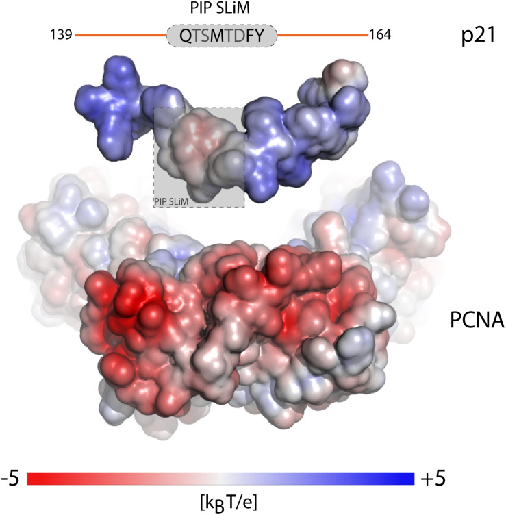
“PCNA has an enormous interactome (Moldovan et al., 2007; Prestel et al., 2019) 1 ), and the APIM motif [KR] (cid:127)(cid:56) 2 [KR] for cytosolic partners (Gilljam et al., 2009; Sebesta et al., 2017).”
BackgroundBoisvert, F., van Koningsbruggen, S., de Navascués, J., & Lamond, A. I. (2007) · Nature Reviews Molecular Cell Biology · doi:10.1038/nrm2184 · Cited by 1,671 · cited 1×
“The nucleolus, a membrane-less compartment, is the site of ribosome biogenesis (Boisvert et al., 2007).”
BackgroundCircadian rhythms from multiple oscillators: lessons from diverse organisms
Bell‐Pedersen, D., Cassone, V. M., Earnest, D. J., Golden, S. S., Hardin, P. E., Thomas, T. L., & Zoran, M. J. (2005) · Nature Reviews Genetics · doi:10.1038/nrg1633 · Cited by 1,523
Molecular architecture of the mammalian circadian clock
Partch, C. L., Green, C. B., & Takahashi, J. S. (2014) · Trends in Cell Biology · doi:10.1016/j.tcb.2013.07.002 · Cited by 1,449 · cited 1×
“Per and Cry dimerize, interact with CLOCK:BMAL1 and inhibit transcriptional activity (Partch et al., 2014) in a negative feedback loop (Gekakis et al.”
BackgroundThe importance of intrinsic disorder for protein phosphorylation
Iakoucheva, L. M. (2004) · Nucleic Acids Research · doi:10.1093/nar/gkh253 · Cited by 1,429
The ensemble nature of allostery
Motlagh, H. N., Wrabl, J. O., Li, J., & Hilser, V. J. (2014) · Nature · doi:10.1038/nature13001 · Cited by 1,247 · cited 1×
“According to this allosteric ensemble model, the flexibility of disorder allows complex allosteric behavior of IDRs in fine-tuning regulatory interactions (Hilser and Thompson, 2007; Motlagh et al., 2014).”
MethodTHE KINETICS OF FORMATION OF NATIVE RIBONUCLEASE DURING OXIDATION OF THE REDUCED POLYPEPTIDE CHAIN
Anfinsen, C. B., Haber, E., Sela, M., & White, F. H. (1961) · Proceedings of the National Academy of Sciences · doi:10.1073/pnas.47.9.1309 · Cited by 1,182 · cited 1×
“…the importance of context in the understanding of proteins was formulated by Christian B. Anfinsen stating “ that the native conformation is determined by the totality of interatomic interactions and hence by the amino acid sequence, in a given environment ” (Anfinsen et al., 1961; Anfinsen, 1973).”
BackgroundValence and patterning of aromatic residues determine the phase behavior of prion-like domains
Martin, E. W., Holehouse, A. S., Peran, I., Farag, M., Incicco, J. J., Bremer, A., Grace, C. R., Soranno, A., Pappu, R. V., & Mittag, T. (2020) · Science · doi:10.1126/science.aaw8653 · Cited by 1,147 · cited 5×
“Sequence properties are an important factor for compaction of disordered chains (Das et al., 2015; Martin et al., 2016, 2020).”
BackgroundInfluentialDas, R. K., & Pappu, R. V. (2013) · Proceedings of the National Academy of Sciences · doi:10.1073/pnas.1304749110 · Cited by 1,008 · cited 1×
“…compositional parameters such as the net charge (Marsh and Forman-kay, 2010; Muller-Spath et al., 2010) or more specifically the distribution of charges (Das and Pappu, 2013), and proline residues (Marsh and Forman-kay, 2010; Martin et al., 2016) as being key determinants for their chain dimensions.”
BackgroundPhase separation in biology; functional organization of a higher order
Mitrea, D. M., & Kriwacki, R. W. (2016) · Cell Communication and Signaling · doi:10.1186/s12964-015-0125-7 · Cited by 727 · cited 1×
“The formation of self-assembled, membrane-less organelles through liquid-liquid phase separation (LLPS) of proteins creates special contexts for IDP-based interactions and partitions with specific functions [for reviews, see Mitrea and Kriwacki (2016), Zaslavsky et al. (2018), Alberti et al. (2019)].”
BackgroundHarmon, T. S., Holehouse, A. S., Rosen, M. K., & Pappu, R. V. (2017) · eLife · doi:10.7554/elife.30294 · Cited by 713
Attributes of short linear motifs
Davey, N. E., Roey, K. V., Weatheritt, R. J., Toedt, G., Uyar, B., Altenberg, B., Budd, A., Diella, F., Dinkel, H., & Gibson, T. J. (2011) · Molecular BioSystems · doi:10.1039/c1mb05231d · Cited by 662 · cited 2×
“Short linear motifs (SLiMs) are more prevalent in IDPs, and the human proteome is estimated to contain > 100.000 – possibly a million – SLiM instances (Davey et al., 2012; Tompa et al., 2014).”
BackgroundInfluentialAlberti, S. (2017) · Current Biology · doi:10.1016/j.cub.2017.08.069 · Cited by 656
Extreme disorder in an ultrahigh-affinity protein complex
Borgia, A., Borgia, M. B., Bugge, K., Kissling, V. M., Heidarsson, P. O., Fernandes, C. B., Sottini, A., Soranno, A., Buholzer, K. J., Nettels, D., et al. (2018) · Nature · doi:10.1038/nature25762 · Cited by 653
Self-citationFlexible nets: disorder and induced fit in the associations of p53 and 14-3-3 with their partners
Oldfield, C. J., Meng, J., Yang, J. Y., Yang, M. Q., Uversky, V. N., & Dunker, A. K. (2008) · BMC Genomics · doi:10.1186/1471-2164-9-s1-s1 · Cited by 608 · cited 1×
“with an intrinsically disordered region (IDR), are key to network fidelity (Dosztányi et al., 2006; Oldfield et al., 2008; Staby et al., 2017; Uversky, 2018), and they exist in ensembles of almost isoenergetic states.”
BackgroundA conserved ER targeting motif in three families of lipid binding proteins and in Opi1p binds VAP
Loewen, C. J. R., Roy, A., & Levine, T. P. (2003) · The EMBO Journal · doi:10.1093/emboj/cdg201 · Cited by 607
Modulation of Intrinsically Disordered Protein Function by Post-translational Modifications
Bah, A., & Forman-Kay, J. D. (2016) · Journal of Biological Chemistry · doi:10.1074/jbc.r115.695056 · Cited by 557
Folding of an intrinsically disordered protein by phosphorylation as a regulatory switch
Bah, A., Vernon, R. M., Siddiqui, Z., Krzeminski, M., Muhandiram, R., Zhao, C. W., Sonenberg, N., Kay, L. E., & Forman-Kay, J. D. (2014) · Nature · doi:10.1038/nature13999 · Cited by 554 · cited 1×
“4EBP2 folds upon phosphorylation of specific sites, and the more folded 4EBP2 becomes, the lower its affinity for eIF4E (Bah et al., 2015).”
BackgroundA Million Peptide Motifs for the Molecular Biologist
Tompa, P., Davey, N. E., Gibson, T. J., & Babu, M. M. (2014) · Molecular Cell · doi:10.1016/j.molcel.2014.05.032 · Cited by 515 · cited 1×
“000 – possibly a million – SLiM instances (Davey et al., 2012; Tompa et al., 2014).”
BackgroundDirect observation of the three regions in α-synuclein that determine its membrane-bound behaviour
Fusco, G., Simone, A. D., Gopinath, T., Vostrikov, V. V., Vendruscolo, M., Dobson, C. M., & Veglia, G. (2014) · Nature Communications · doi:10.1038/ncomms4827 · Cited by 493
Charge interactions can dominate the dimensions of intrinsically disordered proteins
Müller-Späth, S., Soranno, A., Hirschfeld, V., Hofmann, H., Rüegger, S., Reymond, L., Nettels, D., & Schuler, B. (2010) · Proceedings of the National Academy of Sciences · doi:10.1073/pnas.1001743107 · Cited by 491 · cited 1×
“These altogether point toward compositional parameters such as the net charge (Marsh and Formankay, 2010; Muller-Spath et al., 2010) or more specifically the distribution of charges (Das and Pappu, 2013), and proline residues (Marsh and Forman-kay, 2010; Martin et al., 2016) as being key…”
BackgroundRey, G., Cesbron, F., Rougemont, J., Reinke, H., Brunner, M., & Naef, F. (2011) · PLoS Biology · doi:10.1371/journal.pbio.1000595 · Cited by 490
Interactome3D: adding structural details to protein networks
Mosca, R., Céol, A., & Aloy, P. (2013) · Nature Methods · doi:10.1038/nmeth.2289 · Cited by 479
Mitrea, D. M., Cika, J., Guy, C., Ban, D., Banerjee, P. R., Stanley, C., Nourse, A., Deniz, A., & Kriwacki, R. (2016) · eLife · doi:10.7554/elife.13571 · Cited by 453 · cited 3×
“In phase separation, the context of the disordered chain also has implications for interactions as explained by the stickers-and-spacers model of phase separation developed based on studies of RNA-binding proteins with prion-like domains (Wang et al., 2018; Martin et al., 2020).”
BackgroundLocal structural disorder imparts plasticity on linear motifs
Fuxreiter, M., Tompa, P., & Simon, I. (2007) · Bioinformatics · doi:10.1093/bioinformatics/btm035 · Cited by 420
Relating sequence encoded information to form and function of intrinsically disordered proteins
Das, R. K., Ruff, K. M., & Pappu, R. V. (2015) · Current Opinion in Structural Biology · doi:10.1016/j.sbi.2015.03.008 · Cited by 410 · cited 2×
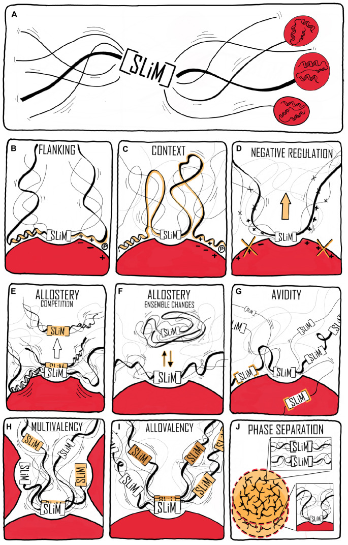
“As most IDPs are polyampholytes (Das et al., 2015), modifications to SLiM flanking regions modulating their charge state (by e.g., phosphorylation) may induce transitions in the conformational ensemble of the disordered chain enabling allosteric regulation of motif accessibility ( Figure 1F ).”
BackgroundIntrinsic disorder as a mechanism to optimize allosteric coupling in proteins
Hilser, V. J., & Thompson, E. B. (2007) · Proceedings of the National Academy of Sciences · doi:10.1073/pnas.0700329104 · Cited by 406
Sequence Determinants of Compaction in Intrinsically Disordered Proteins
Marsh, J. A., & Forman-Kay, J. D. (2010) · Biophysical Journal · doi:10.1016/j.bpj.2010.02.006 · Cited by 382 · cited 2×
“, 2010) or more specifically the distribution of charges (Das and Pappu, 2013), and proline residues (Marsh and Forman-kay, 2010; Martin et al., 2016) as being key determinants for their chain dimensions.”
BackgroundHox protein mutation and macroevolution of the insect body plan
Ronshaugen, M., McGinnis, N., & McGinnis, W. J. (2002) · Nature · doi:10.1038/nature716 · Cited by 367 · cited 1×
“A comparison of five Ubx TF orthologs spanning 540 million years of evolution (Ronshaugen et al., 2002) revealed that the strength of the activation domains changed during evolution, and that the location of the activation domain moved relative to conserved motifs and sub-domain organization (Liu et al.”
BackgroundCrystal Structure of the Heterodimeric CLOCK:BMAL1 Transcriptional Activator Complex
Huang, N. N., Chelliah, Y., Shan, Y., Taylor, C. A., Yoo, S. H., Partch, C. L., Green, C. B., Zhang, H., & Takahashi, J. S. (2012) · Science · doi:10.1126/science.1222804 · Cited by 354
Dynamic equilibrium engagement of a polyvalent ligand with a single-site receptor
Mittag, T., Orlicky, S. M., Choy, W., Tang, X., Lin, H., Sicheri, F., Kay, L. E., Tyers, M., & Forman-Kay, J. D. (2008) · Proceedings of the National Academy of Sciences · doi:10.1073/pnas.0809222105 · Cited by 347 · cited 1×
“The binding is cooperative, with almost no detectable binding until a sixth arbitrary site among ten becomes phosphorylated, producing strong binding (Mittag et al., 2008); a scenario only possible in the context of a flexible, dynamic chain (Locasale, 2008).”
BackgroundDisorder and Sequence Repeats in Hub Proteins and Their Implications for Network Evolution
Dosztányi, Z., Chen, J., Dunker, A. K., Simon, I., & Tompa, P. (2006) · Journal of Proteome Research · doi:10.1021/pr060171o · Cited by 341 · cited 1×
“…whether it is a completely intrinsically disordered protein (IDP) or a protein with an intrinsically disordered region (IDR), are key to network fidelity (Dosztányi et al., 2006; Oldfield et al., 2008; Staby et al., 2017; Uversky, 2018), and they exist in ensembles of almost isoenergetic states.”
BackgroundA structural explanation for the binding of endocytic dileucine motifs by the AP2 complex
Kelly, B. T., McCoy, A. J., Späte, K., Miller, S. E., Evans, P. R., Höning, S., & Owen, D. J. (2008) · Nature · doi:10.1038/nature07422 · Cited by 336
Murphy, S. E., & Levine, T. P. (2016) · Biochimica et Biophysica Acta (BBA) - Molecular and Cell Biology of Lipids · doi:10.1016/j.bbalip.2016.02.009 · Cited by 328
Differential Regulation of Retinoblastoma Protein Function by Specific Cdk Phosphorylation Sites
Knudsen, E. S., & Wang, J. Y. J. (1996) · Journal of Biological Chemistry · doi:10.1074/jbc.271.14.8313 · Cited by 320
Modulation of allostery by protein intrinsic disorder
Ferreon, A. C. M., Ferreon, J. C., Wright, P. E., & Deniz, A. A. (2013) · Nature · doi:10.1038/nature12294 · Cited by 317
Cooperation of GGAs and AP-1 in Packaging MPRs at the Trans-Golgi Network
Doray, B., Ghosh, P., Griffith, J., Geuze, H. J., & Kornfeld, S. (2002) · Science · doi:10.1126/science.1075327 · Cited by 312
Plasticity of an Ultrafast Interaction between Nucleoporins and Nuclear Transport Receptors
Milles, S., Mercadante, D., Aramburu, I. V., Jensen, M. R., Banterle, N., Koehler, C., Tyagi, S., Clarke, J., Shammas, S. L., Blackledge, M. J., et al. (2015) · Cell · doi:10.1016/j.cell.2015.09.047 · Cited by 304
A Conserved Motif Provides Binding Specificity to the PP2A-B56 Phosphatase
Hertz, E. P. T., Kruse, T., Davey, N. E., López-Méndez, B., Sigurðsson, J. O., Montoya, G., Olsen, J. V., & Nilsson, J. (2016) · Molecular Cell · doi:10.1016/j.molcel.2016.06.024 · Cited by 282
Georgieva, E. R., Ramlall, T. F., Borbat, P. P., Freed, J. H., & Eliezer, D. (2008) · Journal of the American Chemical Society · doi:10.1021/ja804517m · Cited by 275 · cited 1×
“A similar behavior was also observed for the IDP α -synuclein (aSN) (Georgieva et al., 2008; Fusco et al., 2014; Cholak et al., 2020).”
BackgroundOptimization of specificity in a cellular protein interaction network by negative selection
Zarrinpar, A., Park, S.-H., & Lim, W. A. (2003) · Nature · doi:10.1038/nature02178 · Cited by 274 · cited 1×
“Similarly, fine-tuning of the interaction of the IDR of the kinase Pbs2 to specifically bind one SH3 domain among a context of many others (Zarrinpar et al., 2003; Kelil et al., 2016) was obtained through evolution.”
BackgroundStructural Basis of FFAT Motif-Mediated ER Targeting
Kaiser, S. E., Brickner, J. H., Reilein, A. R., Fenn, T. D., Walter, P., & Brünger, A. T. (2005) · Structure · doi:10.1016/j.str.2005.04.010 · Cited by 267
Allostery and Intrinsic Disorder Mediate Transcription Regulation by Conditional Cooperativity
Garcia‐Pino, A., Balasubramanian, S., Wyns, L., Gazit, E., Greve, H. D., Magnuson, R. D., Charlier, D., van Nuland, N. A. J., & Loris, R. (2010) · Cell · doi:10.1016/j.cell.2010.05.039 · Cited by 261
Martin, E. W., Holehouse, A. S., Grace, C. R., Hughes, A. C., Pappu, R. V., & Mittag, T. (2016) · Journal of the American Chemical Society · doi:10.1021/jacs.6b10272 · Cited by 248 · cited 2×
“Sequence properties are an important factor for compaction of disordered chains (Das et al., 2015; Martin et al., 2016, 2020).”
BackgroundStructure and Function of the Core Histone N-Termini: More Than Meets the Eye
Hansen, J. C., Tse, C., & Wolffe, A. P. (1998) · Biochemistry · doi:10.1021/bi982409v · Cited by 247 · cited 1×
“Similarly, proteins with high overall positive charge, such as core histones, partially fold when pH increases, affecting DNA binding (Hansen et al., 1998; Munishkina et al., 2004).”
BackgroundShort linear motifs – ex nihilo evolution of protein regulation
Davey, N. E., Cyert, M., & Moses, A. M. (2015) · Cell Communication and Signaling · doi:10.1186/s12964-015-0120-z · Cited by 228 · cited 1×
“For many IDPs, their interactions – and hence their contacts to their partner molecule – are made via short, sequence-embedded motifs of limited information (Sharma et al., 2014; Davey et al., 2015).”
BackgroundStructural Polymorphism and Multifunctionality of Myelin Basic Protein
Harauz, G., Ladizhansky, V., & Boggs, J. M. (2009) · Biochemistry · doi:10.1021/bi901005f · Cited by 216 · cited 1×
“MBP is disordered, but folds into an α -helical structure upon electrostatic interaction with the membrane (Polverini et al., 1999; Harauz et al., 2009), a transition highly regulated by salt and Ca 2 + (Raasakka et al., 2019).”
BackgroundA Proteome-wide Screen for Mammalian SxIP Motif-Containing Microtubule Plus-End Tracking Proteins
Jiang, K., Toedt, G., Montenegro Gouveia, S., Davey, N. E., Hua, S., van der Vaart, B., Grigoriev, I., Larsen, J., Pedersen, L. B., Bezstarosti, K., et al. (2012) · Current Biology · doi:10.1016/j.cub.2012.07.047 · Cited by 209
Structural polymorphism in the N-terminal oligomerization domain of NPM1
Mitrea, D. M., Grace, C. R., Buljan, M., Yun, M., Pytel, N. J., Satumba, J., Nourse, A., Park, C., Babu, M. M., White, S. W., et al. (2014) · Proceedings of the National Academy of Sciences · doi:10.1073/pnas.1321007111 · Cited by 193
The SLC9A-C Mammalian Na + /H + Exchanger Family: Molecules, Mechanisms, and Physiology
Pedersen, S. F., & Counillon, L. (2019) · Physiological Reviews · doi:10.1152/physrev.00028.2018 · Cited by 191
Disorder and residual helicity alter p53-Mdm2 binding affinity and signaling in cells
Borcherds, W. M., Theillet, F., Katzer, A., Finzel, A., Mishall, K. M., Powell, A. T., Wu, H., Manieri, W., Dieterich, C., Selenko, P., et al. (2014) · Nature Chemical Biology · doi:10.1038/nchembio.1668 · Cited by 187
Identification of a novel, widespread, and functionally important PCNA-binding motif
Gilljam, K. M., Feyzi, E., Aas, P. A., de Sousa, M. M. L., Müller, R., Vågbø, C. B., Catterall, T., Liabakk, N., Slupphaug, G., Drabløs, F., et al. (2009) · The Journal of Cell Biology · doi:10.1083/jcb.200903138 · Cited by 179 · cited 2×
“PCNA has an enormous interactome (Moldovan et al., 2007; Prestel et al., 2019) 1 ), and the APIM motif [KR] (cid:127)(cid:56) 2 [KR] for cytosolic partners (Gilljam et al., 2009; Sebesta et al., 2017).”
BackgroundHavens, C. G., & Walter, J. C. (2009) · Molecular Cell · doi:10.1016/j.molcel.2009.05.012 · Cited by 178
The Underappreciated Role of Allostery in the Cellular Network
Nussinov, R., Tsai, C., & Ma, B. (2013) · Annual Review of Biophysics · doi:10.1146/annurev-biophys-083012-130257 · Cited by 176 · cited 1×
“Proteins with energetically frustrated IDRs can mediate allosteric regulation (Garcia-Pino et al., 2010; Ferreon et al., 2013), which play a central role in orchestrating cellular signaling networks (Nussinov et al., 2013).”
BackgroundKDEL-Cargo Regulates Interactions between Proteins Involved in COPI Vesicle Traffic
Majoul, I. V., Straub, M., Hell, S. W., Duden, R., & Söling, H. (2001) · Developmental Cell · doi:10.1016/s1534-5807(01)00004-1 · Cited by 175 · cited 1×
“Retrieval of many endoplasmic reticulum (ER)-resident proteins from post-ER compartments depends on a C-terminal [HKR]DEL motif (Majoul et al., 2001).”
BackgroundStructural basis for acidic-cluster-dileucine sorting-signal recognition by VHS domains
Misra, S., Puertollano, R., Kato, Y., Bonifacino, J. S., & Hurley, J. H. (2002) · Nature · doi:10.1038/415933a · Cited by 170 · cited 1×
“Thus, binding to the GGA proteins requires a precise spacing between the di-Leu and the free C-terminus (Misra et al., 2002) ( Table 1 ).”
BackgroundLowe, R. D., Pountney, D. L., Jensen, P. H., Gai, W., & Voelcker, N. H. (2004) · Protein Science · doi:10.1110/ps.04879704 · Cited by 166
Staller, M. V., Holehouse, A. S., Swain-Lenz, D., Das, R. K., Pappu, R. V., & Cohen, B. A. (2018) · Cell Systems · doi:10.1016/j.cels.2018.01.015 · Cited by 165 · cited 1×
“However, this motif is part of a region with several hydrophobic patches, and GCN4 binds the Mediator co-activator component MED15 via multiple, low-affinity interactions, which additively contribute to activation activity (Drysdale et al., 1995; Warfield et al., 2014; Staller et al., 2018).”
BackgroundContextual Specificity in Peptide-Mediated Protein Interactions
Stein, A., & Aloy, P. (2008) · PLoS ONE · doi:10.1371/journal.pone.0002524 · Cited by 164
Helical Propensity in an Intrinsically Disordered Protein Accelerates Ligand Binding
Iešmantavičius, V., Dogan, J., Jemth, P., Teilum, K., & Kjærgaard, M. (2014) · Angewandte Chemie International Edition · doi:10.1002/anie.201307712 · Cited by 162
Hypersensitive termination of the hypoxic response by a disordered protein switch
Berlow, R. B., Dyson, H. J., & Wright, P. E. (2017) · Nature · doi:10.1038/nature21705 · Cited by 161 · cited 11×
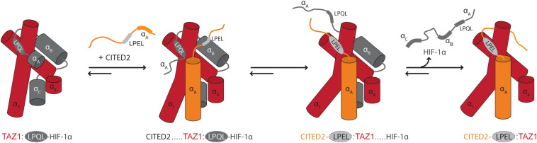
“The dynamics particularly of the N-terminal region of bound HIF-1 α was suggested to allow CITED2 to access a key surface on TAZ1, promoting ternary complex formation and eventually displacement of HIF-1 α .”
BackgroundMethodInfluentialNatively Unfolded Human Prothymosin α Adopts Partially Folded Collapsed Conformation at Acidic pH
Uversky, V. N., Gillespie, J. R., Millett, I. S., Khodyakova, A. V., Vasiliev, A. M., Chernovskaya, T. V., Vasilenko, R. N., Kozlovskaya, G. D., Dolgikh, D. A., Fink, A. L., et al. (1999) · Biochemistry · doi:10.1021/bi990752+ · Cited by 154 · cited 1×
“Prothymosin-α (ProTα), a highly acidic, nuclear IDP, is expanded at neutral pH, but compacts as the pH decreases (Uversky et al., 1999).”
BackgroundDrysdale, C. M., Dueñas, E., Jackson, B. M., Reusser, U., Braus, G. H., & Hinnebusch, A. G. (1995) · Molecular and Cellular Biology · doi:10.1128/mcb.15.3.1220 · Cited by 154
The molecular mechanism of nuclear transport revealed by atomic-scale measurements
Hough, L. E., Dutta, K., Sparks, S., Temel, D. B., Kamal, A., Tetenbaum-Novatt, J. E., Rout, M. P., & Cowburn, D. (2015) · eLife · doi:10.7554/elife.10027 · Cited by 148 · cited 1×
“…through, e.g., the specific expression of isoforms at different developmental stages that differ in their ability to interact with partners, as shown for the Ubx transcription factors (TFs), which are key players in Drosophila embryonic development (López et al., 1996; Hsiao et al., 2014).”
Sørensen, C. S., & Kjærgaard, M. (2019) · Proceedings of the National Academy of Sciences · doi:10.1073/pnas.1904813116 · Cited by 148 · cited 1×
“Interestingly, while hydrophobicity has been suggested to play a marginal role in IDR compaction (Marsh and Forman-kay, 2010), a correlation between the fraction and patterning of aromatic residues and chain compaction was recently established (Sørensen and Kjaergaard, 2019; Martin et al., 2020).”
BackgroundRTX Calcium Binding Motifs Are Intrinsically Disordered in the Absence of Calcium
Chenal, A., Guijarro, J. I., Raynal, B. D. E., Delepierre, M., & Ladant, D. (2008) · Journal of Biological Chemistry · doi:10.1074/jbc.m807312200 · Cited by 144 · cited 1×
“For the disordered region of the adenylate cyclase toxin (Chenal et al., 2009), Ca 2 + binding to > 40 repeats in toxin motifs (RTX) GGxGxDx[ (cid:127) , (cid:57) ] led to folding into a β -roll structure (Sotomayor Pérez et al., 2010) central to its secretion (O’Brien et al., 2018) ( Table 1 ).”
BackgroundStructural basis for pH-dependent retrieval of ER proteins from the Golgi by the KDEL receptor
Bräuer, P., Parker, J. L., Gerondopoulos, A., Zimmermann, I., Seeger, M. A., Barr, F. A., & Newstead, S. L. (2019) · Science · doi:10.1126/science.aaw2859 · Cited by 143
Opposing effects of Elk-1 multisite phosphorylation shape its response to ERK activation
Mylona, A., Theillet, F., Foster, C., Cheng, T. M. K., Miralles, F., Bates, P. A., Selenko, P., & Treisman, R. (2016) · Science · doi:10.1126/science.aad1872 · Cited by 138 · cited 1×
“…have been observed, which may increase or decrease transient helicity in the vicinity of binding motifs, which in turn contributes to selection and deselection of binding partners as well as initiating degradation (Bah and Forman-Kay, 2016; Mylona et al., 2016; Hendus-Altenburger et al., 2017).”
BackgroundDescribing sequence–ensemble relationships for intrinsically disordered proteins
Mao, A. H., Lyle, N., & Pappu, R. V. (2013) · Biochemical Journal · doi:10.1042/bj20121346 · Cited by 135 · cited 1×
“To date, various studies have assessed the sequence-encoded conformational preferences of IDPs, using concepts from polymer physics reviewed in Mao et al. (2013) and by combining several biophysical techniques with molecular simulations.”
BackgroundEukaryotic transcription factors: paradigms of protein intrinsic disorder
Staby, L., O’Shea, C., Willemoës, M., Theisen, F. F., Kragelund, B. B., & Skriver, K. (2017) · Biochemical Journal · doi:10.1042/bcj20160631 · Cited by 134 · cited 2×
“The activation activity of GCN4 depends on the SLiM 88W88LF (Table 1) (Warfield et al., 2014; Staby et al., 2017).”
BackgroundMutations in Disordered Regions Can Cause Disease by Creating Dileucine Motifs
Meyer, K., Kirchner, M., Uyar, B., Cheng, J.-Y., Russo, G., Hernandez-Miranda, L. R., Szymborska, A., Zauber, H., Rudolph, I.-M., Willnow, T. E., et al. (2018) · Cell · doi:10.1016/j.cell.2018.08.019 · Cited by 132
Intrinsic Disorder, Protein–Protein Interactions, and Disease
Uversky, V. N. (2017) · Advances in protein chemistry and structural biology · doi:10.1016/bs.apcsb.2017.06.005 · Cited by 120 · cited 2×
“with an intrinsically disordered region (IDR), are key to network fidelity (Dosztányi et al., 2006; Oldfield et al., 2008; Staby et al., 2017; Uversky, 2018), and they exist in ensembles of almost isoenergetic states.”
BackgroundCryptic sequence features within the disordered protein p27 Kip1 regulate cell cycle signaling
Das, R. K., Huang, Y., Phillips, A. H., Kriwacki, R. W., & Pappu, R. V. (2016) · Proceedings of the National Academy of Sciences · doi:10.1073/pnas.1516277113 · Cited by 119
Krystkowiak, I., & Davey, N. E. (2017) · Nucleic Acids Res. · doi:10.1093/nar/gkx238 · Cited by 116
Predicting the targeting of tail-anchored proteins to subcellular compartments in mammalian cells
Costello, J. L., Castro, I. G., Camões, F., Schrader, T. A., McNeall, D., Yang, J., Giannopoulou, E., Gomes, S. R. A. M., Pogenberg, V., Bonekamp, N. A., et al. (2017) · Journal of Cell Science · doi:10.1242/jcs.200204 · Cited by 115 · cited 1×
“Proteins with FFAT motifs (Loewen et al., 2003) communicate between the ER and other compartments by bridging (Murphy and Levine, 2016; Costello et al., 2017; Slee and Levine, 2019).”
BackgroundThe PCNA interaction motifs revisited: thinking outside the PIP-box
Prestel, A., Wichmann, N., Martins, J. M. D. S., Marabini, R., Kassem, N., Broendum, S. S., Otterlei, M., Nielsen, O., Willemoës, M., Ploug, M., et al. (2019) · Cellular and Molecular Life Sciences · doi:10.1007/s00018-019-03150-0 · Cited by 112 · cited 5×
“PCNA has an enormous interactome (Moldovan et al., 2007; Prestel et al., 2019) and common to its binding partners is the Qxx8xx[FHT][FHY] PIP-box motif of which variations are known, including the PIP-degron for degradation QTD8xx[FHT][FHY]-X4-[KR] (Havens and Walter, 2009) (Table 1), and the APIM motif [KR]82[KR] fo…”
BackgroundInfluentialInteraction of the TAZ1 Domain of the CREB-Binding Protein with the Activation Domain of CITED2
Guzman, R. N. D., Martinez‐Yamout, M. A., Dyson, H. J., & Wright, P. E. (2004) · Journal of Biological Chemistry · doi:10.1074/jbc.m310348200 · Cited by 109 · cited 1×
“The two TFs use the core motif LP [ Q/E ] L for binding to the same site on TAZ1 ( Table 1 ), and bind with close to identical affinities (De Guzman et al., 2004; Berlow et al., 2017).”
BackgroundAffinity and specificity of motif-based protein–protein interactions
Ivarsson, Y., & Jemth, P. (2018) · Current Opinion in Structural Biology · doi:10.1016/j.sbi.2018.09.009 · Cited by 102 · cited 1×
“The SLiM is the central anchoring site for many IDP interactions and a recent review discusses the molecular details of SLiM-based affinity and specificity (Ivarsson and Jemth, 2019).”
BackgroundComputational identification of MoRFs in protein sequences
Malhis, N., & Gsponer, J. A. (2015) · Bioinformatics · doi:10.1093/bioinformatics/btv060 · Cited by 101
Organellar Na+/H+ Exchangers: Novel Players in Organelle pH Regulation and Their Emerging Functions
Ohgaki, R., van IJzendoorn, S. C. D., Matsushita, M., Hoekstra, D., & Kanazawa, H. (2010) · Biochemistry · doi:10.1021/bi101082e · Cited by 101 · cited 1×
“…are brain specific, e.g., NHE6 and NHE9 (Schwede et al., 2014; Zhao et al., 2016), and some are ubiquitously located in the plasma membrane, such as NHE1, while other isoforms are localized in specific organellar membranes (Ohgaki et al., 2011; Prasad and Rao, 2015; Pedersen and Counillon, 2019).”
BackgroundParkinson's Disease-associated α-Synuclein Is a Calmodulin Substrate
Martinez, J., Moeller, I., Erdjument‐Bromage, H., Tempst, P., & Lauring, B. P. (2003) · Journal of Biological Chemistry · doi:10.1074/jbc.m209020200 · Cited by 100 · cited 1×
“For aSN, this meant that sub-states of the aggregation-prone region were relieved of negative coupling, which the authors suggested may explain how calmodulin facilitates aggregation of aSN (Martinez et al., 2003).”
BackgroundLi, J., White, J. T., Saavedra, H. G., Wrabl, J. O., Motlagh, H. N., Liu, K., Sowers, J. R., Schroer, T. A., Thompson, E. B., & Hilser, V. J. (2017) · eLife · doi:10.7554/elife.30688 · Cited by 98 · cited 2×
“The importance of such energetic frustration in IDP interactions has also been demonstrated from work on the glucocorticoid receptor (GR), where the disordered domain allosterically controls function by principles of energetic frustration (Li et al., 2018).”
BackgroundWarfield, L., Tuttle, L. M., Pacheco, D., Klevit, R. E., & Hahn, S. (2014) · Proceedings of the National Academy of Sciences · doi:10.1073/pnas.1412088111 · Cited by 94 · cited 2×
“The activation activity of GCN4 depends on the SLiM W LF ( Table 1 ) (Warfield et al., 2014; Staby et al., 2017).”
BackgroundMethodFuruita, K., Jee, J., Fukada, H., Mishima, M., & Kojima, C. (2010) · Journal of Biological Chemistry · doi:10.1074/jbc.m109.082602 · Cited by 91 · cited 2×
“The core of the FFAT motif, EFFDAxE, found in, e.g., oxysterol-binding protein (OSBP) (Furuita et al., 2010), is an extended region of seven residues ( Table 1 ), the second and fifth of which bind into pockets in the integral ER membrane protein VAMP-associated protein (VAP) (Kaiser et al., 2005).”
BackgroundUyar, B., Weatheritt, R. J., Dinkel, H., Davey, N. E., & Gibson, T. J. (2014) · Molecular BioSystems · doi:10.1039/c4mb00290c · Cited by 90 · cited 2×
“A proteome-wide study found that 22% of all human disease mutations locate to disordered regions (Uyar et al., 2014).”
BackgroundMolecular Recognition by Templated Folding of an Intrinsically Disordered Protein
Toto, A., Camilloni, C., Giri, R., Brunori, M., Vendruscolo, M., & Gianni, S. (2016) · Scientific Reports · doi:10.1038/srep21994 · Cited by 90 · cited 1×
“Conformational heterogeneity, characteristic of many ID-based interactions (Fuxreiter, 2019), is also regulated by residues outside the SLiM-binding site, where they form transient heterogeneous contacts, which may facilitate partner-templated coupled folding and binding (Toto et al., 2016).”
BackgroundEmerging roles of Na+/H+ exchangers in epilepsy and developmental brain disorders
Zhao, H., Carney, K. E., Falgoust, L., Pan, J. W., Sun, D., & Zhang, Z. (2016) · Progress in Neurobiology · doi:10.1016/j.pneurobio.2016.02.002 · Cited by 89
Understanding Pre-Structured Motifs (PreSMos) in Intrinsically Unfolded Proteins
Lee, S., Kim, D.-H., Han, J. J., Cha, E., Lim, J., Cho, Y., Lee, C., & Han, K. (2012) · Current Protein and Peptide Science · doi:10.2174/138920312799277974 · Cited by 88 · cited 1×
“, 2018) or pre-structured motifs (PreSMos) (Lee et al., 2012), the flanking regions can modulate the structural context of the motif and hence interactions.”
BackgroundZheleva, D. I., Zhelev, N., Fischer, P. M., Duff, S. V., Warbrick, E., Blake, D., & Lane, D. P. (2000) · Biochemistry · doi:10.1021/bi992498r · Cited by 86
Long-Range Correlated Dynamics in Intrinsically Disordered Proteins
Parigi, G., Rezaei‐Ghaleh, N., Giachetti, A., Becker, S., Fernández, C. O., Blackledge, M. J., Griesinger, C., Zweckstetter, M., & Luchinat, C. (2014) · Journal of the American Chemical Society · doi:10.1021/ja506820r · Cited by 83 · cited 2×
“Using FFC relaxometry, it was shown for four IDPs that slow reorientations on the several nanosecond time scale occurred in the chain, without specific residue interactions, but rather based on frictionmediated coupling (Parigi et al., 2014).”
MethodJones, R. E., Wegrzyn, R. J., Patrick, D. R., Balishin, N. L., Vuocolo, G. A., Riemen, M. W., Defeo-Jones, D., Garsky, V. M., Heimbrook, D. C., & Oliff, A. I. (1990) · Journal of Biological Chemistry · doi:10.1016/s0021-9258(19)38226-2 · Cited by 82
Fold or not to fold upon binding — does it really matter?
Fuxreiter, M. (2018) · Current Opinion in Structural Biology · doi:10.1016/j.sbi.2018.09.008 · Cited by 79
Conformational Prerequisites for Formation of Amyloid Fibrils from Histones
Munishkina, L. A., Fink, A. L., & Uversky, V. N. (2004) · Journal of Molecular Biology · doi:10.1016/j.jmb.2004.06.094 · Cited by 77 · cited 1×
“Similarly, proteins with high overall positive charge, such as core histones, partially fold when pH increases, affecting DNA binding (Hansen et al., 1998; Munishkina et al., 2004).”
BackgroundPrasad, H., & Rao, R. (2015) · Journal of Biological Chemistry · doi:10.1074/jbc.m114.602219 · Cited by 76 · cited 1×
“, 2016), and some are ubiquitously located in the plasma membrane, such as NHE1, while other isoforms are localized in specific organellar membranes (Ohgaki et al., 2011; Prasad and Rao, 2015; Pedersen and Counillon, 2019).”
BackgroundRapali, P., Radnai, L., Süveges, D., Harmat, V., Tölgyesi, F., Wahlgren, W. Y., Katona, G., Nyitray, L., & Pál, G. (2011) · PLoS ONE · doi:10.1371/journal.pone.0018818 · Cited by 75
Mapping, modeling, and characterization of protein–protein interactions on a proteomic scale
Cafarelli, T. M., Desbuleux, A., Wang, Y., Choi, S. G., Ridder, D. D., & Vidal, M. (2017) · Current Opinion in Structural Biology · doi:10.1016/j.sbi.2017.05.003 · Cited by 75
Polymer effects modulate binding affinities in disordered proteins
Vancraenenbroeck, R., Harel, Y. S., Zheng, W., & Hofmann, H. (2019) · Proceedings of the National Academy of Sciences · doi:10.1073/pnas.1904997116 · Cited by 75 · cited 1×
“However, ions not only affect structure and conformational properties via distinct binding, but their presence can also lead to changes in the ensemble, as shown for five different IDPs, which all expanded due to an increase in salt concentration (Vancraenenbroeck et al., 2019).”
BackgroundWicky, B., Shammas, S. L., & Clarke, J. (2017) · Proceedings of the National Academy of Sciences of the United States of America · doi:10.1073/pnas.1705105114 · Cited by 73 · cited 1×
“Salts also tune disorder-based affinities and binding kinetics, as demonstrated for two different IDPs (Wicky et al., 2017), where the observed effects were dependent on the specific ions and not simply correlated to the ionic strength.”
BackgroundStructural Dynamics and Regulation of the Mammalian SLC9A Family of Na+/H+ Exchangers
Hendus-Altenburger, R., Kragelund, B. B., & Pedersen, S. F. (2014) · Current topics in membranes · doi:10.1016/b978-0-12-800223-0.00002-5 · Cited by 72 · cited 1×
“…and the nine isoforms have similar membrane transport functions, but have IDRs of different lengths with varying SLiM content and sequence (Nørholm et al., 2011; Hendus-Altenburger et al., 2014), suggesting that the mechanisms and regulations underlying their function in the membrane, differ.”
BackgroundA Slow Conformational Switch in the BMAL1 Transactivation Domain Modulates Circadian Rhythms
Gustafson, C. L., Parsley, N. C., Asimgil, H., Lee, H., Ahlbach, C., Michael, A. K., Xu, H., Williams, O. L., Davis, T. L., Liu, A. C., et al. (2017) · Molecular Cell · doi:10.1016/j.molcel.2017.04.011 · Cited by 72 · cited 2×
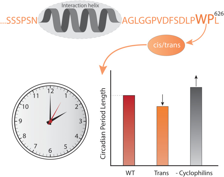
“Locking the switch into the trans conformation results in a shorter circadian period compared to the wild-type, while inhibiting cyclophilins, which accelerate the interconversion between isomers, lengthens the circadian period in a dose-dependent manner (Gustafson et al., 2017).”
BackgroundSotomayor-Pérez, A.-C., Karst, J. C., Davi, M., Guijarro, J. I., Ladant, D., & Chenal, A. (2010) · Journal of Molecular Biology · doi:10.1016/j.jmb.2010.01.031 · Cited by 68
Conformation of bovine myelin basic protein purified with bound lipids
Polverini, E., Fasano, A., Zito, F. A., Riccio, P., & CAVATORTA, P. (1999) · European Biophysics Journal · doi:10.1007/s002490050218 · Cited by 66 · cited 1×
“MBP is disordered, but folds into an α -helical structure upon electrostatic interaction with the membrane (Polverini et al., 1999; Harauz et al., 2009), a transition highly regulated by salt and Ca 2 + (Raasakka et al., 2019).”
BackgroundO’Shea, C., Staby, L., Bendsen, S. K., Tidemand, F. G., Redsted, A., Willemoës, M., Kragelund, B. B., & Skriver, K. (2016) · Journal of Biological Chemistry · doi:10.1074/jbc.m116.753426 · Cited by 65
Genes for endosomal NHE6 and NHE9 are misregulated in autism brains
Schwede, M., Garbett, K. A., Mirnics, K., Geschwind, D. H., & Morrow, E. M. (2013) · Molecular Psychiatry · doi:10.1038/mp.2013.28 · Cited by 61
Nørholm, A., Hendus-Altenburger, R., Bjerre, G., Kjærgaard, M., Pedersen, S. F., & Kragelund, B. B. (2011) · Biochemistry · doi:10.1021/bi1019989 · Cited by 61
Yi, S., Boys, B. L., Brickenden, A. M., Konermann, L., & Choy, W. (2007) · Biochemistry · doi:10.1021/bi7014822 · Cited by 59
Structural insights into the function of ZRANB3 in replication stress response
Šebesta, M., Cooper, C. D. O., Ariza, A., Carnie, C. J., & Ahel, D. (2017) · Nature Communications · doi:10.1038/ncomms15847 · Cited by 58
Multivalency regulates activity in an intrinsically disordered transcription factor
Clark, S. J., Myers, J. B., King, A., Fiala, R., Nováček, J., Pearce, F. G., Heierhorst, J., Reichow, S., & Barbar, E. J. (2018) · eLife · doi:10.7554/elife.36258 · Cited by 56 · cited 6×
“Cooperativity driven by flanking regions is elegantly demonstrated by the IDRs constituting the activation domain of the zinc finger TF ataxia telangiectasia mutated substrate Chk2-interacting Zn 2 + -finger protein (ASCIZ) (Clark et al., 2018).”
BackgroundInfluentialSharma, R., Bayarjargal, M., Tsunoda, T., Patil, A., & Sharma, A. K. (2017) · Journal of Theoretical Biology · doi:10.1016/j.jtbi.2017.10.015 · Cited by 55 · cited 1×
“Finally, when SLiMs are placed in pre-structured contexts, defined as molecular recognition features (MoRFs) (Malhis and Gsponer, 2015; Sharma et al., 2018) or pre-structured motifs (PreSMos) (Lee et al., 2012), the flanking regions can modulate the structural context of the motif and hence…”
BackgroundThe solvent side of proteinaceous membrane-less organelles in light of aqueous two-phase systems
Zaslavsky, B. Y., Ferreira, L. A., Darling, A. L., & Uversky, V. N. (2018) · International Journal of Biological Macromolecules · doi:10.1016/j.ijbiomac.2018.06.030 · Cited by 55
Slee, J. A., & Levine, T. P. (2019) · Contact · doi:10.1177/2515256419883136 · Cited by 54 · cited 1×
“Proteins with FFAT motifs (Loewen et al., 2003) communicate between the ER and other compartments by bridging (Murphy and Levine, 2016; Costello et al., 2017; Slee and Levine, 2019).”
BackgroundElucidation of the binding preferences of peptide recognition modules: SH3 and PDZ domains
Teyra, J., Sidhu, S. S., & Kim, P. M. (2012) · FEBS Letters · doi:10.1016/j.febslet.2012.05.043 · Cited by 50
Random coil chemical shifts for serine, threonine and tyrosine phosphorylation over a broad pH range
Hendus-Altenburger, R., Fernandes, C. B., Bugge, K., Kunze, M. B. A., Boomsma, W., & Kragelund, B. B. (2019) · Journal of Biomolecular NMR · doi:10.1007/s10858-019-00283-z · Cited by 50
Li, J., Motlagh, H. N., Chakuroff, C., Thompson, E. B., & Hilser, V. J. (2012) · Journal of Biological Chemistry · doi:10.1074/jbc.m112.355651 · Cited by 48 · cited 1×
“For GR, signaling is tuned by changing the length of the intrinsically disordered context through translational isoforms, resulting in modulation of the degree of energetic frustration (Li et al., 2012, 2018).”
BackgroundMajava, V., Petoukhov, M. V., Hayashi, N., Pirilä, P., Svergun, D. I., & Kursula, P. (2008) · BMC Structural Biology · doi:10.1186/1472-6807-8-10 · Cited by 46 · cited 1×
“Another example is the highly positively charged myelin basic protein (MBP), essential for the formation and stability of the myelin sheath in the central nervous system (Majava et al., 2008; De Avila et al., 2014).”
BackgroundXiang, W., Menges, S., Schlachetzki, J. C. M., Meixner, H., Hoffmann, A.-C., Schlötzer-Schrehardt, U. M., Becker, C., Winkler, J., & Klucken, J. (2015) · Molecular Neurodegeneration · doi:10.1186/s13024-015-0004-0 · Cited by 44 · cited 1×
“Some disease-related mutations are SLiM-conserving, leaving affinity unperturbed, but with mutations located in the context resulting in altered specificity, cross-reactivity, and self-association as in the case of some neurodegenerative diseases (Uyar et al., 2014; Xiang et al., 2015).”
BackgroundHendus-Altenburger, R., Lambrughi, M., Terkelsen, T., Pedersen, S. F., Papaleo, E., Lindorff‐Larsen, K., & Kragelund, B. B. (2017) · Cellular Signalling · doi:10.1016/j.cellsig.2017.05.015 · Cited by 44
Avidity within the N‐terminal anchor drives α‐synuclein membrane interaction and insertion
Cholak, E., Bugge, K., Khondker, A., Gauger, K., Pedraz‐Cuesta, E., Pedersen, M. E., Bucciarelli, S., Vestergaard, B., Pedersen, S. F., Rheinstadter, M. C., et al. (2020) · The FASEB Journal · doi:10.1096/fj.202000107r · Cited by 44
Multivalent IDP assemblies: Unique properties of LC8‐associated, IDP duplex scaffolds
Clark, S. A., Jespersen, N. E., Woodward, C. K., & Barbar, E. J. (2015) · FEBS Letters · doi:10.1016/j.febslet.2015.07.032 · Cited by 43
Berlow, R. B., Martinez‐Yamout, M. A., Dyson, H. J., & Wright, P. E. (2019) · Biochemistry · doi:10.1021/acs.biochem.8b01290 · Cited by 43
Evolution of domain–peptide interactions to coadapt specificity and affinity to functional diversity
Kelil, A., Levy, E. D., & Michnick, S. W. (2016) · Proceedings of the National Academy of Sciences · doi:10.1073/pnas.1518469113 · Cited by 40
Palópoli, N., Foutel, N. S. G., Gibson, T. J., & Chemes, L. B. (2018) · Protein Engineering Design and Selection · doi:10.1093/protein/gzx068 · Cited by 40 · cited 3×
“Here, negative charges in the flanking regions act as affinity and specificity modulators (Figure 1B) (Palopoli et al., 2018), conversely regulated by introduction of negative charge from phosphorylation of binding regions on the Rbs (Knudsen and Wang, 1996).”
BackgroundD-cycloserine uses an active transport mechanism in the human intestinal cell line Caco 2
Ranaldi, G., Islam, K., & Sambuy, Y. (1994) · Antimicrobial Agents and Chemotherapy · doi:10.1128/aac.38.6.1239 · Cited by 39 · cited 5×
“The multivalency of ASCIZ and Chia allows both positive and negative cooperativity in its interaction with LC8.”
BackgroundInfluentialBeyond KDEL: The Role of Positions 5 and 6 in Determining ER Localization
Alanen, H. I., Raykhel, I. B., Luukas, M. J., Salo, K. E. H., & Ruddock, L. W. (2011) · Journal of Molecular Biology · doi:10.1016/j.jmb.2011.03.070 · Cited by 35 · cited 2×
“However, the flanking regions may also have a highly hydrophobic character, as for the flanks of the [HKR]DEL SLiM (Alanen et al., 2011).”
BackgroundBugge, K., Staby, L., Kemplen, K. R., O’Shea, C., Bendsen, S. K., Jensen, M. K., Olsen, J. G., Skriver, K., & Kragelund, B. B. (2018) · Structure · doi:10.1016/j.str.2018.03.013 · Cited by 35 · cited 2×
“For the plant TFs dehydration response element binding proteins 2A (DREB2A) and ANAC013, which both use the SLiM for binding to the cellular hub protein Radical Induced Cell Death1 (Bugge et al., 2018), disordered regions surrounding the binding motif exert positive and negative allosteric effects on binding, respectiv…”
BackgroundCragnell, C., Staby, L., Lenton, S., Kragelund, B. B., & Skepö, M. (2019) · Biomolecules · doi:10.3390/biom9050168 · Cited by 35 · cited 2×
“Chain compaction may further be affected by metal ions, as observed for Zn 2 + binding to ProT α , and to histatin, a small peptide of the mouth, here promoting formation of higher order structures (Cragnell et al., 2019).”
BackgroundLee, C., Kalmár, L., Xue, B., Tompa, P., Daughdrill, G. W., Uversky, V. N., & Han, K. (2013) · Biochimica et Biophysica Acta (BBA) - General Subjects · doi:10.1016/j.bbagen.2013.10.042 · Cited by 34 · cited 1×
“, been shown that when a proline was mutated in an N-terminal flanking region, helicity was significantly decreased, whereas when mutated in the C-terminal flanking region, the helical content increased (Lee et al., 2014).”
BackgroundThe Anchored Flexibility Model in LC8 Motif Recognition: Insights from the Chica Complex
Clark, S. A., Nyarko, A., Löhr, F., Karplus, P. A., & Barbar, E. J. (2015) · Biochemistry · doi:10.1021/acs.biochem.5b01099 · Cited by 31
Kurzbach, D., Vanas, A., Flamm, A. G., Tarnoczi, N., Kontaxis, G., Maltar‐Strmečki, N., Widder, K., Hinderberger, D., & Konrat, R. (2015) · Physical Chemistry Chemical Physics · doi:10.1039/c5cp04858c · Cited by 30
Mei, M., Zhai, C., Li, X., Zhou, Y., Peng, W., Ma, L., Wang, Q., Iverson, B. L., Zhang, G., & Li, Y. (2017) · Journal of Biological Chemistry · doi:10.1074/jbc.m117.812107 · Cited by 30
O'Brien, D. P., Pérez, A. C. S., Karst, J. C., Cannella, S. E., Enguéné, V. Y. N., Hessel, A., Raoux‐Barbot, D., Voegele, A., Subrini, O., Davi, M., et al. (2018) · Toxicon · doi:10.1016/j.toxicon.2018.01.007 · Cited by 26
Beier, A., Schwarz, T. C., Kurzbach, D., Platzer, G., Tribuzio, F., & Konrat, R. (2018) · Journal of Molecular Biology · doi:10.1016/j.jmb.2018.04.035 · Cited by 26 · cited 3×
“Both proteins exhibited both correlated, anti-correlated and uncorrelated long-range chain fluctuations (Beier et al., 2018), with two residues being correlated if they display concerted motions or undergo simultaneous transitions between different conformational states, anti-correlated if they have…”
BackgroundMethodInfluentialIntrinsically Disordered Proteins
Kragelund, B. B., & Skriver, K. (2020) · Methods in molecular biology · doi:10.1007/978-1-0716-0524-0 · Cited by 25
Kurzbach, D., Beier, A., Vanas, A., Flamm, A. G., Platzer, G., Schwarz, T. C., & Konrat, R. (2017) · Physical Chemistry Chemical Physics · doi:10.1039/c7cp00430c · Cited by 24 · cited 1×
“Providing further details, a method based on PRE NMR, termed paramagnetic relaxation interference (PRI), allowed detection of correlated motions through covariance analysis of the effect of adding two paramagnetic labels to the same chain (Kurzbach et al., 2016, 2017).”
MethodChristensen, L. F., Staby, L., Bugge, K., O’Shea, C., Kragelund, B. B., & Skriver, K. (2019) · Scientific Reports · doi:10.1038/s41598-019-55385-3 · Cited by 24
Hsiao, H., Gonzalez, K. L., Catanese, D. J., Jordy, K. E., Matthews, K. S., & Bondos, S. E. (2014) · PLoS ONE · doi:10.1371/journal.pone.0108217 · Cited by 21
Noval, M. G., Gallo, M. L., Perrone, S., Salvay, A. G., Chemes, L. B., & Gay, G. D. P. (2013) · PLoS ONE · doi:10.1371/journal.pone.0072760 · Cited by 20 · cited 1×
“A similar effect was seen for pocket proteins of the retinoblastoma protein (Rb) family, that bind the SLiM LxCxE, present in host and viral interactors of the Rb family (Jones et al., 1990; Noval et al., 2013).”
BackgroundSharma, A. K., Dehzangi, A., Lyons, J., Imoto, S., Miyano, S., Nakai, K., & Patil, A. (2014) · PLoS ONE · doi:10.1371/journal.pone.0089890 · Cited by 20 · cited 2×
“For many IDPs, their interactions – and hence their contacts to their partner molecule – are made via short, sequence-embedded motifs of limited information (Sharma et al., 2014; Davey et al., 2015).”
BackgroundGorelik, M., & Davidson, A. R. (2012) · Journal of Biological Chemistry · doi:10.1074/jbc.m111.330753 · Cited by 20
Allovalency revisited: An analysis of multisite phosphorylation and substrate rebinding
Locasale, J. W. (2008) · The Journal of Chemical Physics · doi:10.1063/1.2841124 · Cited by 19 · cited 2×
“The binding is cooperative, with almost no detectable binding until a sixth arbitrary site among ten becomes phosphorylated, producing strong binding (Mittag et al., 2008); a scenario only possible in the context of a flexible, dynamic chain (Locasale, 2008).”
BackgroundMapping multivalency in the CLIP-170–EB1 microtubule plus-end complex
Chen, Y., Wang, P., & Slep, K. C. (2018) · Journal of Biological Chemistry · doi:10.1074/jbc.ra118.006125 · Cited by 19
Raasakka, A., Jones, N. C., Hoffmann, S. V., & Kursula, P. (2019) · Biochemical and Biophysical Research Communications · doi:10.1016/j.bbrc.2019.02.025 · Cited by 19 · cited 1×
“, 2009), a transition highly regulated by salt and Ca2+ (Raasakka et al., 2019).”
BackgroundL�pez, A. J., Artero, R. D., & P�rez-Alonso, M. (1996) · Development Genes and Evolution · doi:10.1007/bf00377226 · Cited by 17
Avila, M. D., Vassall, K. A., Smith, G. S. T., Bamm, V. V., & Harauz, G. (2014) · Bioscience Reports · doi:10.1042/bsr20140149 · Cited by 16
Stender, E. G. P., O’Shea, C., & Skriver, K. (2015) · Plant Signaling & Behavior · doi:10.1080/15592324.2015.1010967 · Cited by 15 · cited 1×
“, 2017) (Figure 1G and Table 1), possibly reflected in the function-related conservation of the disorder-order profiles for the NAC transcription factors (Stender et al., 2015).”
BackgroundGarg, A., Orrù, R., Ye, W., Distler, U., Chojnacki, J. E., Köhn, M., Tenzer, S., Sönnichsen, C., & Wolf, E. (2019) · Journal of Biological Chemistry · doi:10.1074/jbc.ra119.009845 · Cited by 15
Organizing Junctions at the Cell-Cell Interface
Akhmanova, A. S., & Yap, A. S. (2008) · Cell · doi:10.1016/j.cell.2008.11.002 · Cited by 12
Evolution of the activation domain in a Hox transcription factor
Liu, Y., Huang, A., Booth, R. M., Mendes, G. G., Merchant, Z., Matthews, K. S., & Bondos, S. E. (2018) · The International Journal of Developmental Biology · doi:10.1387/ijdb.180151sb · Cited by 6 · cited 1×
“, 2002) revealed that the strength of the activation domains changed during evolution, and that the location of the activation domain moved relative to conserved motifs and sub-domain organization (Liu et al., 2018).”
BackgroundCorrection to: Intrinsically Disordered Proteins
Kragelund, B. B., & Skriver, K. (2021) · Methods in molecular biology · doi:10.1007/978-1-0716-0524-0_47 · Cited by 4
Li, J., White, J. T., Saavedra, H. G., Wrabl, J. O., Motlagh, H. N., Liu, K., Sowers, J. R., Schroer, T. A., Thompson, E. B., & Hilser, V. J. (2018) · eLife · doi:10.7554/elife.35768 · Cited by 2 · cited 2×
“The importance of such energetic frustration in IDP interactions has also been demonstrated from work on the glucocorticoid receptor (GR), where the disordered domain allosterically controls function by principles of energetic frustration (Li et al., 2018).”
BackgroundRichardson, M. (1999) · doi:10.1002/(sici)1521-1878(199907)21:7
Cited by
Sorted by citations
The molecular basis for cellular function of intrinsically disordered protein regions
Holehouse, A. S., & Kragelund, B. B. (2023) · Nature Reviews Molecular Cell Biology · doi:10.1038/s41580-023-00673-0 · Cited by 741
“…presence of osmolytes or ions; (ii) the chemical context of the IDR, namely, PTMs and changes in pH leading to protonation and deprotonation effects 41 ; and (iii) the structural context, that is, the presence or absence Intrinsically disordered regions (IDRs) have critical cellular roles across…”
AlphaFold and Implications for Intrinsically Disordered Proteins
Ruff, K. M., & Pappu, R. V. (2021) · Journal of Molecular Biology · doi:10.1016/j.jmb.2021.167208 · Cited by 635
Abyzov, A., Blackledge, M., & Zweckstetter, M. (2022) · Chemical Reviews · doi:10.1021/acs.chemrev.1c00774 · Cited by 215
Brocca, S., Grandori, R., Longhi, S., & Uversky, V. N. (2020) · International Journal of Molecular Sciences · doi:10.3390/ijms21239045 · Cited by 193
ELM—the Eukaryotic Linear Motif resource—2024 update
Kumar, M., Michael, S., Alvarado-Valverde, J., Zeke, A., Lázár, T., Glavina, J., Nagy‐Kanta, E., Donagh, J. M., Kálmán, Z. E., Pascarelli, S., et al. (2023) · Nucleic Acids Research · doi:10.1093/nar/gkad1058 · Cited by 185
“SLiMs bind partner protein domains with moderate affinity and therefore in many contexts they exhibit co-operativity to achieve their functional output ( 22–24 ).”
Background
Metadata
- Authors
- Bugge, K., Brakti, I., Fernandes, C. B., Dreier, J. E., Lundsgaard, J. E., Olsen, J. G., Skriver, K., & Kragelund, B. B.
- Affiliations
- University of Copenhagen (DK)
- Corresponding author
- Skriver, Karen
- Kragelund, Birthe B.
- ORCID
- Venue
- Frontiers in Molecular Biosciences
- Publisher
- Frontiers Media SA
- Published
- 2020-06-16
- Volume
- 7
- Pages
- 110-110
- ISSN
- 2296-889X (ISSN-L)
- PMC
- PMC7308724
- OpenAlex
- W3036355682
- Cited by
- 155
- References
- 159
- Citations by year
- 2026: 16 · 2025: 21 · 2024: 20 · 2023: 31 · 2022: 34 · 2021: 24 · 2020: 9
- FWCI
- 2.35
- Type
- journal article
- Language
- en
- Open access
- gold
- doi.org
- Licence
- Locations
- Frontiers in Molecular Biosciences (publishedVersion, cc-by)
- PubMed Central (publishedVersion, cc-by)
- PubMed (publishedVersion)
- Research at the University of Copenhagen (University of Copenhagen) (publishedVersion, cc-by)
- DOAJ (DOAJ: Directory of Open Access Journals) (submittedVersion, cc-by-sa)
- Funders
- Novo Nordisk Fonden
- Topics
- Life Sciences › Biochemistry, Genetics and Molecular Biology › Molecular Biology › Protein Structure and Dynamics
- Life Sciences › Biochemistry, Genetics and Molecular Biology › Molecular Biology › Protein Interaction Studies and Fluorescence Analysis
- Life Sciences › Biochemistry, Genetics and Molecular Biology › Molecular Biology › 14-3-3 protein interactions
- Keywords
- protein-protein interaction, intrinsically disordered proteins, structural disorder, flanking regions, context-dependent interactions, short linear motifs, multivalency, charge clusters, hydrophobic patch, allosteric regulation, feedback regulation, protein-protein interaction network, drug discovery
About these checks
Registry, retraction, Cites and statistics results are computed, not judged, with one exception: a printed reference that no index lists is read once by a model before it counts as a citation error. Claims are extracted by a model and kept only when their quote appears verbatim in the text; each cited source is then checked, with a model’s help, for whether it says what the claim relies on. Those verdicts can be wrong — read the claim’s quote and the cited source before relying on one.
Statistics and arithmetic are recomputed, not judged.
Confidence bands are shown only for fields calibrated on our annotated test set. None is calibrated yet, so no band is shown.
What counts as a claim ›How we measure confidence ›How we check this ›
Are you an author, or do you see a problem with this page? Request a review
