

The DOI matches its Crossref record; 13 of the 13 references it cites were found in a registry or an index.
Differential spatio-temporal regulation of MMPs in the 5xFAD mouse model of Alzheimer’s disease: evidence for a pro-amyloidogenic role of MT1-MMP
Overview
The claims and statistics of this paper haven’t been checked yet.
Run the check — free, no account needed. It reads the open-access text and checks the sources it cites.
Integrity signals
Registry
Cites
Statistics
Figures
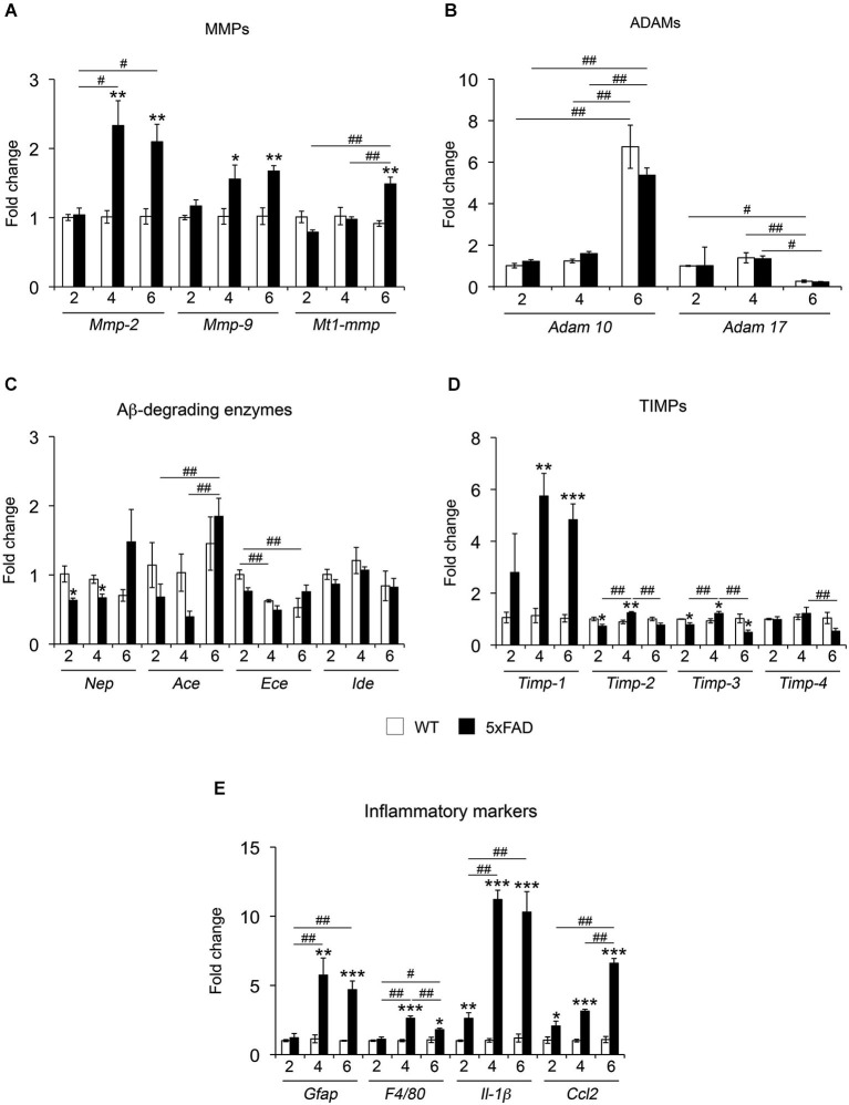
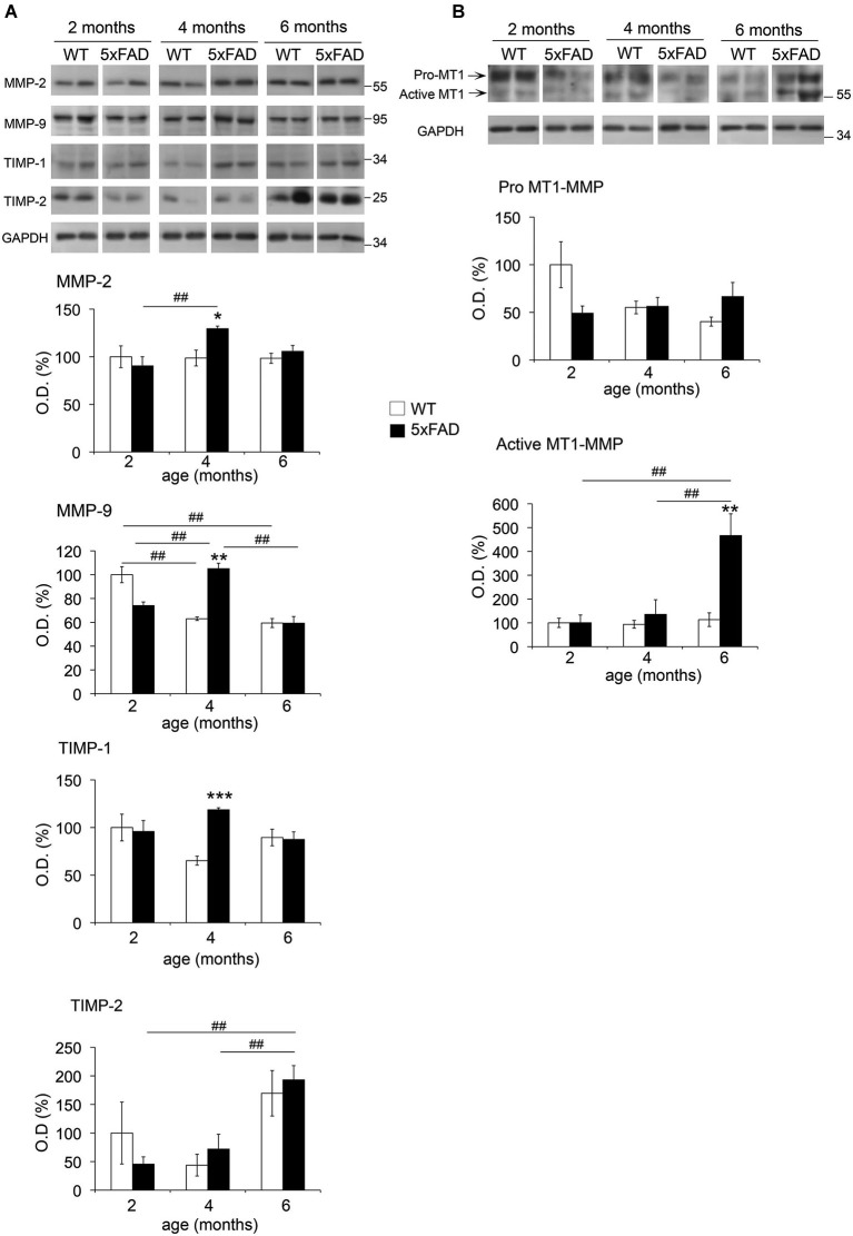
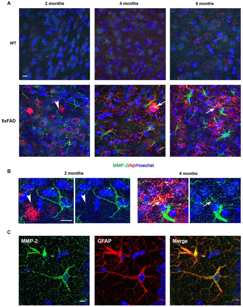
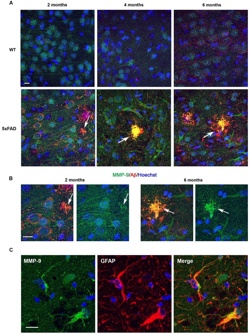
Verification
Claims the paper makes
The claims and statistics of this paper haven’t been checked yet.
Run the check — free, no account needed. It reads the open-access text and checks the sources it cites.
Claims other papers make about this one
Each is checked against this paper’s own text: does it say what the citing paper says it does?
The source backs part of the claim, not all of it — or backs it for a different population, condition or comparison.
In the citing paper
“all close MT5-MMP homologues, also involved in the control of APP metabolism and amyloidogenesis [33, 34, 54, 55]”
From MT5-MMP promotes neuroinflammation, neuronal excitability and Aβ production in primary neuron/astrocyte cultures from the 5xFAD mouse model of Alzheimer’s disease (2022) · cites this paper as [34]
Experimental — accuracy not yet measuredIn the citing paper
“Enhanced expression of MMP-2 and MMP-9 has been found in astrocytes surrounding amyloid plaques and in the neurons and glia of the hippocampus after intracerebroventricular Ab injection (25)(26)(27)(28).”
From Differential protective effects of connective tissue growth factor against Aβ neurotoxicity on neurons and glia (2017) · cites this paper as [25]
In the citing paper
“and later at 2 months, along with the onset of Aβ accumulation [34].”
In the citing paper
“C99 did not accumulate in developing neural cells.”
Self-check
Percentages, sums and reported differences re-computed from the paper’s own numbers.
The self-check needs the paper’s full text.
Citations
Cites
1 retracted
RETRACTED ARTICLE: A specific amyloid-β protein assembly in the brain impairs memory
Lesné, S., Koh, M. T., Kotilinek, L., Kayed, R., Glabe, C. G., Yang, A., Gallagher, M., & Ashe, K. H. (2006) · Nature · doi:10.1038/nature04533 · Cited by 2,769 · cited 1×
“In this context, the β-secretase-derived CTF of APP, C99 (βCTF) and small Aβ oligomers (dimers and trimers) have been shown to be particularly neurotoxic (Walsh et al., 2002; Cleary et al., 2005; Lesné et al., 2006; Townsend et al., 2006; Shankar et al., 2008; Lauritzen et al., 2012).”
BackgroundRetractedShankar, G. M., Li, S., Mehta, T. H., Garcia-Munoz, A., Shepardson, N. E., Smith, I., Brett, F. M., Farrell, M. A., Rowan, M. J., Lemere, C. A., et al. (2008) · Nature Medicine · doi:10.1038/nm1782 · Cited by 3,685
Oakley, H., Cole, S. L., Logan, S., Maus, E., Shao, P., Craft, J., Guillozet-Bongaarts, A., Ohno, M., Disterhoft, J., Van Eldik, L., et al. (2006) · The Journal of Neuroscience · doi:10.1523/jneurosci.1202-06.2006 · Cited by 3,530 · cited 5×
“(2005). Astrocyte reactivity to Fas activation is attenuated in TIMP-1”
BackgroundInfluentialKarran, E., Mercken, M., & Strooper, B. D. (2011) · Nature Reviews Drug Discovery · doi:10.1038/nrd3505 · Cited by 2,188 · cited 1×
“Histopathologically, AD is characterized by the accumulation of intracellular and extracellular deposits of amyloid beta peptide (Aβ), which according to the amyloid cascade hypothesis is considered as a crucial process in AD pathogenesis (Hardy and Higgins, 1992; Karran et al., 2011).”
BackgroundYang, Y., Estrada, E. Y., Thompson, J. F., Liu, W., & Rosenberg, G. A. (2007) · Journal of Cerebral Blood Flow & Metabolism · doi:10.1038/sj.jcbfm.9600375 · Cited by 1,060
Jawhar, S., Trawicka, A., Jenneckens, C., Bayer, T. A., & Wirths, O. (2012) · Neurobiology of Aging · doi:10.1016/j.neurobiolaging.2010.05.027 · Cited by 623 · cited 2×
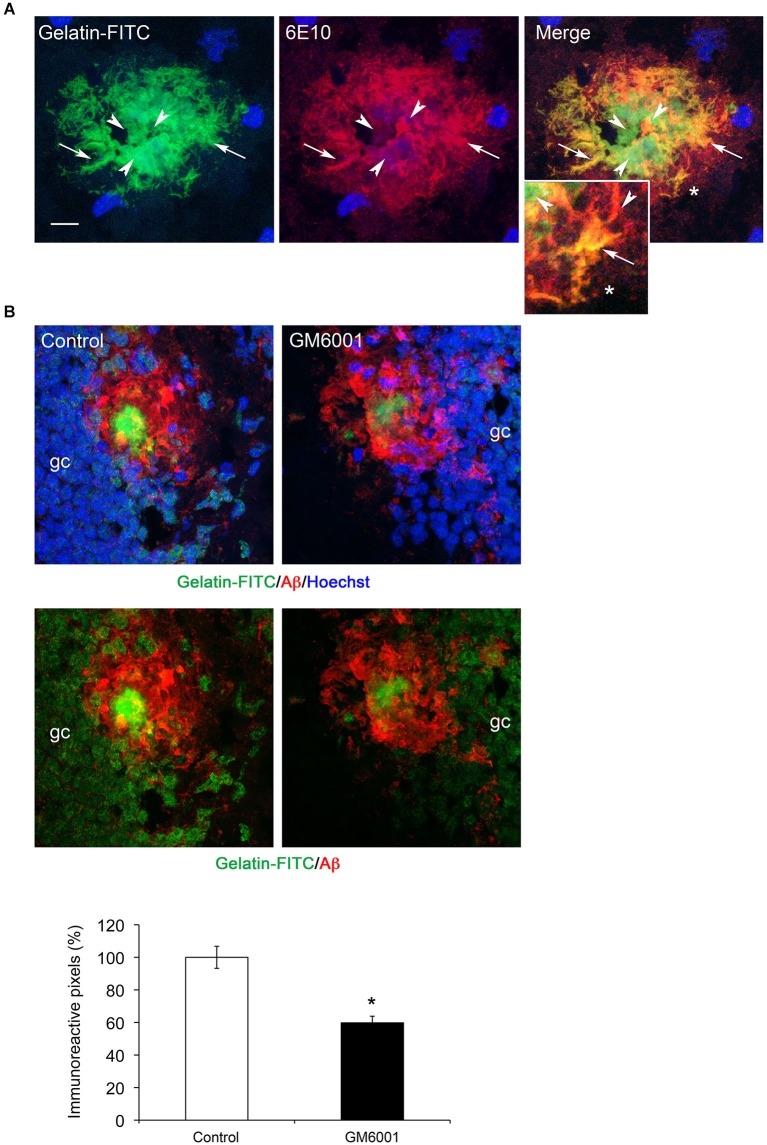
“IN SITU ZYMOGRAPHY In situ zymography was performed as previously described, with slight modifications (Jourquin et al., 2005).”
BackgroundMethodProteases and Proteolysis in Alzheimer Disease: A Multifactorial View on the Disease Process
De Strooper, B. (2010) · Physiological Reviews · doi:10.1152/physrev.00023.2009 · Cited by 455
Matrix Metalloproteinase-9 Degrades Amyloid-β Fibrils in Vitro and Compact Plaques in Situ
Yan, P., Hu, X., Song, H., Yin, K., Bateman, R. J., Cirrito, J. R., Xiao, Q., Hsu, F. F., Turk, J. W., Xu, J., et al. (2006) · Journal of Biological Chemistry · doi:10.1074/jbc.m602440200 · Cited by 380 · cited 1×
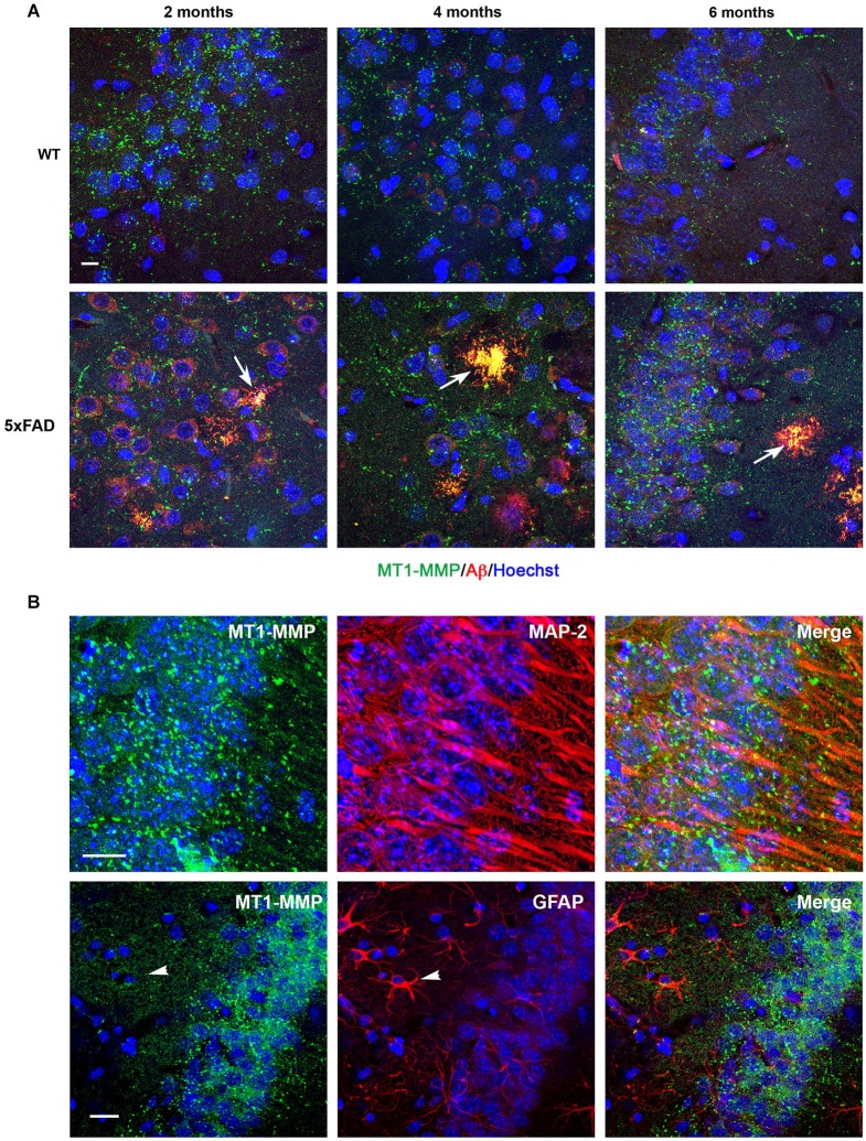
“…MMP-9 and MT1MMP immunostaining were particularly prominent in amyloid plaques, it is possible that they contribute to gelatinolysis in a significant manner, which would be consistent with the singular ability of both MMPs to degrade fibrillar Aβ (Yan et al., 2006; Liao and Van Nostrand, 2010).”
ResultBackstrom, J. R., Lim, G. P., Cullen, M. J., & Tökés, Z. A. (1996) · The Journal of Neuroscience · doi:10.1523/jneurosci.16-24-07910.1996 · Cited by 331 · cited 2×
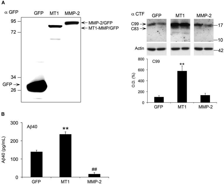
“Several MMPs, including MMP-2, MMP-9 and membranetype 1-MMP (MT1-MMP), a principal activator of MMP-2, may degrade Aβ (Roher et al., 1994; Backstrom et al., 1996; Yin et al., 2006; Liao and Van Nostrand, 2010).”
BackgroundLauritzen, I., Pardossi-Piquard, R., Bauer, C., Brigham, E., Abraham, J.-D., Ranaldi, S., Fraser, P., St-George-Hyslop, P., Le Thuc, O., Espin, V., et al. (2012) · The Journal of Neuroscience · doi:10.1523/jneurosci.2775-12.2012 · Cited by 201 · cited 2×
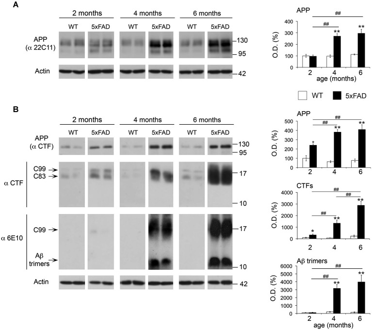
“This finding may be particularly relevant, since the β-secretase derived fragment C99 has been described as an early neurotoxic metabolite of APP that accumulates much earlier than Aβ species (Lauritzen et al., 2012).”
BackgroundSelf-citation
Research works printed but not linked: 46
- Ahmad, 2006, Cleavage of amyloid-beta precursor protein (APP) by membrane-type matrix metalloproteinases, J. Biochem., 139, 517, 10.1093/jb/mvj054 · DOI · Paper page
- Buggia-Prevot, 2008, NFkappaB-dependent control of BACE1 promoter transactivation by Abeta42, J. Biol. Chem., 283, 10037, 10.1074/jbc.M706579200 · DOI
- Candelario-Jalil, 2009, Diverse roles of matrix metalloproteinases and tissue inhibitors of metalloproteinases in neuroinflammation and cerebral ischemia, Neuroscience, 158, 983, 10.1016/j.neuroscience.2008.06.025 · DOI
- Chaillan, 2006, Involvement of tissue inhibition of metalloproteinases-1 in learning and memory in mice, Behav. Brain Res., 173, 191, 10.1016/j.bbr.2006.06.020 · DOI
- Chevallier, 1997, Examination of the role of endopeptidase 3.4.24.15 in A beta secretion by human transfected cells, Br. J. Pharmacol., 121, 556, 10.1038/sj.bjp.0701151 · DOI
- Cleary, 2005, Natural oligomers of the amyloid-beta protein specifically disrupt cognitive function, Nat. Neurosci., 8, 79, 10.1038/nn1372 · DOI
- Crouzin, 2013, Area-specific alterations of synaptic plasticity in the 5XFAD mouse model of Alzheimer’s disease: dissociation between somatosensory cortex and hippocampus, PLoS One, 8, e74667, 10.1371/journal.pone.0074667 · DOI
- Dafnis, 2012, An apolipoprotein E4 fragment affects matrix metalloproteinase 9, tissue inhibitor of metalloproteinase 1 and cytokine levels in brain cell lines, Neuroscience, 210, 21, 10.1016/j.neuroscience.2012.03.013 · DOI · Paper page
- Deb, 2003, Beta-amyloid induces the production of active, matrix-degrading proteases in cultured rat astrocytes, Brain Res., 970, 205, 10.1016/s0006-8993(03)02344-8 · DOI · Paper page
- Fanjul-Fernández, 2010, Matrix metalloproteinases: evolution, gene regulation and functional analysis in mouse models, Biochim. Biophys. Acta, 1803, 3, 10.1016/j.bbamcr.2009.07.004 · DOI
- Giannoni, 2013, Early administration of RS 67333, a specific 5-HT4 receptor agonist, prevents amyloidogenesis and behavioral deficits in the 5XFAD mouse model of Alzheimer’s disease, Front. Aging Neurosci., 5, 96, 10.3389/fnagi.2013.00096 · DOI
- Gonthier, 2007, Functional interaction between matrix metalloproteinase-3 and semaphorin-3C during cortical axonal growth and guidance, Cereb. Cortex, 17, 1712, 10.1093/cercor/bhl082 · DOI
- Hardy, 1992, Alzheimer’s disease: the amyloid cascade hypothesis, Science, 256, 184, 10.1126/science.1566067 · DOI · Paper page
- Higashi, 2003, Novel processing of beta-amyloid precursor protein catalyzed by membrane type 1 matrix metalloproteinase releases a fragment lacking the inhibitor domain against gelatinase A, Biochemistry, 42, 6514, 10.1021/bi020643m · DOI · Paper page
- Hongpaisan, 2011, PKC ε activation prevents synaptic loss, Aβ elevation and cognitive deficits in Alzheimer’s disease transgenic mice, J. Neurosci., 31, 630, 10.1523/JNEUROSCI.5209-10.2011 · DOI
- Hu, 2007, Matrix metalloproteinase inhibitors as therapy for inflammatory and vascular diseases, Nat. Rev. Drug Discov., 6, 480, 10.1038/nrd2308 · DOI
- Imai, 1996, Membrane-type matrix metalloproteinase 1 is a gelatinolytic enzyme and is secreted in a complex with tissue inhibitor of metalloproteinases 2, Cancer Res., 56, 2707
- Jourquin, 2005, Tissue inhibitor of metalloproteinases-1 (TIMP-1) modulates neuronal death, axonal plasticity and learning and memory, Eur. J. Neurosci., 22, 2569, 10.1111/j.1460-9568.2005.04426.x · DOI
- Jourquin, 2003, Neuronal activity-dependent increase of net matrix metalloproteinase activity is associated with MMP-9 neurotoxicity after kainate, Eur. J. Neurosci., 18, 1507, 10.1046/j.1460-9568.2003.02876.x · DOI · Paper page
- Jung, 2003, Pathogenic A beta induces the expression and activation of matrix metalloproteinase-2 in human cerebrovascular smooth muscle cells, J. Neurochem., 85, 1208, 10.1046/j.1471-4159.2003.01745.x · DOI
- Kaliszewska, 2012, Experience-dependent plasticity of the barrel cortex in mice observed with 2-DG brain mapping and c-Fos: effects of MMP-9 KO, Cereb. Cortex, 22, 2160, 10.1093/cercor/bhr303 · DOI · Paper page
- Kimura, 2009, Impairments in remote memory stabilization precede hippocampal synaptic and cognitive failures in 5XFAD Alzheimer mouse model, Neurobiol. Dis., 33, 229, 10.1016/j.nbd.2008.10.006 · DOI · Paper page
- Langenfurth, 2013, Membrane-type 1 metalloproteinase is upregulated in microglia/brain macrophages in neurodegenerative and neuroinflammatory diseases, J. Neurosci. Res., 92, 275, 10.1002/jnr.23288 · DOI
- LePage, 1995, Gelatinase a possesses a beta-secretase-like activity in cleaving the amyloid protein precursor of Alzheimer’s disease, FEBS Lett., 377, 267, 10.1016/0014-5793(95)01358-x · DOI
- Liao, 2010, Degradation of soluble and fibrillar amyloid beta-protein by matrix metalloproteinase (MT1-MMP) in vitro, Biochemistry, 49, 1127, 10.1021/bi901994d · DOI · Paper page
- Mazzone, 2004, Intracellular processing and activation of membrane type 1 matrix metalloprotease depends on its partitioning into lipid domains, J. Cell Sci., 117, 6275, 10.1242/jcs.01563 · DOI
- Meighan, 2006, Effects of extracellular matrix-degrading proteases matrix metalloproteinases 3 and 9 on spatial learning and synaptic plasticity, J. Neurochem., 96, 1227, 10.1111/j.1471-4159.2005.03565.x · DOI
- Mizoguchi, 2009, Matrix metalloprotease-9 inhibition improves amyloid beta-mediated cognitive impairment and neurotoxicity in mice, J. Pharmacol. Exp. Ther., 331, 14, 10.1124/jpet.109.154724 · DOI
- Moon, 2012, Intracellular amyloid-beta accumulation in calcium-binding protein-deficient neurons leads to amyloid-beta plaque formation in animal model of Alzheimer’s disease, J. Alzheimers Dis., 29, 615, 10.3233/JAD-2011-111778 · DOI · Paper page
- Nagy, 2006, Matrix metalloproteinase-9 is required for hippocampal late-phase long-term potentiation and memory, J. Neurosci., 26, 1923, 10.1523/jneurosci.4359-05.2006 · DOI · Paper page
- Ogier, 2006, Matrix metalloproteinase-2 (MMP-2) regulates astrocyte motility in connection with the actin cytoskeleton and integrins, Glia, 54, 272, 10.1002/glia.20349 · DOI
- Ogier, 2005, Astrocyte reactivity to Fas activation is attenuated in TIMP-1 deficient mice, an in vitro study, BMC Neurosci., 6, 68, 10.1186/1471-2202-6-68 · DOI · Paper page
- Ohno, 2007, BACE1 gene deletion prevents neuron loss and memory deficits in 5XFAD APP/PS1 transgenic mice, Neurobiol. Dis., 26, 134, 10.1016/j.nbd.2006.12.008 · DOI · Paper page
- Ould-yahoui, 2009, A new role for TIMP-1 in modulating neurite outgrowth and morphology of cortical neurons, PLoS One, 4, e8289, 10.1371/journal.pone.0008289 · DOI
- Rivera, 2010, Metzincin proteases and their inhibitors, foes or friends in nervous system physiology?, J. Neurosci., 30, 15337, 10.1523/JNEUROSCI.3467-10.2010 · DOI · Paper page
- Rivera, 2002, Gelatinase B and TIMP-1 are regulated in a cell- and time dependent manner in association with neuronal death and glial reactivity after global forebrain ischemia, Eur. J. Neurosci., 15, 19, 10.1046/j.0953-816x.2001.01838.x · DOI · Paper page
- Rivera, 1997, Tissue inhibitor of metalloproteinases-1 (TIMP-1) is differentially induced in neurons and astrocytes after seizures: evidence for developmental, immediate early gene and lesion response, J. Neurosci., 17, 4223, 10.1523/JNEUROSCI.17-11-04223.1997 · DOI · Paper page
- Roher, 1994, Proteolysis of A beta peptide from Alzheimer disease brain by gelatinase A, Biochem. Biophys. Res. Commun., 205, 1755, 10.1006/bbrc.1994.2872 · DOI
- Sbai, 2008, Vesicular trafficking and secretion of matrix metalloproteinases-2, -9 and tissue inhibitor of metalloproteinases-1 in neuronal cells, Mol. Cell. Neurosci., 39, 549, 10.1016/j.mcn.2008.08.004 · DOI
- Sbai, 2010, Differential vesicular distribution and trafficking of MMP-2, MMP-9 and their inhibitors in astrocytes, Glia, 58, 344, 10.1002/glia.20927 · DOI
- Soreghan, 1994, Surfactant properties of Alzheimer’s A beta peptides and the mechanism of amyloid aggregation, J. Biol. Chem., 269, 28551, 10.1016/S0021-9258(19)61939-3 · DOI
- Strongin, 1995, Mechanism of cell surface activation of 72-kDa type IV collagenase. Isolation of the activated form of the membrane metalloprotease, J. Biol. Chem., 270, 5331, 10.1074/jbc.270.10.5331 · DOI
- Talamagas, 2007, Abeta(1–40)-induced secretion of matrix metalloproteinase-9 results in sAPPalpha release by association with cell surface APP, Neurobiol. Dis., 28, 304, 10.1016/j.nbd.2007.07.016 · DOI
- Townsend, 2006, Effects of secreted oligomers of amyloid beta-protein on hippocampal synaptic plasticity: a potent role for trimers, J. Physiol., 572, 477, 10.1113/jphysiol.2005.103754 · DOI · Paper page
- Walsh, 2002, Naturally secreted oligomers of amyloid beta protein potently inhibit hippocampal long-term potentiation in vivo, Nature, 416, 535, 10.1038/416535a · DOI · Paper page
- Yin, 2006, Matrix metalloproteinases expressed by astrocytes mediate extracellular amyloid-beta peptide catabolism, J. Neurosci., 26, 10939, 10.1523/jneurosci.2085-06.2006 · DOI · Paper page
Cited by
Sorted by citations
MMPs in learning and memory and neuropsychiatric disorders
Beroun, A., Mitra, S., Michaluk, P., Pijet, B., Stefaniuk, M., & Kaczmarek, L. (2019) · Cellular and Molecular Life Sciences · doi:10.1007/s00018-019-03180-8 · Cited by 207
Matrix Metalloproteinases as New Targets in Alzheimer’s Disease: Opportunities and Challenges
Zipfel, P., Rochais, C., Baranger, K., Rivera, S., & Dallemagne, P. (2020) · Journal of Medicinal Chemistry · doi:10.1021/acs.jmedchem.0c00352 · Cited by 94
Self-citationEFAD transgenic mice as a human APOE relevant preclinical model of Alzheimerʼns disease
Tai, L. M., Balu, D., Avila-Munoz, E., Abdullah, L., Thomas, R., Collins, N., Valencia-Olvera, A. C., & LaDu, M. J. (2017) · Journal of Lipid Research · doi:10.1194/jlr.r076315 · Cited by 93
García-González, L., Pilat, D., Baranger, K., & Rivera, S. (2019) · Frontiers in Aging Neuroscience · doi:10.3389/fnagi.2019.00244 · Cited by 88
Self-citationKim, W., Ma, L., Lomoio, S., Willen, R., Lombardo, S., Dong, J., Haydon, P. G., & Tesco, G. (2018) · Molecular Neurodegeneration · doi:10.1186/s13024-018-0239-7 · Cited by 32
Pilat, D., Paumier, J.-M., García-González, L., Louis, L., Stephan, D., Manrique, C., Khrestchatisky, M., Di Pasquale, E., Baranger, K., & Rivera, S. (2022) · Journal of Neuroinflammation · doi:10.1186/s12974-022-02407-z · Cited by 28
Self-citationYang, C.-N., Wu, M.-F., Liu, C.-C., Jung, W.-H., Chang, Y.-C., Lee, W.-P., Shiao, Y.-J., Wu, C.-L., Liou, H.-H., Lin, S.-K., et al. (2017) · Human Molecular Genetics · doi:10.1093/hmg/ddx278 · Cited by 27
MT1-MMP inhibition rejuvenates ageing brain and rescues cognitive deficits in obesity
Asthana, P., Li, L., Lu, L., Wu, J., Zhang, S., Li, N., Wong, S. K. K., Gurung, S., Zhang, Y., Lin, Y., et al. (2025) · Cell Discovery · doi:10.1038/s41421-025-00825-w · Cited by 4
Metadata
- Authors
- Py, N. A., Bonnet, A. E., Bernard, A., Marchalant, Y., Charrat, E., Checler, F., Khrestchatisky, M., Baranger, K., & Rivera, S.
- Venue
- Frontiers in Aging Neuroscience
- Publisher
- Frontiers Media SA
- Published
- 2014-09-18
- Volume
- 6
- ISSN
- 1663-4365
- PMC
- PMC4166961
- Cited by
- 76
- References
- 13
About these checks
Registry, retraction, Cites and statistics results are computed, not judged, with one exception: a printed reference that no index lists is read once by a model before it counts as a citation error. Claims are extracted by a model and kept only when their quote appears verbatim in the text; each cited source is then checked, with a model’s help, for whether it says what the claim relies on. Those verdicts can be wrong — read the claim’s quote and the cited source before relying on one.
Statistics and arithmetic are recomputed, not judged.
Confidence bands are shown only for fields calibrated on our annotated test set. None is calibrated yet, so no band is shown.
What counts as a claim ›How we measure confidence ›How we check this ›
Are you an author, or do you see a problem with this page? Request a review
