

The DOI matches its Crossref record; 73 of the 73 references it cites were found in a registry or an index.
Involvement of the precuneus/posterior cingulate cortex is significant for the development of Alzheimer’s disease: a pet (THK5351, PiB) and resting fMRI study.
Open accessAbstract
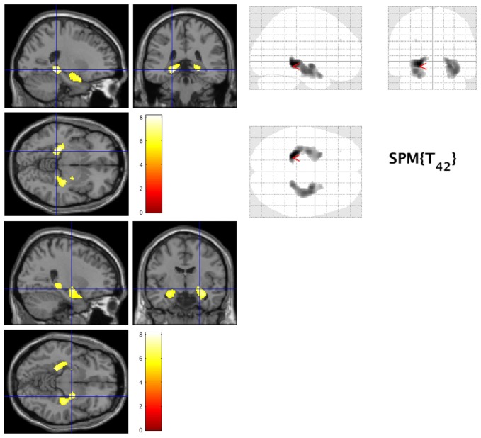
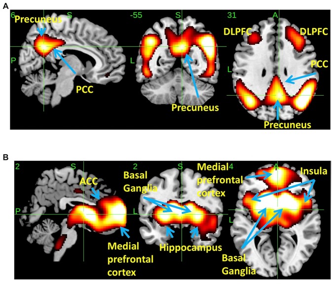
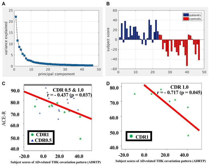
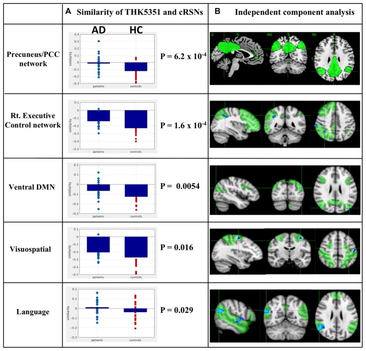
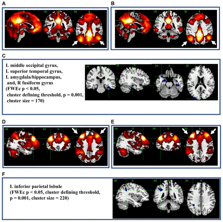
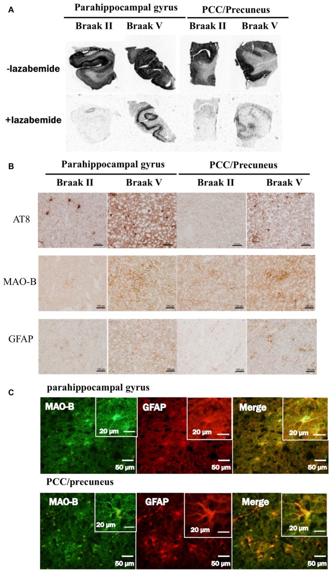
Background: Imaging studies in Alzheimer’s disease have yet to answer the underlying questions concerning the relationship among tau retention, neuroinflammation, network disruption, and cognitive decline. We compared the spatial retention patterns of 18F-THK5351 and resting state network disruption in patients with early Alzheimer’s disease and healthy controls. Methods: We enrolled 23 11C-Pittsburgh compound B (PiB)-positive patients with early Alzheimer’s disease and 24 11C-PiB-negative participants as healthy controls. All participants underwent resting state functional MRI and 18F-THK5351 PET scans. We used scaled subprofile modeling/principal component analysis (SSM/PCA) to reduce the complexity of multivariate data and to identify patterns that exhibited the largest statistical effects (variances) in THK5351 concentration in Alzheimer’s disease and healthy controls. Findings: SSM/PCA identified a significant spatial THK5351 pattern composed by mainly three clusters including precuneus/posterior cingulate cortex, right and left dorsolateral prefrontal cortex which accounted for 23.6% of the total subject voxel variance of the data and had 82.6% sensitivity and 79.1% specificity in discriminating Alzheimer’s disease from healthy controls. There was a significant relationship between the intensity of the 18F-THK5351 covariation pattern and cognitive scores in Alzheimer’s disease. The spatial patterns of 18F-THK5351 uptake showed significant similarity with intrinsic functional connectivity, especially in the precuneus/posterior cingulate cortex network. Seed-based connectivity analysis from the precuneus/posterior cingulate cortex showed significant decrease in connectivity over widespread brain regions in Alzheimer’s disease patients. An evaluation of an autopsied Alzheimer’s disease patient with Braak V showed that 18F-THK5351 retention corresponded to tau deposition, monoamine oxidase-B and astrogliosis in the precuneus/posterior cingulate cortex. Interpretation: We identified an Alzheimer’s disease-specific spatial pattern of 18F-THK5351 retention in the precuneus/posterior cingulate cortex, an important connectivity hub region in the brain. Disruption of the functional connections of this important network hub may play an important role in developing dementia in Alzheimer’s disease.
Overview
The claims and statistics of this paper haven’t been checked yet.
Run the check — free, no account needed. It reads the open-access text and checks the sources it cites.
Integrity signals
Registry
Cites
Statistics
Figures
Verification
Claims the paper makes
The claims and statistics of this paper haven’t been checked yet.
Run the check — free, no account needed. It reads the open-access text and checks the sources it cites.
Claims other papers make about this one
Each is checked against this paper’s own text: does it say what the citing paper says it does?
The cited source says what the claim attributes to it.
In the citing paper
“In a previous THK study with early AD patients, the precuneus was the most prominent AD-specific THK retention area, and the spatial pattern of THK uptake showed significant similarity with functional connectivity (Yokoi et al., 2018).”
From Effects of Alzheimer’s and Vascular Pathologies on Structural Connectivity in Early- and Late-Onset Alzheimer’s Disease (2021) · cites this paper as Yokoi et al., 2018
Self-check
Percentages, sums and reported differences re-computed from the paper’s own numbers.
The self-check needs the paper’s full text.
Citations
Cites
Hirono, N., Mori, E., Ikejiri, Y., Imamura, T., Shimomura, T., Hashimoto, M., Yamashita, H., & Ikeda, M. (1997) · PubMed · Cited by 21,012
McKhann, G. M., Knopman, D. S., Chertkow, H., Hyman, B. T., Jack, C. R., Kawas, C. H., Klunk, W. E., Koroshetz, W. J., Manly, J. J., Mayeux, R., et al. (2011) · Alzheimer's & Dementia · doi:10.1016/j.jalz.2011.03.005 · Cited by 18,657
Jenkinson, M., Bannister, P., Brady, M., & Smith, S. (2002) · NeuroImage · doi:10.1006/nimg.2002.1132 · Cited by 11,888
Jenkinson, M., Beckmann, C. F., Behrens, T. E. J., Woolrich, M. W., & Smith, S. M. (2012) · NeuroImage · doi:10.1016/j.neuroimage.2011.09.015 · Cited by 11,314
Fast robust automated brain extraction
Smith, S. M. (2002) · Human Brain Mapping · doi:10.1002/hbm.10062 · Cited by 10,496
The Brain's Default Network: Anatomy, Function, and Relevance to Disease
Buckner, R. L., Andrews‐Hanna, J. R., & Schacter, D. L. (2008) · Annals of the New York Academy of Sciences · doi:10.1196/annals.1440.011 · Cited by 9,761 · cited 10×
“However, in the patient group, a shift towards a more positive association, i.e., higher connectivity, higher THK5351 concentration, was observed and was significant in some canonical RSNs. Two-sample t-test comparisons of the similarity between the spatial distribution patterns of THK5351 retention and withinnetwork…”
BackgroundInfluentialA fast diffeomorphic image registration algorithm
Ashburner, J. (2007) · NeuroImage · doi:10.1016/j.neuroimage.2007.07.007 · Cited by 8,122
Mild cognitive impairment as a diagnostic entity
Petersen, R. C. (2004) · Journal of Internal Medicine · doi:10.1111/j.1365-2796.2004.01388.x · Cited by 7,864
Nonparametric permutation tests for functional neuroimaging: A primer with examples
Nichols, T. E., & Holmes, A. P. (2002) · Human Brain Mapping · doi:10.1002/hbm.1058 · Cited by 6,256
SMITH, S., & NICHOLS, T. (2009) · NeuroImage · doi:10.1016/j.neuroimage.2008.03.061 · Cited by 5,250
Cited by
Sorted by citations
Neuroinflammation and microglial activation in Alzheimer disease: where do we go from here?
Leng, F., & Edison, P. (2021) · Nature Reviews Neurology · doi:10.1038/s41582-020-00435-y · Cited by 3,179
Ibrahim, B., Suppiah, S., Ibrahim, N., Mohamad, M., Hassan, H. A., Nasser, N. S., & Saripan, M. I. (2021) · Human Brain Mapping · doi:10.1002/hbm.25369 · Cited by 272
Precuneus magnetic stimulation for Alzheimer’s disease: a randomized, sham-controlled trial
Koch, G., Casula, E. P., Bonnı̀, S., Borghi, I., Assogna, M., Minei, M., Pellicciari, M. C., Motta, C., D’Acunto, A., Porrazzini, F., et al. (2022) · Brain · doi:10.1093/brain/awac285 · Cited by 277
McCluskey, S. P., Plisson, C., Rabiner, E. A., & Howes, O. D. (2019) · European Journal of Nuclear Medicine and Molecular Imaging · doi:10.1007/s00259-019-04488-0 · Cited by 127
Anatomy and white-matter connections of the precuneus
Tanglay, O., Young, I. M., Dadario, N. B., Briggs, R. G., Fonseka, R. D., Dhanaraj, V., Hormovas, J., Lin, Y., & Sughrue, M. E. (2021) · Brain Imaging and Behavior · doi:10.1007/s11682-021-00529-1 · Cited by 101
Metadata
- Authors
- Yokoi, T., Watanabe, H., Yamaguchi, H., Bagarinao, E., Masuda, M., Imai, K., Ogura, A., Ohdake, R., Kawabata, K., Hara, K., Riku, Y., Ishigaki, S., Katsuno, M., Miyao, S., Kato, K., Naganawa, S., Harada, R., Okamura, N., Yanai, K., Yoshida, M., & Sobue, G.
- Affiliations
- Nagoya University (JP)
- Brain & Mind Research Center, Nagoya University (JP)
- Meitetsu Hospital (JP)
- Tohoku University (JP)
- Tohoku Medical and Pharmaceutical University (JP)
- Aichi Medical University (JP)
- Corresponding author
- Sobue, Gen
- ORCID
- Venue
- Frontiers in Aging Neuroscience
- Publisher
- Frontiers Media SA
- Published
- 2018-10-05
- Volume
- 10
- Pages
- 304-304
- ISSN
- 1663-4365 (ISSN-L)
- PMC
- PMC6182068
- OpenAlex
- W2895455599
- Cited by
- 116
- References
- 73
- Citations by year
- 2026: 5 · 2025: 20 · 2024: 18 · 2023: 14 · 2022: 24 · 2021: 15 · 2020: 8 · 2019: 10 · 2018: 1
- FWCI
- 6.69
- Type
- journal article
- Language
- en
- Open access
- gold
- doi.org
- Locations
- Frontiers in Aging Neuroscience (publishedVersion, cc-by)
- Europe PMC (PubMed Central) (publishedVersion, cc-by)
- PubMed Central (publishedVersion, cc-by)
- PubMed (publishedVersion)
- Institutional Repositories DataBase (IRDB) (submittedVersion, cc-by-nc-nd)
- Nagoya University Repository (NAGOYA Repository) (submittedVersion, other-oa)
- DOAJ (DOAJ: Directory of Open Access Journals) (submittedVersion, cc-by-sa)
- Funders
- Ministry of Education, Culture, Sports, Science and Technology (26117002, 80569781)
- Ministry of Health, Labour and Welfare
- Japan Society for the Promotion of Science (18K07367, 18H02771, 26117002, 26117001, 26117004)
- Topics
- Health Sciences › Medicine › Psychiatry and Mental health › Dementia and Cognitive Impairment Research
- Life Sciences › Neuroscience › Cellular and Molecular Neuroscience › Alzheimer's disease research and treatments
- Life Sciences › Neuroscience › Cognitive Neuroscience › Functional Brain Connectivity Studies
- Keywords
- Alzheimer's disease, precuneus, posterior cingulate cortex, 18F-THK5351, tau PET, [11C]PIB, resting-state functional connectivity, intrinsic functional connectivity, seed-based functional connectivity, early Alzheimer's disease, tau deposition, dorsolateral prefrontal cortex, Braak staging, astrocyte activation
About these checks
Registry, retraction, Cites and statistics results are computed, not judged, with one exception: a printed reference that no index lists is read once by a model before it counts as a citation error. Claims are extracted by a model and kept only when their quote appears verbatim in the text; each cited source is then checked, with a model’s help, for whether it says what the claim relies on. Those verdicts can be wrong — read the claim’s quote and the cited source before relying on one.
Statistics and arithmetic are recomputed, not judged.
Confidence bands are shown only for fields calibrated on our annotated test set. None is calibrated yet, so no band is shown.
What counts as a claim ›How we measure confidence ›How we check this ›
Are you an author, or do you see a problem with this page? Request a review
