

The DOI matches its Crossref record; 115 of the 116 references it cites were found in a registry or an index, and 1 are not checked yet.
Intrinsically Photosensitive Retinal Ganglion Cells of the Human Retina
Open accessAbstract
Light profoundly affects our mental and physical health. In particular, light, when not delivered at the appropriate time, may have detrimental effects. In mammals, light is perceived not only by rods and cones but also by a subset of retinal ganglion cells that express the photopigment melanopsin that renders them intrinsically photosensitive (ipRGCs). ipRGCs participate in contrast detection and play critical roles in non-image-forming vision, a set of light responses that include circadian entrainment, pupillary light reflex (PLR), and the modulation of sleep/alertness, and mood. ipRGCs are also found in the human retina, and their response to light has been characterized indirectly through the suppression of nocturnal melatonin and PLR. However, until recently, human ipRGCs had rarely been investigated directly. This gap is progressively being filled as, over the last years, an increasing number of studies provided descriptions of their morphology, responses to light, and gene expression. Here, I review the progress in our knowledge of human ipRGCs, in particular, the different morphological and functional subtypes described so far and how they match the murine subtypes. I also highlight questions that remain to be addressed. Investigating ipRGCs is critical as these few cells play a major role in our well-being. Additionally, as ipRGCs display increased vulnerability or resilience to certain disorders compared to conventional RGCs, a deeper knowledge of their function could help identify therapeutic approaches or develop diagnostic tools. Overall, a better understanding of how light is perceived by the human eye will help deliver precise light usage recommendations and implement light-based therapeutic interventions to improve cognitive performance, mood, and life quality.
Overview
The claims and statistics of this paper haven’t been checked yet.
Run the check — free, no account needed. It reads the open-access text and checks the sources it cites.
Integrity signals
Registry
Cites
Statistics
Figures
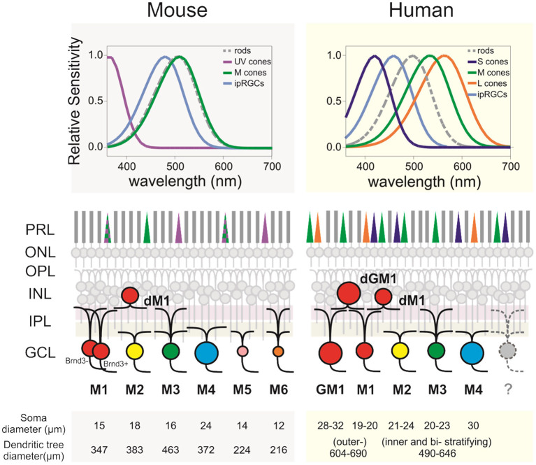
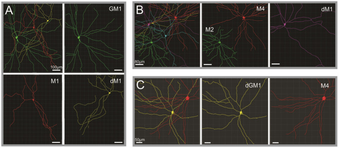
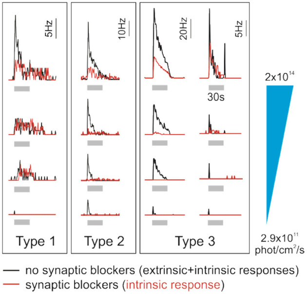
Verification
Claims the paper makes
The claims and statistics of this paper haven’t been checked yet.
Run the check — free, no account needed. It reads the open-access text and checks the sources it cites.
Claims other papers make about this one
Each is checked against this paper’s own text: does it say what the citing paper says it does?
The source backs part of the claim, not all of it — or backs it for a different population, condition or comparison.
In the citing paper
“When light enters the eye, it is detected by intrinsically photosensitive retinal ganglion cells, which then send signals to the suprachiasmatic nuclei in the hypothalamus, the brain's master clock that coordinates daily physiological rhythms [60].”
From Ocular light exposure interventions for sleep, circadian rhythms, rest-activity cycles, mood, and cognitive function in older adults: An Overview of Cochrane and non-Cochrane Systematic Reviews (2025) · cites this paper as [60]
Experimental — accuracy not yet measuredThe cited source says what the claim attributes to it.
In the citing paper
“The mechanism behind ocular light exposure interventions lies in the non‐image‐forming effects of light, mediated by specialised, intrinsically photosensitive retinal ganglion cell photoreceptors in the eye [60].”
Self-check
Percentages, sums and reported differences re-computed from the paper’s own numbers.
The self-check needs the paper’s full text.
Citations
Cites
Phototransduction by retinal ganglion cells that set the circadian clock
Berson, D. M., Dunn, F. A., & Takao, M. (2002) · Science · doi:10.1126/science.1067262 · Cited by 3,629
Action Spectrum for Melatonin Regulation in Humans: Evidence for a Novel Circadian Photoreceptor
Brainard, G. C., Hanifin, J. P., Greeson, J. M., Byrne, B., Glickman, G., Gerner, E., & Rollag, M. D. (2001) · The Journal of Neuroscience · doi:10.1523/jneurosci.21-16-06405.2001 · Cited by 1,930
Topography of ganglion cells in human retina
Curcio, C. A., & Allen, K. A. (1990) · The Journal of Comparative Neurology · doi:10.1002/cne.903000103 · Cited by 1,849
Chang, A.-M., Aeschbach, D., Duffy, J. F., & Czeisler, C. A. (2015) · Proceedings of the National Academy of Sciences · doi:10.1073/pnas.1418490112 · Cited by 1,430 · cited 1×
“Even low-level light exposure from light-emitting devices, smartphones, or tablets may disrupt sleep (5, 6).”
BackgroundDacey, D. M., Liao, H.-W., Peterson, B. B., Robinson, F. R., Smith, V. C., Pokorny, J., Yau, K.-W., & Gamlin, P. D. (2005) · Nature · doi:10.1038/nature03387 · Cited by 1,333
Thapan, K., Arendt, J., & Skene, D. J. (2001) · The Journal of Physiology · doi:10.1111/j.1469-7793.2001.t01-1-00261.x · Cited by 1,320
A Novel Human Opsin in the Inner Retina
Provencio, I., Rodriguez, I. R., Jiang, G., Hayes, W. P., Moreira, E. F., & Rollag, M. D. (2000) · The Journal of Neuroscience · doi:10.1523/jneurosci.20-02-00600.2000 · Cited by 1,290
Central projections of melanopsin‐expressing retinal ganglion cells in the mouse
Hattar, S., Kumar, M., Park, A., Tong, P., Tung, J., Yau, K., & Berson, D. M. (2006) · Journal of Comparative Neurology · doi:10.1002/cne.20970 · Cited by 996
Melanopsin: An opsin in melanophores, brain, and eye
Provencio, I., Jiang, G., De Grip, W. J., Hayes, W. P., & Rollag, M. D. (1998) · Proceedings of the National Academy of Sciences · doi:10.1073/pnas.95.1.340 · Cited by 975
Artificially lit surface of Earth at night increasing in radiance and extent
Kyba, C. C. M., Kuester, T., de Miguel, A. S., Baugh, K., Jechow, A., Hölker, F., Bennie, J., Elvidge, C. D., Gaston, K. J., & Guanter, L. (2017) · Science Advances · doi:10.1126/sciadv.1701528 · Cited by 958
Cited by
Sorted by citations
Blue Light Exposure: Ocular Hazards and Prevention—A Narrative Review
Cougnard‐Grégoire, A., Merle, B. M. J., Aslam, T. M., Seddon, J. M., Aknin, I., Klaver, C. C. W., Garhöfer, G., García‐Layana, A., Minnella, A. M., da Silva, R. M., et al. (2023) · Ophthalmology and Therapy · doi:10.1007/s40123-023-00675-3 · Cited by 217
Reducing nighttime light exposure in the urban environment to benefit human health and society
Zielińska-Dąbkowska, K. M., Schernhammer, E. S., Hanifin, J. P., & Brainard, G. C. (2023) · Science · doi:10.1126/science.adg5277 · Cited by 158
Understanding light pollution: Recent advances on its health threats and regulations
Cao, M., Xu, T., & Yin, D. (2022) · Journal of Environmental Sciences · doi:10.1016/j.jes.2022.06.020 · Cited by 131
The cognitive impact of light: illuminating ipRGC circuit mechanisms
Mahoney, H. L., & Schmidt, T. M. (2024) · Nature reviews. Neuroscience · doi:10.1038/s41583-023-00788-5 · Cited by 83
Spitschan, M., Kervezee, L., Lok, R., McGlashan, E., Najjar, R. P., Allen, A. E., Andersen, M., Bará, S., Blattner, P., Blume, C., et al. (2023) · eBioMedicine · doi:10.1016/j.ebiom.2023.104889 · Cited by 60
Self-citation
Metadata
- Authors
- Mure, L. S.
- Affiliations
- University of Bern (CH)
- Corresponding author
- Mure, Ludovic S.
- ORCID
- Mure, Ludovic S.
- Venue
- Frontiers in Neurology
- Publisher
- Frontiers Media SA
- Published
- 2021-03-25
- Volume
- 12
- Pages
- 636330-636330
- ISSN
- 1664-2295 (ISSN-L)
- PMC
- PMC8027232
- OpenAlex
- W3151649762
- Cited by
- 151
- References
- 116
- Citations by year
- 2026: 32 · 2025: 31 · 2024: 27 · 2023: 37 · 2022: 19 · 2021: 5
- FWCI
- 2.81
- Type
- journal article
- Language
- en
- Open access
- gold
- doi.org
- Licence
- Locations
- Frontiers in Neurology (publishedVersion, cc-by)
- PubMed Central (publishedVersion, cc-by)
- PubMed (publishedVersion)
- BORIS (University Library Bern) (submittedVersion, cc-by)
- DOAJ (DOAJ: Directory of Open Access Journals) (submittedVersion, cc-by-sa)
- Open Access CRIS of the University of Bern
- Funders
- University of Bern
- Topics
- Life Sciences › Neuroscience › Endocrine and Autonomic Systems › Circadian rhythm and melatonin
- Health Sciences › Medicine › Ophthalmology › Retinal Development and Disorders
- Life Sciences › Neuroscience › Cognitive Neuroscience › Visual perception and processing mechanisms
- Keywords
- intrinsically photosensitive retinal ganglion cells, melanopsin, human retina, non-image-forming vision, circadian entrainment, pupillary light reflex, melatonin suppression, light perception, phototherapy
About these checks
Registry, retraction, Cites and statistics results are computed, not judged, with one exception: a printed reference that no index lists is read once by a model before it counts as a citation error. Claims are extracted by a model and kept only when their quote appears verbatim in the text; each cited source is then checked, with a model’s help, for whether it says what the claim relies on. Those verdicts can be wrong — read the claim’s quote and the cited source before relying on one.
Statistics and arithmetic are recomputed, not judged.
Confidence bands are shown only for fields calibrated on our annotated test set. None is calibrated yet, so no band is shown.
What counts as a claim ›How we measure confidence ›How we check this ›
Are you an author, or do you see a problem with this page? Request a review
