

The DOI matches its Crossref record; 168 of the 171 references it cites were found in a registry or an index.
The role of the striatum in social behavior
Open accessAbstract
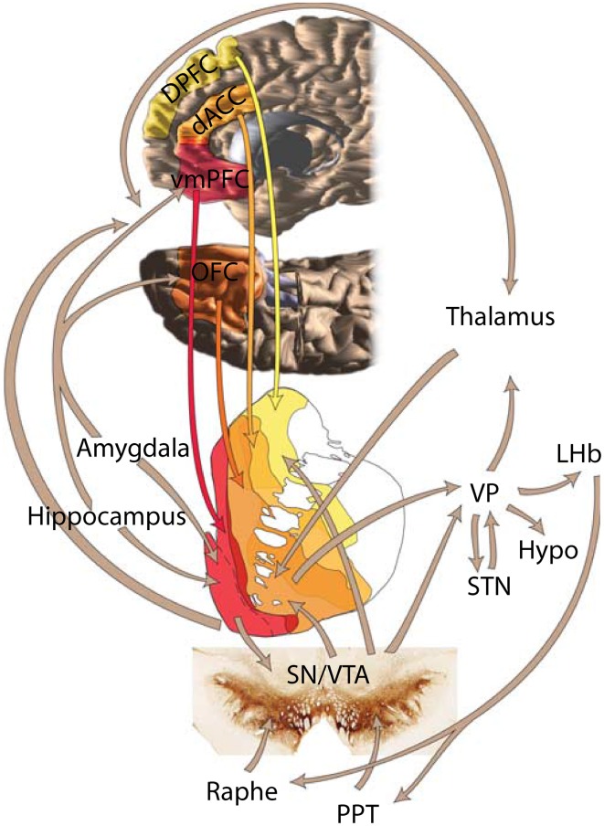
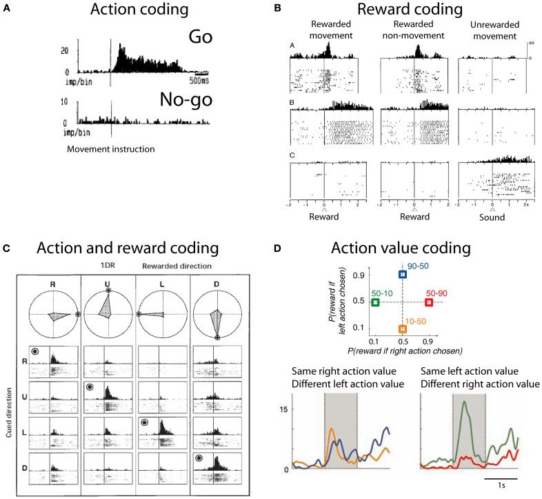
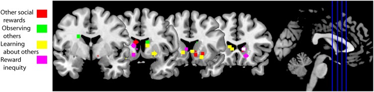
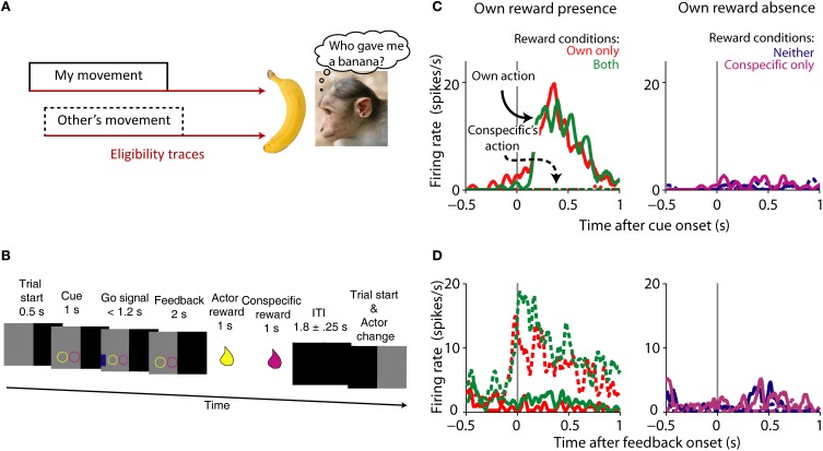
Where and how does the brain code reward during social behavior? Almost all elements of the brain's reward circuit are modulated during social behavior. The striatum in particular is activated by rewards in social situations. However, its role in social behavior is still poorly understood. Here, we attempt to review its participation in social behaviors of different species ranging from voles to humans. Human fMRI experiments show that the striatum is reliably active in relation to others' rewards, to reward inequity and also while learning about social agents. Social contact and rearing conditions have long-lasting effects on behavior, striatal anatomy and physiology in rodents and primates. The striatum also plays a critical role in pair-bond formation and maintenance in monogamous voles. We review recent findings from single neuron recordings showing that the striatum contains cells that link own reward to self or others' actions. These signals might be used to solve the agency-credit assignment problem: the question of whose action was responsible for the reward. Activity in the striatum has been hypothesized to integrate actions with rewards. The picture that emerges from this review is that the striatum is a general-purpose subcortical region capable of integrating social information into coding of social action and reward.
Overview
The claims and statistics of this paper haven’t been checked yet.
Run the check — free, no account needed. It reads the open-access text and checks the sources it cites.
Integrity signals
Registry
Cites
Statistics
Figures
Verification
Claims the paper makes
The claims and statistics of this paper haven’t been checked yet.
Run the check — free, no account needed. It reads the open-access text and checks the sources it cites.
Claims other papers make about this one
Each is checked against this paper’s own text: does it say what the citing paper says it does?
The source backs part of the claim, not all of it — or backs it for a different population, condition or comparison.
In the citing paper
“This activation is mediated by the release of dopamine, a neurotransmitter associated with pleasure and reward (Báez-Mendoza and Schultz, 2013).”
From The impact of digital technology, social media, and artificial intelligence on cognitive functions: a review (2023) · cites this paper as Báez-Mendoza and Schultz, 2013
Experimental — accuracy not yet measuredThe source backs part of the claim, not all of it — or backs it for a different population, condition or comparison.
In the citing paper
“Research has shown that excessive digital use, particularly related to activities such as social media and gaming, can trigger addictive behaviors by activating the brain's reward circuitry.”
From The impact of digital technology, social media, and artificial intelligence on cognitive functions: a review (2023) · cites this paper as Báez-Mendoza and Schultz, 2013
Experimental — accuracy not yet measured
Self-check
Percentages, sums and reported differences re-computed from the paper’s own numbers.
The self-check needs the paper’s full text.
Citations
Cites
Reinforcement Learning: An Introduction
Sutton, R. S., & Barto, A. G. (1998) · IEEE Transactions on Neural Networks · doi:10.1109/tnn.1998.712192 · Cited by 27,225
Reinforcement Learning: An Introduction
Sutton, R. S., & Barto, A. (2005) · IEEE Transactions on Neural Networks · doi:10.1109/tnn.2004.842673 · Cited by 25,268
Axelrod, R., & Hamilton, W. D. (1981) · Science · doi:10.1126/science.7466396 · Cited by 20,065
Theory of games and economic behavior
Garcia, S. R., & Miller, S. J. (2019) · American Mathematical Society eBooks · doi:10.1090/mbk/121/32 · Cited by 16,777
A Theory of Fairness, Competition, and Cooperation
Fehr, E., & Schmidt, K. M. (1999) · The Quarterly Journal of Economics · doi:10.1162/003355399556151 · Cited by 11,264
Five Rules for the Evolution of Cooperation
Nowak, M. A. (2006) · Science · doi:10.1126/science.1133755 · Cited by 5,894
ERC: A Theory of Equity, Reciprocity, and Competition
Bolton, G. E., & Ockenfels, A. (2000) · American Economic Review · doi:10.1257/aer.90.1.166 · Cited by 5,399
An experimental analysis of ultimatum bargaining
Güth, W., Schmittberger, R., & Schwarze, B. (1982) · Journal of Economic Behavior & Organization · doi:10.1016/0167-2681(82)90011-7 · Cited by 4,868
Incorporating Fairness into Game Theory and Economics
Rabin, M. (2011) · Princeton University Press eBooks · doi:10.2307/j.ctvcm4j8j.15 · Cited by 4,614
The reward circuit: linking primate anatomy and human imaging
Haber, S. N., & Knutson, B. (2010) · Neuropsychopharmacology · doi:10.1038/npp.2009.129 · Cited by 3,718
Research works printed but not linked: 3
- Báez-Mendoza R. Hoffman K. L. (2009). Object ontology in temporal lobe ensembles, in Cortical Mechanisms of Vision, 1st Edn., eds Jenkin M. Harris L. (Cambridge: Cambridge University Press), 237– 253
- Dawkins R. (2006). The Selfish Gene: – with a New Introduction by the Author. Oxford: University Press
- Hall F. S. Wilkinson L. S. Humby T. Robbins T. W. (1999). Maternal deprivation of neonatal rats produces enduring changes in dopamine function. Synapse 32, 37– 43 10188636
Cited by
Sorted by citations
Items 101–200 of 313
Schindler, S., Kruse, O., Stark, R. M., & Kissler, J. M. (2018) · Cognitive Affective & Behavioral Neuroscience · doi:10.3758/s13415-018-00660-5 · Cited by 23
The cortico-striatal circuitry in autism-spectrum disorders: a balancing act
Soghomonian, J. (2024) · Frontiers in Cellular Neuroscience · doi:10.3389/fncel.2023.1329095 · Cited by 21
Arslan, N., Esin, K., & Ayyıldız, F. (2025) · BMC Public Health · doi:10.1186/s12889-025-22507-8 · Cited by 22
Dopamine manipulations modulate paranoid social inferences in healthy people
Barnby, J. M., Bell, V., Deeley, Q., & Mehta, M. A. (2020) · Translational Psychiatry · doi:10.1038/s41398-020-00912-4 · Cited by 22
Lee, S.-J., Kim, T. K., Choi, J. E., Choi, Y., You, M., Ryu, J., Chun, Y. L., Ham, S., Hyeon, S. J., Ryu, H., et al. (2022) · Theranostics · doi:10.7150/thno.68439 · Cited by 22
Manfré, G., Novati, A., Faccini, I., Rossetti, A. C., Bosch, K., Molteni, R., Riva, M. A., van der Harst, J. E., Nguyen, H. P., & Homberg, J. R. (2018) · PLoS ONE · doi:10.1371/journal.pone.0192289 · Cited by 25
Exploring the role of microglia in mood disorders associated with experimental multiple sclerosis
Gentile, A., Vito, F. D., Fresegna, D., Musella, A., Buttari, F., Bullitta, S., Mandolesi, G., & Centonze, D. (2015) · Frontiers in Cellular Neuroscience · doi:10.3389/fncel.2015.00243 · Cited by 22
Mother brain is wired for social moments
Shimon-Raz, O., Salomon, R., Bloch, M., Romano, G. A., Hendler, T., Yeshurun, Y., Ulmer-Yaniv, A., Zagoory‐Sharon, O., & Feldman, R. (2021) · eLife · doi:10.7554/elife.59436 · Cited by 20
Hirter, K. N., Miller, E. N., Stimpson, C. D., Phillips, K. A., Hopkins, W. D., Hof, P. R., Sherwood, C. C., Lovejoy, C. O., & Raghanti, M. A. (2021) · Brain Structure and Function · doi:10.1007/s00429-021-02300-0 · Cited by 23
Personality from a cognitive-biological perspective
Neuman, Y. (2014) · Physics of Life Reviews · doi:10.1016/j.plrev.2014.09.002 · Cited by 21
Czekóová, K., Shaw, D. J., Saxunová, K., Dufek, M., Mareček, R., Vaníček, J., & Brázdil, M. (2019) · Frontiers in Neurology · doi:10.3389/fneur.2019.00525 · Cited by 22
Effect of nanoplastic intake on the dopamine system during the development of male mice
Kim, N., Choo, H.-I., & Lee, Y. (2024) · Neuroscience · doi:10.1016/j.neuroscience.2024.07.018 · Cited by 20
Blázquez, G., Castañé, A., Saavedra, A., Masana, M., Alberch, J., & Pérez‐Navarro, E. (2019) · Frontiers in Behavioral Neuroscience · doi:10.3389/fnbeh.2018.00317 · Cited by 22
Sosa, P. M., Souza, M. A., & Mello‐Carpes, P. B. (2018) · Neural Plasticity · doi:10.1155/2018/5158724 · Cited by 20
Neural reward-related reactions to monetary gains for self and charity
Spaans, J., Peters, S., & Crone, E. A. (2018) · Cognitive Affective & Behavioral Neuroscience · doi:10.3758/s13415-018-00672-1 · Cited by 21
Tian, X., Richard, A. D., El-Saadi, M. W., Bhandari, A., Latimer, B., Savage, I. V., Holmes, K., Klein, R. L., Dwyer, D. S., Goeders, N. E., et al. (2019) · Molecular Psychiatry · doi:10.1038/s41380-019-0492-3 · Cited by 20
Synapsin IIb as a functional marker of submissive behavior
Nesher, E., Koman, I. E., Gross, M., Tikhonov, T., Bairachnaya, M., Salmon‐Divon, M., Levin, Y., Gerlitz, G., Michaelevski, I., Yadid, G., et al. (2015) · Scientific Reports · doi:10.1038/srep10287 · Cited by 20
Tully, J., Cross, B., Gerrie, B., Griem, J., Blackwood, N., Blair, J., & McCutcheon, R. A. (2023) · Nature Mental Health · doi:10.1038/s44220-023-00032-0 · Cited by 22
Bortolini, T., Laport, M. C., Latgé-Tovar, S., Fischer, R. D., Zahn, R., de Oliveira‐Souza, R., & Moll, J. (2024) · Neuroscience & Biobehavioral Reviews · doi:10.1016/j.neubiorev.2024.105584 · Cited by 22
Sosa, P. M., Schimidt, H. L., Altermann, C. D. C., Vieira, A. S., Cibin, F. W. S., Carpes, F. P., & Mello‐Carpes, P. B. (2015) · Brazilian Journal of Medical and Biological Research · doi:10.1590/1414-431x20154429 · Cited by 19
Dworkin, S., Auden, A., Partridge, D. D., Daglas, M., Medcalf, R. L., Mantamadiotis, T., Georgy, S. R., Darido, C., Jane, S. M., & Ting, S. B. (2016) · Developmental Neurobiology · doi:10.1002/dneu.22469 · Cited by 19
Kays, J. S., & Yamamoto, B. K. (2019) · Brain Sciences · doi:10.3390/brainsci9120340 · Cited by 18
What Is Social about Autism? The Role of Allostasis-Driven Learning
Djerassi, M., Ophir, S., & Atzil, S. (2021) · Brain Sciences · doi:10.3390/brainsci11101269 · Cited by 22
Altered brain activation during reward anticipation in bipolar disorder
Long, X., Wang, X., Tian, F., Cao, Y., Xie, H., & Jia, Z. (2022) · Translational Psychiatry · doi:10.1038/s41398-022-02075-w · Cited by 22
Akinmoladun, A. C., Saliu, I. O., Olowookere, B. D., Ojo, O. B., Olaleye, M. T., Farombi, E. O., & Akindahunsi, A. A. (2017) · Annals of Neurosciences · doi:10.1159/000484517 · Cited by 18
Stanisavljević, A., Perić, I., Gass, P., Inta, D., Lang, U. E., Borgwardt, S., & Filipović, D. (2020) · Brain Research Bulletin · doi:10.1016/j.brainresbull.2020.07.021 · Cited by 18
Self-competence increases the willingness to pay for social influence
Hertz, U., Tyropoulou, E., Traberg, C. S., & Bahrami, B. (2020) · Scientific Reports · doi:10.1038/s41598-020-74857-5 · Cited by 20
Neural signatures of cooperation enforcement and violation: a coordinate-based meta-analysis
Yang, Z., Zheng, Y., Yang, G., Li, Q., & Liu, X. (2019) · Social Cognitive and Affective Neuroscience · doi:10.1093/scan/nsz073 · Cited by 19
McKenna, M., Filteau, J. R., Butler, B., Sluis, K., Chungyoun, M., Schimek, N., & Nance, E. A. (2022) · Journal of Biological Engineering · doi:10.1186/s13036-022-00293-w · Cited by 19
Binge-Like Exposure to Ethanol Enhances Morphine's Anti-nociception in B6 Mice
Chang, S. L., Huang, W., Han, H., & Sariyer, I. K. (2019) · Frontiers in Psychiatry · doi:10.3389/fpsyt.2018.00756 · Cited by 17
A Scope of the Social Brain in Multiple Sclerosis: Insights From Neuroimaging Studies
Chalah, M. A., & Ayache, S. S. (2020) · Cognitive and Behavioral Neurology · doi:10.1097/wnn.0000000000000227 · Cited by 17
Lin, S., Chen, Z., Zhao, Y., & Gong, Q. (2025) · Journal of Psychiatry and Neuroscience · doi:10.1503/jpn.240112 · Cited by 16
Faster but Less Careful Prehension in Presence of High, Rather than Low, Social Status Attendees
Fantoni, C., Rigutti, S., Piccoli, V., Sommacal, E., & Carnaghi, A. (2016) · PLoS ONE · doi:10.1371/journal.pone.0158095 · Cited by 18
Cao, C. C., & Reimann, M. (2020) · Frontiers in Psychology · doi:10.3389/fpsyg.2020.550204 · Cited by 20
Ru, F.-X., Kong, F., Ren, C., He, Y.-S., Xia, S.-Y., Yu-ning, L., Liang, Y., Feng, J.-J., Wei, Z.-Y., & Chen, J. (2022) · Frontiers in Psychiatry · doi:10.3389/fpsyt.2022.896794 · Cited by 16
Reward and decision processes in the brains of humans and nonhuman primates
Sirigu, A., & Duhamel, J. (2016) · Dialogues in Clinical Neuroscience · doi:10.31887/dcns.2016.18.1/asirigu · Cited by 17
Martin-Söelch, C., Guillod, M., Gaillard, C., Recabarren, R. E., Federspiel, A., Mueller‐Pfeiffer, C., Homan, P., Hasler, G., Schoebi, D., Horsch, A., et al. (2021) · Frontiers in Psychiatry · doi:10.3389/fpsyt.2020.563475 · Cited by 14
Neural correlates of recursive thinking during interpersonal strategic interactions
Zhen, S., & Yu, R. (2021) · Human Brain Mapping · doi:10.1002/hbm.25355 · Cited by 14
Social processing by the primate medial frontal cortex
Putnam, P. T., & Chang, S. W. C. (2021) · International review of neurobiology · doi:10.1016/bs.irn.2020.12.003 · Cited by 19
Covid-19 and Mental Health: Could Visual Art Exposure Help?
Gallo, L. M. H., Giampietro, V., Zunszain, P. A., & Tan, K. S. (2021) · Frontiers in Psychology · doi:10.3389/fpsyg.2021.650314 · Cited by 19
Hippocampal and striatal volumes correlate with spatial memory impairment in Huntington’s disease
Glikmann‐Johnston, Y., Mercieca, E., Carmichael, A. M., Alexander, B., Harding, I. H., & Stout, J. C. (2021) · Journal of Neuroscience Research · doi:10.1002/jnr.24966 · Cited by 15
The overlooked potential for social factors to improve effectiveness of brain-computer interfaces
Sexton, C. A. (2015) · Frontiers in Systems Neuroscience · doi:10.3389/fnsys.2015.00070 · Cited by 16
Neural bases of loss aversion when choosing for oneself versus known or unknown others
Arioli, M., Basso, G., Baud‐Bovy, G., Mattioni, L., Poggi, P., & Canessa, N. (2023) · Cerebral Cortex · doi:10.1093/cercor/bhad025 · Cited by 16
Emotional contagion and prosocial behaviour in fish: An evolutionary and mechanistic approach
Kareklas, K., & Oliveira, R. F. (2024) · Neuroscience & Biobehavioral Reviews · doi:10.1016/j.neubiorev.2024.105780 · Cited by 14
The Role of Social Reward and Corticostriatal Connectivity in Substance Use
Sazhin, D., Frazier, A. M., Haynes, C. R., Johnston, C. R., Chat, I. K.-Y., Dennison, J. B., Bart, C. P., McCloskey, M., Chein, J. M., Fareri, D. S., et al. (2020) · Journal of Psychiatry and Brain Science · doi:10.20900/jpbs.20200024 · Cited by 15
HIV-Proteins-Associated CNS Neurotoxicity, Their Mediators, and Alternative Treatments
Saro, A., Gao, Z., Kambey, P. A., Pielnaa, P., Marcellin, D. F. H., Luo, A., Zheng, R., Huang, Z., Liao, L., Zhao, M., et al. (2021) · Cellular and Molecular Neurobiology · doi:10.1007/s10571-021-01151-x · Cited by 14
Ehrman, J. M., Merchan-Sala, P., Ehrman, L. A., Chen, B., Lim, H.-W., Waclaw, R. R., & Campbell, K. (2022) · Journal of Neuroscience · doi:10.1523/jneurosci.2291-21.2022 · Cited by 14
Beaumont, A., Granon, S., Godefroy, O., Lelard, T., & Mouras, H. (2021) · Experimental Brain Research · doi:10.1007/s00221-021-06102-y · Cited by 13
Stölting, G., Dinh, H. A., Volkert, M., Hellmig, N., Schewe, J., Hennicke, L., Seidel, E., Oberacher, H., Zhang, J., Lifton, R. P., et al. (2023) · JCI Insight · doi:10.1172/jci.insight.162468 · Cited by 13
Otsuka, A., Shimomura, K., Niwa, H., & Kagawa, N. (2020) · Journal of Fish Biology · doi:10.1111/jfb.14293 · Cited by 14
Neuroanatomical Correlates of Social Intelligence Measured by the Guilford Test
Myznikov, A., Zheltyakova, M., Korotkov, A. D., Kireev, M. V., Masharipov, R. S., Jagmurov, O. D., Habel, U., & Votinov, M. (2021) · Brain Topography · doi:10.1007/s10548-021-00837-1 · Cited by 16
Sugimoto, H., Shigemune, Y., & Tsukiura, T. (2016) · NeuroImage · doi:10.1016/j.neuroimage.2016.02.020 · Cited by 12
Effects of psychostimulants on social interaction in adult male rats
Šlamberová, R., Mikulecká, A., Macuchova, E., HREBÍČKOVÁ, I., Ševčíková, M., Nohejlová, K., & Pometlová, M. (2015) · Behavioural Pharmacology · doi:10.1097/fbp.0000000000000148 · Cited by 13
Programming social behavior by the maternal fragile X protein
Zupan, B., Sharma, A., Frazier, A., Klein, S. L., & Tóth, M. (2016) · Genes Brain & Behavior · doi:10.1111/gbb.12298 · Cited by 14
Studer, B., & Knecht, S. (2016) · Progress in brain research · doi:10.1016/bs.pbr.2016.07.001 · Cited by 13
A Review of MDMA-Assisted Therapy for Posttraumatic Stress Disorder
Lewis, B. R., & Byrne, K. (2023) · FOCUS The Journal of Lifelong Learning in Psychiatry · doi:10.1176/appi.focus.20220088 · Cited by 14
Forcelli, P. A., Wellman, L. L., & Málková, L. (2017) · Behavioral Neuroscience · doi:10.1037/bne0000187 · Cited by 14
Zaccarelli‐Magalhães, J., Sandini, T. M., Abreu, G., Petrocelli, B. M., Moreira, N. C. D. S., Reis-Silva, T. M., Lebrun, I., Flório, J. C., Ricci, E. L., Fukushima, A. R., et al. (2019) · Pharmacology Biochemistry and Behavior · doi:10.1016/j.pbb.2019.03.009 · Cited by 12
Faruk, M. O., Tsuboi, D., Yamahashi, Y., Funahashi, Y., Lin, Y., Ahammad, R. U., Hossen, E., Amano, M., Nishioka, T., Tzingounis, A. V., et al. (2021) · Journal of Neurochemistry · doi:10.1111/jnc.15555 · Cited by 12
Patients with basal ganglia damage show preserved learning in an economic game
Zhu, L., Jiang, Y., Scabini, D., Knight, R. T., & Hsu, M. (2019) · Nature Communications · doi:10.1038/s41467-019-08766-1 · Cited by 15
Neural Basis of Professional Pride in the Reaction to Uniform Wear
Hong, Y.-J., Park, S., Kyeong, S.-H., & Kim, J. (2019) · Frontiers in Human Neuroscience · doi:10.3389/fnhum.2019.00253 · Cited by 14
Rae, M. B., Gomes, I., Spelta, L. E. W., Bailey, A., Marcourakis, T., Devi, L. A., & Camarini, R. (2024) · Neuropharmacology · doi:10.1016/j.neuropharm.2024.109971 · Cited by 11
An ensemble recruited by α2a-adrenergic receptors is engaged in a stressor-specific manner in mice
Brown, J. A., Petersen, N., Centanni, S. W., Jin, A., Yoon, H. J., Cajigas, S. A., Bedenbaugh, M. N., Luchsinger, J. R., Patel, S., Calipari, E. S., et al. (2022) · Neuropsychopharmacology · doi:10.1038/s41386-022-01442-x · Cited by 11
Rat models of major neurodegenerative disorders
Novati, A., Singer, E., Yu-Taeger, L., Clemensson, E. K. H., & Nguyen, H. P. (2022) · Ageing and Neurodegenerative Diseases · doi:10.20517/and.2022.19 · Cited by 10
Gonzalez, M. Z., Puglia, M. H., Morris, J. P., & Connelly, J. J. (2017) · Social Neuroscience · doi:10.1080/17470919.2017.1403954 · Cited by 10
The Neural Basis of Social Influence in a Dictator Decision
Wei, Z., Zhao, Z., & Zheng, Y. (2017) · Frontiers in Psychology · doi:10.3389/fpsyg.2017.02134 · Cited by 12
Gonçalves, C., Santos, D. B., Portilho, S. S., Lopes, M. W., Ghizoni, H., de Souza, V., Mack, J. M., Naime, A. A., Dafré, A. L., Brocardo, P. S., et al. (2018) · Neurochemical Research · doi:10.1007/s11064-018-2477-z · Cited by 10
Characterizing affiliative touch in humans and its role in advancing haptic design
Kryklywy, J. H., Vyas, P., MacLean, K. E., & Todd, R. M. (2023) · Annals of the New York Academy of Sciences · doi:10.1111/nyas.15056 · Cited by 11
Donnelly, B. M., Hsu, D. T., Gardus, J. D., Wang, J., Yang, J., Parsey, R. V., & DeLorenzo, C. (2023) · NeuroImage Clinical · doi:10.1016/j.nicl.2023.103553 · Cited by 10
Calvey, T. (2019) · Neuroscience · doi:10.1016/j.neuroscience.2019.09.013 · Cited by 11
Maunder, L., Schoemaker, D., & Pruessner, J. C. (2016) · Archives of Sexual Behavior · doi:10.1007/s10508-016-0890-4 · Cited by 10
The Cerebellum and Autism: More than Motor Control
Fernández, M. C., Sierra-Arregui, T., & Peñagarikano, O. (2019) · IntechOpen eBooks · doi:10.5772/intechopen.85897 · Cited by 10
Vázquez, D., Schneider, K. N., & Roesch, M. R. (2022) · Frontiers in Systems Neuroscience · doi:10.3389/fnsys.2022.926388 · Cited by 11
Kim, H.-J., Woo, R., Yang, E.-J., Kim, H.-B., Jo, E. H., Lee, S., Kim, H.-S., Kim, S., & Kim, H.-S. (2022) · Translational Psychiatry · doi:10.1038/s41398-022-02056-z · Cited by 13
Scientometric portraits of recognized scientists: a structured literature review
Serenko, A., Marrone, M., & Dumay, J. (2022) · Scientometrics · doi:10.1007/s11192-022-04466-8 · Cited by 9
HREBÍČKOVÁ, I., Ševčíková, M., Macuchova, E., & Šlamberová, R. (2017) · Physiology & Behavior · doi:10.1016/j.physbeh.2017.07.009 · Cited by 9
Ackerley, R., Croy, I., Olausson, H., & Badre, G. (2019) · Chemosensory Perception · doi:10.1007/s12078-019-09269-5 · Cited by 10
Furukawa, E., Bado, P., da Costa, R. Q. M., Melo, B. R. P., Erthal, P., de Oliveira, I. P., Wickens, J. R., Moll, J., Tripp, G., & Mattos, P. (2022) · Psychiatry Research Neuroimaging · doi:10.1016/j.pscychresns.2022.111561 · Cited by 8
Larger striatal volume is associated with increased adult psychopathy
Choy, O., Raine, A., & Schug, R. A. (2022) · Journal of Psychiatric Research · doi:10.1016/j.jpsychires.2022.03.006 · Cited by 8
Huebschman, J. L., Monterrey, C. A., Foster, D. M., Omoregie, C. C., Cakir, A. E., Sevilla-Gutierrez, A., Chow, E. C., Essoh, A., Guo, Y., & Smith, L. N. (2022) · Brain Research · doi:10.1016/j.brainres.2022.148060 · Cited by 6
N ω -nitro-L-arginine, a nitric oxide synthase inhibitor, attenuates nickel-induced neurotoxicity
Ijomone, O. M., Aluko, O. M., Okoh, C. O. A., & Ebokaiwe, A. P. (2021) · Drug and Chemical Toxicology · doi:10.1080/01480545.2021.1917382 · Cited by 8
Leszkowicz, E., Maio, G. R., Linden, D. E. J., & Ihssen, N. (2021) · Social Neuroscience · doi:10.1080/17470919.2021.1953582 · Cited by 22
Turnover in Japanese IT Professionals: Antecendants and Nuances
Serenko, A., Sasaki, H., Palvia, P., & Sato, O. (2022) · AJIS. Australasian journal of information systems/AJIS. Australian journal of information systems/Australian journal of information systems · doi:10.3127/ajis.v26i0.3037 · Cited by 9
The neural basis of shared preference learning
Farmer, H., Hertz, U., & Hamilton, A. F. D. C. (2019) · Social Cognitive and Affective Neuroscience · doi:10.1093/scan/nsz076 · Cited by 11
Bansal, P., Roitman, M. F., & Jung, E. E. (2023) · Physiology & Behavior · doi:10.1016/j.physbeh.2023.114144 · Cited by 7
Lantos, D., Costa, C., Briglia, M., Molenberghs, P., Kanske, P., & Singer, T. (2023) · Neuroimage Reports · doi:10.1016/j.ynirp.2023.100180 · Cited by 10
Follower-Centric Influences on Sexual Decision Making in a Pentecostal Church Faith Community
Mpofu, E. (2016) · Religions · doi:10.3390/rel7060066 · Cited by 7
Effect of sex on aging-related decline of dopamine transporter in healthy subjects
Shin, S., Nam, H., Lee, M. J., Pak, K., Kim, K., & Kim, I. J. (2020) · Annals of Nuclear Medicine · doi:10.1007/s12149-020-01538-8 · Cited by 7
Etchegaray, E., Baas, D., Naville, M., Haftek-Terreau, Z., & Volff, J.-N. (2022) · Molecular Biology and Evolution · doi:10.1093/molbev/msac173 · Cited by 7
Lee, J. C. (2017) · Brain and Language · doi:10.1016/j.bandl.2017.07.006 · Cited by 7
Effects of docosahexaenoic acid on locomotor activity in ethanol-treated HIV-1 transgenic rats
He, J., Huang, W., Zheng, S., Vigorito, M., & Chang, S. L. (2017) · Journal of NeuroVirology · doi:10.1007/s13365-017-0597-x · Cited by 7
Domesticated species: It takes one to know one
Raghanti, M. A. (2019) · Proceedings of the National Academy of Sciences · doi:10.1073/pnas.1908964116 · Cited by 7
Learning under social versus nonsocial uncertainty: A meta‐analytic approach
Martinez‐Saito, M., & Gorina, E. (2022) · Human Brain Mapping · doi:10.1002/hbm.25948 · Cited by 6
Characterization of social behavior in young and middle-aged ChAT-IRES-Cre mouse
Lhopitallier, C., Perrault, C., Chauveau, F., Saurini, F., Berrard, S., Granon, S., & Faure, A. (2022) · PLoS ONE · doi:10.1371/journal.pone.0272141 · Cited by 7
Yoo, T., Joshi, S. D., Prajapati, S., Cho, Y. S., Kim, J.-K., Park, P., Bae, Y. C., Kim, E., & Kim, S. Y. (2022) · Frontiers in Molecular Neuroscience · doi:10.3389/fnmol.2022.938590 · Cited by 6
Barbetti, M., Mancabelli, L., Vacondio, F., Longhi, G., Ferlenghi, F., Viglioli, M., Turroni, F., Carnevali, L., Mor, M., Ventura, M., et al. (2024) · Progress in Neuro-Psychopharmacology and Biological Psychiatry · doi:10.1016/j.pnpbp.2024.110963 · Cited by 6
Bradshaw, J. L., Wilson, E. N., Mabry, S., Shrestha, P., Gardner, J. J., & Cunningham, R. L. (2024) · Frontiers in Endocrinology · doi:10.3389/fendo.2024.1420144 · Cited by 6
Wang, A., Chao, O. Y.-H., Nikolaus, S., Lamounier‐Zepter, V., Hollenberg, C. P., Lübec, G., Trossbach, S. V., Korth, C., & Huston, J. P. (2022) · Neuroscience · doi:10.1016/j.neuroscience.2022.04.013 · Cited by 6
The Centrencephalic Space of Functional Integration: a Model for Complex Intelligent Systems
(2018) · Acta Polytechnica Hungarica · doi:10.12700/aph.15.5.2018.5.10 · Cited by 6
Baudou, É., Péran, P., Tensaouti, F., Arribarat, G., Pariente, J., Courbieres, N., Pollidoro, L., Bertozzi, A., Gambart, M., Sévely, A., et al. (2023) · Radiotherapy and Oncology · doi:10.1016/j.radonc.2023.110073 · Cited by 6
Metadata
- Authors
- Báez-Mendoza, R., & Schultz, W.
- Affiliations
- University of Cambridge (GB)
- Corresponding author
- Báez-Mendoza, Raymundo
- Venue
- Frontiers in Neuroscience
- Publisher
- Frontiers Media SA
- Published
- 2013
- Volume
- 7
- Pages
- 233-233
- ISSN
- 1662-453X (ISSN-L)
- PMC
- PMC3857563
- OpenAlex
- W2116794070
- Cited by
- 312
- References
- 171
- Citations by year
- 2026: 18 · 2025: 22 · 2024: 23 · 2023: 27 · 2022: 38 · 2021: 36 · 2020: 26 · 2019: 32 · 2018: 21 · 2017: 22 · 2016: 17 · 2015: 19 · 2014: 11
- FWCI
- 1.59
- Type
- journal article
- Language
- en
- Open access
- gold
- doi.org
- Licence
- cc-by
- Locations
- Frontiers in Neuroscience (publishedVersion, cc-by)
- Europe PMC (PubMed Central) (publishedVersion, cc-by)
- PubMed Central (publishedVersion, cc-by)
- PubMed (publishedVersion)
- DOAJ (DOAJ: Directory of Open Access Journals) (submittedVersion, cc-by-sa)
- Funders
- Wellcome Trust (095495)
- Topics
- Life Sciences › Neuroscience › Cellular and Molecular Neuroscience › Neurotransmitter Receptor Influence on Behavior
- Life Sciences › Neuroscience › Endocrine and Autonomic Systems › Neuroendocrine regulation and behavior
- Life Sciences › Neuroscience › Cognitive Neuroscience › Neural and Behavioral Psychology Studies
- Keywords
- striatum, social behavior, reward coding, social reward, reward inequity, social learning, pair bonding, social contact, rearing conditions, single-unit recording, reward systems
About these checks
Registry, retraction, Cites and statistics results are computed, not judged, with one exception: a printed reference that no index lists is read once by a model before it counts as a citation error. Claims are extracted by a model and kept only when their quote appears verbatim in the text; each cited source is then checked, with a model’s help, for whether it says what the claim relies on. Those verdicts can be wrong — read the claim’s quote and the cited source before relying on one.
Statistics and arithmetic are recomputed, not judged.
Confidence bands are shown only for fields calibrated on our annotated test set. None is calibrated yet, so no band is shown.
What counts as a claim ›How we measure confidence ›How we check this ›
Are you an author, or do you see a problem with this page? Request a review
