

The DOI matches its Crossref record; 205 of the 217 references it cites were found in a registry or an index; none of the 204 sources its claims cite is checked for support yet.
Interactions between Muscle and Bone—Where Physics Meets Biology
Open accessAbstract
Muscle and bone interact via physical forces and secreted osteokines and myokines. Physical forces are generated through gravity, locomotion, exercise, and external devices. Cells sense mechanical strain via adhesion molecules and translate it into biochemical responses, modulating the basic mechanisms of cellular biology such as lineage commitment, tissue formation, and maturation. This may result in the initiation of bone formation, muscle hypertrophy, and the enhanced production of extracellular matrix constituents, adhesion molecules, and cytoskeletal elements. Bone and muscle mass, resistance to strain, and the stiffness of matrix, cells, and tissues are enhanced, influencing fracture resistance and muscle power. This propagates a dynamic and continuous reciprocity of physicochemical interaction. Secreted growth and differentiation factors are important effectors of mutual interaction. The acute effects of exercise induce the secretion of exosomes with cargo molecules that are capable of mediating the endocrine effects between muscle, bone, and the organism. Long-term changes induce adaptations of the respective tissue secretome that maintain adequate homeostatic conditions. Lessons from unloading, microgravity, and disuse teach us that gratuitous tissue is removed or reorganized while immobility and inflammation trigger muscle and bone marrow fatty infiltration and propagate degenerative diseases such as sarcopenia and osteoporosis. Ongoing research will certainly find new therapeutic targets for prevention and treatment.
Overview
AI snapshot
Written from the paper’s full text.
TL;DR
This review synthesizes how muscle and bone communicate through mechanical forces and secreted factors, integrating mechanobiology with endocrine/paracrine signaling. It argues that loading maintains musculoskeletal homeostasis while unloading and chronic inflammation drive fatty infiltration and degenerative diseases such as sarcopenia and osteoporosis.
Key findings
- Mechanical impacts ranging from 0.1 to 100 kPa are sensed by adhesion molecules, ion channels, the primary cilium, and other mechanotransduction machinery, leading to bone formation, muscle hypertrophy, and enhanced ECM/cytoskeletal stiffness, fracture resistance, and muscle power.
- Muscle and bone act as endocrine organs via myokines/osteokines and extracellular vesicles; exosomes are 50–150 nm and microvesicles 50–1000 nm (up to 10 µm), carrying miRNA, proteins, and organelles that may mediate exercise-induced cross-talk.
- Unloading causes rapid loss: a meta-analysis of 75 simulated microgravity studies found moderate muscle changes within 7–14 days and large effects on muscle force, mass, and peak power after 35 days; 90-day 6° head-down tilt bed rest decreased muscle fiber cross-sectional area by 26%, with increased SOST and myostatin that was reversible upon reloading.
- Chronic inflammation and disuse promote muscle fatty infiltration and bone marrow adipose expansion; in the EFFECT study of 40 female acute hip fracture patients and 55 controls, soft tissue parameters discriminated hip fractures as well as trabecular BMD plus cortical thickness.
Methods
- Narrative review; no primary experimental data are reported. It synthesizes mechanistic, developmental, clinical, imaging, and spaceflight/bed-rest literature.
- Cites specific studies and meta-analyses, including 75 simulated microgravity studies, a 90-day bed-rest study, the EFFECT cross-sectional study of 40 female hip fracture patients and 55 controls, and imaging methods such as DXA, BIA, CT, MRI/Dixon, and MRS.
- Main analysis is conceptual integration of mechanotransduction, secretome/extracellular vesicle biology, immune and endothelial interactions, epigenetics, and disease mechanisms.
Limitations
- The paper states that more research is needed to dissect molecular events, define the borders between beneficial and harmful mechanical stimuli, and understand exercise resistance in disease; it also says it cannot provide an in-depth review of any pathology.
- Evident from the text: as a narrative review, no systematic search strategy or risk-of-bias assessment is described, and conclusions rely on selected cited literature rather than new data.
- Evident from the text: many mechanistic claims derive from mouse or in vitro studies and are not fully validated in humans; imaging lacks guidelines for which parameters to quantify, and direct comparison of CT muscle density with MRI Dixon fat fraction has not been published.
Model-written summary. Check it against the paper before relying on it.
Integrity signals
Registry
Cites
Statistics
Figures
Verification
Claims the paper makes
102 Not checked yet · 24 Own interpretation · 19 No basis given
The paper states this without a source or data behind it here, so there is nothing to check it against. The quote shows it is in the paper.
In the paper
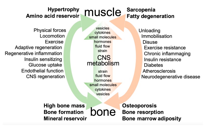
“Many interactions are not only bilateral but follow a multilateral or even organismic relationship, involving metabolism and the nervous system (Figure 1).”
§ Interactions between Muscle and Bone—Where Physics Meets Biology > 1. Introduction
plaincorrelational- Asserted: passes — The authors put it forward as true.
- Checkable: passes — It can be checked against evidence.
- Load-bearing: unclear — It is unclear whether the paper’s conclusions depend on it.
- Based: passes — The paper gives a reason why it holds.
A conclusion the paper draws from its own results — it cites no other source for it, so there is none to check. The quote shows it is in the paper.
In the paper
“The acute effects of exercise induce the secretion of exosomes with cargo molecules that are capable of mediating the endocrine effects between muscle, bone, and the organism.”
§ Interactions between Muscle and Bone—Where Physics Meets Biology > Abstract
plaindirect- Asserted: passes — The authors put it forward as true.
- Checkable: passes — It can be checked against evidence.
- Load-bearing: passes — The paper’s conclusions depend on it.
- Based: unclear — It is unclear why the paper says it holds.
The paper states this without a source or data behind it here, so there is nothing to check it against. The quote shows it is in the paper.
In the paper
“Cells sense mechanical strain via adhesion molecules and translate it into biochemical responses, modulating the basic mechanisms of cellular biology such as lineage commitment, tissue formation, and maturation.”
§ Interactions between Muscle and Bone—Where Physics Meets Biology > Abstract
plaindirect- Asserted: passes — The authors put it forward as true.
- Checkable: passes — It can be checked against evidence.
- Load-bearing: passes — The paper’s conclusions depend on it.
- Based: unclear — It is unclear why the paper says it holds.
The paper states this without a source or data behind it here, so there is nothing to check it against. The quote shows it is in the paper.
In the paper
“Lessons from unloading, microgravity, and disuse teach us that gratuitous tissue is removed or reorganized”
§ Interactions between Muscle and Bone—Where Physics Meets Biology > Abstract
plaindirect- Asserted: passes — The authors put it forward as true.
- Checkable: passes — It can be checked against evidence.
- Load-bearing: passes — The paper’s conclusions depend on it.
- Based: unclear — It is unclear why the paper says it holds.
The paper states this without a source or data behind it here, so there is nothing to check it against. The quote shows it is in the paper.
In the paper
“Secreted growth and differentiation factors are important effectors of mutual interaction.”
§ Interactions between Muscle and Bone—Where Physics Meets Biology > Abstract
plaindirect- Asserted: passes — The authors put it forward as true.
- Checkable: passes — It can be checked against evidence.
- Load-bearing: passes — The paper’s conclusions depend on it.
- Based: unclear — It is unclear why the paper says it holds.
None of the sources cited for this claim have been checked yet.
In the paper
“Muscle and bone interact via physical forces and secreted osteokines and myokines.”
§ Interactions between Muscle and Bone—Where Physics Meets Biology > Abstract
What the sources say
- Not checked yetcites [1] Felsenthal, 2017, Mechanical regulation of musculoskeletal system development, Dev. source
- Not checked yetcites [2] Mikolajewicz, 2019, Transmission of Mechanical Information by Purinergic Signaling, Biophys. J. source
- Not checked yetcites [3] Vogel, 2018, Unraveling the Mechanobiology of Extracellular Matrix, Annu. Rev. Physiol. source
- Not checked yetcites [4] Ayad, 2019, Tissue mechanics, an important regulator of development and disease, Philos. Trans. R. Soc. B: Boil. Sci. source
plaindirect- Asserted: passes — The authors put it forward as true.
- Checkable: passes — It can be checked against evidence.
- Load-bearing: passes — The paper’s conclusions depend on it.
- Based: passes — The paper gives a reason why it holds.
A conclusion the paper draws from its own results — it cites no other source for it, so there is none to check. The quote shows it is in the paper.
In the paper
“and propagate degenerative diseases such as sarcopenia and osteoporosis.”
§ Interactions between Muscle and Bone—Where Physics Meets Biology > Abstract
plaindirect- Asserted: passes — The authors put it forward as true.
- Checkable: passes — It can be checked against evidence.
- Load-bearing: passes — The paper’s conclusions depend on it.
- Based: unclear — It is unclear why the paper says it holds.
None of the sources cited for this claim have been checked yet.
In the paper
“Recent information indicates that the reciprocity of mechanochemical interaction is extremely dynamic, including the cellular, the tissue, and the organismic level [1,2,3,4].”
§ Interactions between Muscle and Bone—Where Physics Meets Biology > 1. Introduction
What the sources say
- Not checked yetcites [1] Felsenthal, 2017, Mechanical regulation of musculoskeletal system development, Dev. source
- Not checked yetcites [2] Mikolajewicz, 2019, Transmission of Mechanical Information by Purinergic Signaling, Biophys. J. source
- Not checked yetcites [3] Vogel, 2018, Unraveling the Mechanobiology of Extracellular Matrix, Annu. Rev. Physiol. source
- Not checked yetcites [4] Ayad, 2019, Tissue mechanics, an important regulator of development and disease, Philos. Trans. R. Soc. B: Boil. Sci. source
hedgedcorrelational- Asserted: passes — The authors put it forward as true.
- Checkable: passes — It can be checked against evidence.
- Load-bearing: unclear — It is unclear whether the paper’s conclusions depend on it.
- Based: passes — The paper gives a reason why it holds.
The paper states this without a source or data behind it here, so there is nothing to check it against. The quote shows it is in the paper.
In the paper
“The physical forces generated in the context of motion are extremely variable from very low to high impact.”
§ Interactions between Muscle and Bone—Where Physics Meets Biology > 2. Principles of Interaction between Muscle and Bone > 2.1. Physical Forces
plain- Asserted: passes — The authors put it forward as true.
- Checkable: passes — It can be checked against evidence.
- Load-bearing: unclear — It is unclear whether the paper’s conclusions depend on it.
- Based: passes — The paper gives a reason why it holds.
In the paper
“Connective tissues physically joining muscle and bone, especially tendons and ligaments, naturally play a critical role in this interaction, as addressed in a recent review article in detail [5].”
§ Interactions between Muscle and Bone—Where Physics Meets Biology > 1. Introduction
What the sources say
- Not checked yetcites [5] Nassari, 2017, Non-myogenic Contribution to Muscle Development and Homeostasis: The Role of Connective Tissues, Front. Cell Dev. Boil.
plaindirect- Asserted: passes — The authors put it forward as true.
- Checkable: passes — It can be checked against evidence.
- Load-bearing: unclear — It is unclear whether the paper’s conclusions depend on it.
- Based: passes — The paper gives a reason why it holds.
- It may state two separate claims as one. 59
- Probably not a substantial claim. 15
- The quote only partly states this claim. 2
In the paper
“After adjustment for BMD and cortical thickness, the hip fracture group was characterized by the lower relative adipose tissue volume of the upper thigh and some of the texture parameters discriminated the osteoporotic hip fracture [204].”
§ Interactions between Muscle and Bone—Where Physics Meets Biology > 4. Muscle and Bone Interactions in Disease—Lessons and Backflashes from Pathology > 4.2. Inflammation and Fatty Degeneration—Lessons from Imaging and Structural Analyses
What the sources say
- Not checked yetcites [204] Museyko, 2019, Three-dimensional Distribution of Muscle and Adipose Tissue of the Thigh at CT: Association with Acute Hip Fracture, Radiol. source
plaincorrelationaln = 40- Asserted: passes — The authors put it forward as true.
- Checkable: passes — It can be checked against evidence.
- Load-bearing: passes — The paper’s conclusions depend on it.
- Based: passes — The paper gives a reason why it holds.
It may state two separate claims as one.
The paper states this without a source or data behind it here, so there is nothing to check it against. The quote shows it is in the paper.
In the paper
“Physical forces are generated through gravity, locomotion, exercise, and external devices.”
§ Interactions between Muscle and Bone—Where Physics Meets Biology > Abstract
plaindirect- Asserted: passes — The authors put it forward as true.
- Checkable: passes — It can be checked against evidence.
- Load-bearing: passes — The paper’s conclusions depend on it.
- Based: fails — The paper may give no reason why it holds.
Probably not a substantial claim. The paper may give no reason why it holds.
A conclusion the paper draws from its own results — it cites no other source for it, so there is none to check. The quote shows it is in the paper.
In the paper
“This may result in the initiation of bone formation, muscle hypertrophy, and the enhanced production of extracellular matrix constituents, adhesion molecules, and cytoskeletal elements.”
§ Interactions between Muscle and Bone—Where Physics Meets Biology > Abstract
hedgeddirect- Asserted: passes — The authors put it forward as true.
- Checkable: passes — It can be checked against evidence.
- Load-bearing: passes — The paper’s conclusions depend on it.
- Based: passes — The paper gives a reason why it holds.
It may state two separate claims as one.
The paper states this without a source or data behind it here, so there is nothing to check it against. The quote shows it is in the paper.
In the paper
“immobility and inflammation trigger muscle and bone marrow fatty infiltration”
§ Interactions between Muscle and Bone—Where Physics Meets Biology > Abstract
plaindirect- Asserted: passes — The authors put it forward as true.
- Checkable: passes — It can be checked against evidence.
- Load-bearing: passes — The paper’s conclusions depend on it.
- Based: fails — The paper may give no reason why it holds.
Probably not a substantial claim. The paper may give no reason why it holds.
A conclusion the paper draws from its own results — it cites no other source for it, so there is none to check. The quote shows it is in the paper.
In the paper
“Bone and muscle mass, resistance to strain, and the stiffness of matrix, cells, and tissues are enhanced, influencing fracture resistance and muscle power.”
§ Interactions between Muscle and Bone—Where Physics Meets Biology > Abstract
plaindirect- Asserted: passes — The authors put it forward as true.
- Checkable: passes — It can be checked against evidence.
- Load-bearing: passes — The paper’s conclusions depend on it.
- Based: passes — The paper gives a reason why it holds.
It may state two separate claims as one.
In the paper
“The former prevail in muscle while bone strongly (but not exclusively) responds to fluid flow in the osteocyte canalicular network [9].”
§ Interactions between Muscle and Bone—Where Physics Meets Biology > 2. Principles of Interaction between Muscle and Bone > 2.1. Physical Forces
What the sources say
- Not checked yetcites [9] Nachury, 2019, Establishing and regulating the composition of cilia for signal transduction, Nat. Rev. Mol. Cell Boil. source
boosteddirect- Asserted: passes — The authors put it forward as true.
- Checkable: passes — It can be checked against evidence.
- Load-bearing: passes — The paper’s conclusions depend on it.
- Based: passes — The paper gives a reason why it holds.
It may state two separate claims as one.
In the paper
““virtually every step of cell and tissue biology depends upon mechanochemical events””
§ Interactions between Muscle and Bone—Where Physics Meets Biology > 2. Principles of Interaction between Muscle and Bone > 2.1. Physical Forces
What the sources say
- Not checked yetcites [18] Storm, 2017, Mechanoreciprocity in cell migration, Nat.
boosteddirect- Asserted: passes — The authors put it forward as true.
- Checkable: passes — It can be checked against evidence.
- Load-bearing: fails — The paper’s conclusions may not depend on it.
- Based: passes — The paper gives a reason why it holds.
Probably not a substantial claim. The paper’s conclusions may not depend on it.
A conclusion the paper draws from its own results — it cites no other source for it, so there is none to check. The quote shows it is in the paper.
In the paper
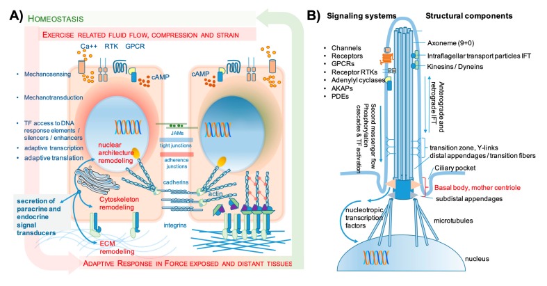
“The clinical result of physical impact is high bone and muscle mass, resulting in enhanced fracture resistance and muscle power (see also Figure 2A,B).”
§ Interactions between Muscle and Bone—Where Physics Meets Biology > 2. Principles of Interaction between Muscle and Bone > 2.1. Physical Forces
plaindirect- Asserted: passes — The authors put it forward as true.
- Checkable: passes — It can be checked against evidence.
- Load-bearing: passes — The paper’s conclusions depend on it.
- Based: passes — The paper gives a reason why it holds.
It may state two separate claims as one.
The paper states this without a source or data behind it here, so there is nothing to check it against. The quote shows it is in the paper.
In the paper
“Mechanical stimuli can be transmitted through deformation (stretching and compression) and/or shear strain applied by fluids that pass by.”
§ Interactions between Muscle and Bone—Where Physics Meets Biology > 2. Principles of Interaction between Muscle and Bone > 2.1. Physical Forces
plaindirect- Asserted: passes — The authors put it forward as true.
- Checkable: passes — It can be checked against evidence.
- Load-bearing: passes — The paper’s conclusions depend on it.
- Based: passes — The paper gives a reason why it holds.
It may state two separate claims as one.
A conclusion the paper draws from its own results — it cites no other source for it, so there is none to check. The quote shows it is in the paper.
In the paper
“There is no doubt that external mechanical vibration may variably substitute for gravity and add up to active muscle contraction both in space missions and as dedicated types of exercise.”
§ Interactions between Muscle and Bone—Where Physics Meets Biology > 2. Principles of Interaction between Muscle and Bone > 2.1. Physical Forces
boostedconditional- Asserted: passes — The authors put it forward as true.
- Checkable: passes — It can be checked against evidence.
- Load-bearing: passes — The paper’s conclusions depend on it.
- Based: passes — The paper gives a reason why it holds.
It may state two separate claims as one.
In the paper
“In human myoblasts, Cav3 deficiency was associated with the hyperactivation of IL-6-STAT3 signaling (interleukin-6, signal transducer and activator of transcription 3), which is a pathway that in the presence of WT Cav3 turned out to be regulated by mechanical strain in a Cav3-dependent manner [35].”
§ Interactions between Muscle and Bone—Where Physics Meets Biology > 2. Principles of Interaction between Muscle and Bone > 2.1. Physical Forces
What the sources say
- Not checked yetcites [35] Dewulf, 2019, Dystrophy-associated caveolin-3 mutations reveal that caveolae couple IL6/STAT3 signaling with mechanosensing in human muscle cells, Nat. Commun. source
plaincorrelational- Asserted: passes — The authors put it forward as true.
- Checkable: passes — It can be checked against evidence.
- Load-bearing: unclear — It is unclear whether the paper’s conclusions depend on it.
- Based: passes — The paper gives a reason why it holds.
It may state two separate claims as one.
None of the sources cited for this claim have been checked yet.
In the paper
“Mechanical bending/deflection of the cilium was reported to enhance prostaglandin-endoperoxide synthase 2 (PTGS2 alias COX2) expression and prostaglandin production as well as bone morphogenetic protein 2 (BMP 2) and osteopontin expression.”
§ Interactions between Muscle and Bone—Where Physics Meets Biology > 2. Principles of Interaction between Muscle and Bone > 2.1. Physical Forces
plaindirect- Asserted: passes — The authors put it forward as true.
- Checkable: passes — It can be checked against evidence.
- Load-bearing: unclear — It is unclear whether the paper’s conclusions depend on it.
- Based: passes — The paper gives a reason why it holds.
It may state two separate claims as one.
The paper states this without a source or data behind it here, so there is nothing to check it against. The quote shows it is in the paper.
In the paper
“Secreted proteins and signaling molecules are the basis of mutual interaction between muscle and bone both in a paracrine and also an endocrine setting.”
§ Interactions between Muscle and Bone—Where Physics Meets Biology > 2. Principles of Interaction between Muscle and Bone > 2.2. Systemic Mutual Interactions of Skeletal Muscle and Bone
plaindirect- Asserted: passes — The authors put it forward as true.
- Checkable: passes — It can be checked against evidence.
- Load-bearing: passes — The paper’s conclusions depend on it.
- Based: fails — The paper may give no reason why it holds.
Probably not a substantial claim. The paper may give no reason why it holds.
In the paper
“In muscle, mechanosensing is mainly transduced via deformation strains, but still, the primary cilium has been related to satellite cell self-renewal and muscle cell differentiation in mice [42].”
§ Interactions between Muscle and Bone—Where Physics Meets Biology > 2. Principles of Interaction between Muscle and Bone > 2.1. Physical Forces
What the sources say
- Not checked yetcites [42] Marican, 2016, Asymmetric Distribution of Primary Cilia Allocates Satellite Cells for Self-Renewal, Stem Cell Rep. source
plaincorrelational- Asserted: passes — The authors put it forward as true.
- Checkable: passes — It can be checked against evidence.
- Load-bearing: unclear — It is unclear whether the paper’s conclusions depend on it.
- Based: passes — The paper gives a reason why it holds.
It may state two separate claims as one.
The paper states this without a source or data behind it here, so there is nothing to check it against. The quote shows it is in the paper.
In the paper
“The primary cilium is a unique subcellular organelle composed of nine (+0) axonema structures (Figure 2B).”
§ Interactions between Muscle and Bone—Where Physics Meets Biology > 2. Principles of Interaction between Muscle and Bone > 2.1. Physical Forces
plain- Asserted: passes — The authors put it forward as true.
- Checkable: passes — It can be checked against evidence.
- Load-bearing: fails — The paper’s conclusions may not depend on it.
- Based: fails — The paper may give no reason why it holds.
Probably not a substantial claim. The paper’s conclusions may not depend on it. The paper may give no reason why it holds.
In the paper
“In bone, the primary cilium appears to be an important mediator of gravity and consecutive osteogenic response, because in simulated microgravity, the cilium gradually shrinks and inhibition of this shrinkage using cytochalasin D maintains osteoblast differentiation capacity (reviewed in [40]).”
§ Interactions between Muscle and Bone—Where Physics Meets Biology > 2. Principles of Interaction between Muscle and Bone > 2.1. Physical Forces
What the sources say
- Not checked yetcites [40] Shi, 2019, Novel functions of the primary cilium in bone disease and cancer, Cytoskelet. source
hedgeddirect- Asserted: passes — The authors put it forward as true.
- Checkable: passes — It can be checked against evidence.
- Load-bearing: unclear — It is unclear whether the paper’s conclusions depend on it.
- Based: passes — The paper gives a reason why it holds.
It may state two separate claims as one.
A conclusion the paper draws from its own results — it cites no other source for it, so there is none to check. The quote shows it is in the paper.
In the paper
“IGF-1 and also FGF2 are growth factors that are actively secreted at the muscle and bone interface and if delivered into the circulation may have distant effects on bone formation and remodeling.”
§ Interactions between Muscle and Bone—Where Physics Meets Biology > 2. Principles of Interaction between Muscle and Bone > 2.2. Systemic Mutual Interactions of Skeletal Muscle and Bone
hedgedconditional- Asserted: passes — The authors put it forward as true.
- Checkable: passes — It can be checked against evidence.
- Load-bearing: unclear — It is unclear whether the paper’s conclusions depend on it.
- Based: passes — The paper gives a reason why it holds.
It may state two separate claims as one.
None of the sources cited for this claim have been checked yet.
In the paper
“EVs may transport myokines/osteokines, miRNA, and organelles and are involved in the regeneration of both bone and muscle tissue after exercise and injury [70,71,72,73,74].”
§ Interactions between Muscle and Bone—Where Physics Meets Biology > 2. Principles of Interaction between Muscle and Bone > 2.2. Systemic Mutual Interactions of Skeletal Muscle and Bone
What the sources say
- Not checked yetcites [70] Bittel, 2019, Contribution of Extracellular Vesicles in Rebuilding Injured Muscles, Front. Physiol. source
- Not checked yetcites [71] Lauritzen, 2013, Contraction and AICAR Stimulate IL-6 Vesicle Depletion From Skeletal Muscle Fibers In Vivo, Diabetes source
- Not checked yetcites [72] Morrell, 2018, Mechanically induced Ca2+ oscillations in osteocytes release extracellular vesicles and enhance bone formation, Bone Res. source
- Not checked yetcites [73] Murray, 2018, Concise Review: Intercellular Communication Via Organelle Transfer in the Biology and Therapeutic Applications of Stem Cells, STEM CELLS source
- Not checked yetcites [74] Trovato, 2019, Extracellular Vesicles: Delivery Vehicles of Myokines, Front. Physiol. source
hedgedconditional- Asserted: passes — The authors put it forward as true.
- Checkable: passes — It can be checked against evidence.
- Load-bearing: passes — The paper’s conclusions depend on it.
- Based: passes — The paper gives a reason why it holds.
It may state two separate claims as one.
None of the sources cited for this claim have been checked yet.
In the paper
“Muscle cells, very much like bone, do not express the molecular components of the regulated secretory pathway (RSP), and the vesicle release upon exercise may be the relevant mechanism to deliver IL-6 into the circulation.”
§ Interactions between Muscle and Bone—Where Physics Meets Biology > 2. Principles of Interaction between Muscle and Bone > 2.2. Systemic Mutual Interactions of Skeletal Muscle and Bone
hedgedconditional- Asserted: passes — The authors put it forward as true.
- Checkable: passes — It can be checked against evidence.
- Load-bearing: unclear — It is unclear whether the paper’s conclusions depend on it.
- Based: passes — The paper gives a reason why it holds.
It may state two separate claims as one.
None of the sources cited for this claim have been checked yet.
In the paper
“Exercise was reported to downregulate both SOST and DKK-1 serum levels, which marks an important link between muscle and bone via physical impact [78,79].”
§ Interactions between Muscle and Bone—Where Physics Meets Biology > 2. Principles of Interaction between Muscle and Bone > 2.2. Systemic Mutual Interactions of Skeletal Muscle and Bone
What the sources say
- Not checked yetcites [78] Davoodi, 2019, Improved carotid intima-media thickness-induced high-intensity interval training associated with decreased serum levels of Dkk-1 and sclerostin in type 2 diabetes, J. Diabetes its Complicat.
- Not checked yetcites [79] Picke, 2018, Differential effects of high-fat diet and exercise training on bone and energy metabolism, Bone source
plaindirect- Asserted: passes — The authors put it forward as true.
- Checkable: passes — It can be checked against evidence.
- Load-bearing: passes — The paper’s conclusions depend on it.
- Based: passes — The paper gives a reason why it holds.
It may state two separate claims as one.
In the paper
“One example is the immediate rise of muscle insulin-like growth factor 1 (IGF-1) mRNA after an exercise bout and the significant rise of peripheral blood IGF-1 protein levels after 24 h (not yet significant after 2 h) post exercise, which was gone after 48 h [75].”
§ Interactions between Muscle and Bone—Where Physics Meets Biology > 2. Principles of Interaction between Muscle and Bone > 2.2. Systemic Mutual Interactions of Skeletal Muscle and Bone
What the sources say
- Not checked yetcites [75] Annibalini, 2019, Muscle and Systemic Molecular Responses to a Single Flywheel Based Iso-Inertial Training Session in Resistance-Trained Men, Front. Physiol. source
plaindirect- Asserted: passes — The authors put it forward as true.
- Checkable: passes — It can be checked against evidence.
- Load-bearing: unclear — It is unclear whether the paper’s conclusions depend on it.
- Based: passes — The paper gives a reason why it holds.
It may state two separate claims as one.
In the paper
“Small vesicles in muscle, which contain immunoreactive IL-6 and are depleted during exercise, have indeed been described in mice [71].”
§ Interactions between Muscle and Bone—Where Physics Meets Biology > 2. Principles of Interaction between Muscle and Bone > 2.2. Systemic Mutual Interactions of Skeletal Muscle and Bone
What the sources say
- Not checked yetcites [71] Lauritzen, 2013, Contraction and AICAR Stimulate IL-6 Vesicle Depletion From Skeletal Muscle Fibers In Vivo, Diabetes source
boosteddirect- Asserted: passes — The authors put it forward as true.
- Checkable: passes — It can be checked against evidence.
- Load-bearing: unclear — It is unclear whether the paper’s conclusions depend on it.
- Based: passes — The paper gives a reason why it holds.
It may state two separate claims as one.
None of the sources cited for this claim have been checked yet.
In the paper
“Similarly, the Hippo pathway has gained attention as an important pathway in mechanotransduction and skeletal physiology. Downstream YAP/TAZ regulation is in fact tightly involved in mechanosensing and mechanotransduction both in bone and muscle as well as in many other cells, especially endothelial cells (see Section 3.3).”
§ Interactions between Muscle and Bone—Where Physics Meets Biology > 3. Common Effector Mechanisms in Muscle and Bone from Development to Postnatal Regeneration > 3.2. Common Effectors of Muscle and Bone Interaction Modulate Tissue Formation and Remodeling
What the sources say
- Not checked yetcites [55] Dasgupta, 2019, Control of cellular responses to mechanical cues through YAP/TAZ regulation, J. Boil. Chem. source
- Not checked yetcites [115] Meng, 2018, RAP2 mediates mechanoresponses of the Hippo pathway, Nat. source
- Not checked yetcites [116] Xiang, 2018, The versatile hippo pathway in oral-maxillofacial development and bone remodeling, Dev. Boil. source
boostedcorrelational- Asserted: passes — The authors put it forward as true.
- Checkable: passes — It can be checked against evidence.
- Load-bearing: unclear — It is unclear whether the paper’s conclusions depend on it.
- Based: passes — The paper gives a reason why it holds.
It may state two separate claims as one.
A conclusion the paper draws from its own results — it cites no other source for it, so there is none to check. The quote shows it is in the paper.
In the paper
“In conclusion, there is convincing evidence that during development, the interaction between physics and biology is boosted by EMT and the acquisition of migratory capacity, and every step of interaction causes and supports biological consequences in terms of lineage commitment and maturation.”
§ Interactions between Muscle and Bone—Where Physics Meets Biology > 3. Common Effector Mechanisms in Muscle and Bone from Development to Postnatal Regeneration > 3.1. Role of Common Musculoskeletal Precursor’s Mechanotransduction during Development
boosteddirect- Asserted: passes — The authors put it forward as true.
- Checkable: passes — It can be checked against evidence.
- Load-bearing: passes — The paper’s conclusions depend on it.
- Based: unclear — It is unclear why the paper says it holds.
It may state two separate claims as one.
In the paper
“Irisin, a cleaved product of fibronectin type III domain containing (FNDC5) with stimulating effects on bone formation, is also delivered from muscle into the circulation after exercise but may have a dual mode of delivery, shedding of preformed polypeptides and de novo synthesis [81].”
§ Interactions between Muscle and Bone—Where Physics Meets Biology > 2. Principles of Interaction between Muscle and Bone > 2.2. Systemic Mutual Interactions of Skeletal Muscle and Bone
What the sources say
- Not checked yetcites [81] Li, 2019, Muscle-bone crosstalk and potential therapies for sarco-osteoporosis, J. Cell. Biochem. source
hedgedconditional- Asserted: passes — The authors put it forward as true.
- Checkable: passes — It can be checked against evidence.
- Load-bearing: unclear — It is unclear whether the paper’s conclusions depend on it.
- Based: passes — The paper gives a reason why it holds.
It may state two separate claims as one.
In the paper
“Above all, the PTH1R signaling cascade is especially sensitive to mechanical loading. Anabolic PTH1R signals elicited through bone anabolic treatment using intermittent 1-34 PTH (teriparatide) are almost ineffective under conditions of unloading [113].”
§ Interactions between Muscle and Bone—Where Physics Meets Biology > 3. Common Effector Mechanisms in Muscle and Bone from Development to Postnatal Regeneration > 3.2. Common Effectors of Muscle and Bone Interaction Modulate Tissue Formation and Remodeling
What the sources say
- Not checked yetcites [113] Tu, 2016, Control of Bone Anabolism in Response to Mechanical Loading and PTH by Distinct Mechanisms Downstream of the PTH Receptor, J. Bone Miner. Res.
plaindirect- Asserted: passes — The authors put it forward as true.
- Checkable: passes — It can be checked against evidence.
- Load-bearing: passes — The paper’s conclusions depend on it.
- Based: passes — The paper gives a reason why it holds.
It may state two separate claims as one.
In the paper
“TSP-4 is expressed in muscle and bone, has different functions compared to TSP-1 (antiangiogenic), and recently has been assigned to angiogenesis and matrix remodeling [94].”
§ Interactions between Muscle and Bone—Where Physics Meets Biology > 2. Principles of Interaction between Muscle and Bone > 2.2. Systemic Mutual Interactions of Skeletal Muscle and Bone
What the sources say
- Not checked yetcites [94] Stenina-Adognravi, O., and Plow, E.F. (2019). Thrombospondin-4 in tissue remodeling. Matrix Boil., 300–313. source
plaincorrelational- Asserted: passes — The authors put it forward as true.
- Checkable: passes — It can be checked against evidence.
- Load-bearing: unclear — It is unclear whether the paper’s conclusions depend on it.
- Based: passes — The paper gives a reason why it holds.
It may state two separate claims as one.
None of the sources cited for this claim have been checked yet.
In the paper
“Joint structures as important features of the musculoskeletal functional unit develop under the influence of respective core signaling cascades of osteogenic, chondrogenic, and ligamentogenic pathways. This process is fundamentally influenced by mechanical forces already in utero, directed by movement of the fetus [45,101].”
§ Interactions between Muscle and Bone—Where Physics Meets Biology > 3. Common Effector Mechanisms in Muscle and Bone from Development to Postnatal Regeneration > 3.1. Role of Common Musculoskeletal Precursor’s Mechanotransduction during Development
What the sources say
plaindirect- Asserted: passes — The authors put it forward as true.
- Checkable: passes — It can be checked against evidence.
- Load-bearing: unclear — It is unclear whether the paper’s conclusions depend on it.
- Based: passes — The paper gives a reason why it holds.
It may state two separate claims as one.
A conclusion the paper draws from its own results — it cites no other source for it, so there is none to check. The quote shows it is in the paper.
In the paper
“These data demonstrate that there are genes whose expression is relevant for tissue formation in both bone and muscle, and others cause antagonistic effects in the brother tissues.”
§ Interactions between Muscle and Bone—Where Physics Meets Biology > 3. Common Effector Mechanisms in Muscle and Bone from Development to Postnatal Regeneration
boosteddirect- Asserted: passes — The authors put it forward as true.
- Checkable: passes — It can be checked against evidence.
- Load-bearing: passes — The paper’s conclusions depend on it.
- Based: passes — The paper gives a reason why it holds.
It may state two separate claims as one.
None of the sources cited for this claim have been checked yet.
In the paper
“Notch signaling has also been reported to be involved in skeletal physiology. We and others recently reported that this pathway is mechanosensitive in skeletal precursors [58,114].”
§ Interactions between Muscle and Bone—Where Physics Meets Biology > 3. Common Effector Mechanisms in Muscle and Bone from Development to Postnatal Regeneration > 3.2. Common Effectors of Muscle and Bone Interaction Modulate Tissue Formation and Remodeling
What the sources say
- Not checked yetcites [58] Ziouti, F., Ebert, R., Rummler, M., Krug, M., Müller-Deubert, S., Lüdemann, M., Jakob, F., Willie, B.M., and Jundt, F. (2019). NOTCH Signaling Is Activated through Mechanical Strain in Human Bone Marrow-Derived Mesenchymal Stromal Cells. Stem Cells Int., 2019. source
- Not checked yetcites [114] Canalis, 2018, Notch in skeletal physiology and disease, Osteoporos. Int. source
plaincorrelational- Asserted: passes — The authors put it forward as true.
- Checkable: passes — It can be checked against evidence.
- Load-bearing: passes — The paper’s conclusions depend on it.
- Based: passes — The paper gives a reason why it holds.
It may state two separate claims as one.
None of the sources cited for this claim have been checked yet.
In the paper
“Interestingly, the early genes such as proinflammatory cytokines IL-1b, IL-6, IL-8, the toll-like receptor 4 (TLR4) ligands serum amyloid A1 (SAA1) and SAA2, CYR61/CCN1, connective tissue growth factor (CTGF/CCN2), angiopoietin-like 4 (ANGPTL4) and ANGPTL2 very closely resemble genes that are also relevant in the initial phase of osteogenic differentiation and contribute to angiogenesis in fract”
§ Interactions between Muscle and Bone—Where Physics Meets Biology > 2. Principles of Interaction between Muscle and Bone > 2.2. Systemic Mutual Interactions of Skeletal Muscle and Bone
What the sources say
- Not checked yetcites [91] Ebert, 2015, Acute phase serum amyloid A induces proinflammatory cytokines and mineralization via toll-like receptor 4 in mesenchymal stem cells, Stem Cell Res. source
- Not checked yetcites [92] Loi, 2016, Inflammation, fracture and bone repair, Bone source
- Not checked yetcites [93] Sass, F.A., Fuchs, M., Pumberger, M., Geissler, S., Duda, G.N., Perka, C., and Schmidt-Bleek, K. (2018). Immunology Guides Skeletal Muscle Regeneration. Int. J. Mol. Sci., 19. source
plaincorrelational- Asserted: passes — The authors put it forward as true.
- Checkable: passes — It can be checked against evidence.
- Load-bearing: passes — The paper’s conclusions depend on it.
- Based: passes — The paper gives a reason why it holds.
It may state two separate claims as one.
In the paper
“The lists of muscle-related transcripts and secreted proteins comprised 149 proteins of unassigned functions as putative interactors with other tissues and 123 transcripts that corresponded to putative myokines including BMPs, chemokines, cytokines, growth/differentiation factors (GDFs), and members of the tumor necrosis factor (TNF) family of secreted proteins and ligands for Wnt signaling (Suppl”
§ Interactions between Muscle and Bone—Where Physics Meets Biology > 2. Principles of Interaction between Muscle and Bone > 2.2. Systemic Mutual Interactions of Skeletal Muscle and Bone
What the sources say
- Not checked yetcites [90] Zhang, 2018, Transcriptional profiling reveals extraordinary diversity among skeletal muscle tissues, eLife source
hedged- Asserted: passes — The authors put it forward as true.
- Checkable: passes — It can be checked against evidence.
- Load-bearing: unclear — It is unclear whether the paper’s conclusions depend on it.
- Based: passes — The paper gives a reason why it holds.
It may state two separate claims as one.
None of the sources cited for this claim have been checked yet.
In the paper
“Bone formation and bone resorption are both part of the adaptive refinement and remodeling processes, which are mechanically guided and where both osteoblasts and osteoclasts respond to physical forces [117,118].”
§ Interactions between Muscle and Bone—Where Physics Meets Biology > 3. Common Effector Mechanisms in Muscle and Bone from Development to Postnatal Regeneration > 3.2. Common Effectors of Muscle and Bone Interaction Modulate Tissue Formation and Remodeling
What the sources say
- Not checked yetcites [117] Amarasekara, D.S., Yun, H., Kim, S., Lee, N., Kim, H., and Rho, J. (2018). Regulation of Osteoclast Differentiation by Cytokine Networks. Immune Netw., 18. source
- Not checked yetcites [118] Ma, 2019, The role of physical forces in osteoclastogenesis, J. Cell. Physiol. source
plaindirect- Asserted: passes — The authors put it forward as true.
- Checkable: passes — It can be checked against evidence.
- Load-bearing: passes — The paper’s conclusions depend on it.
- Based: passes — The paper gives a reason why it holds.
It may state two separate claims as one.
In the paper
“However, there are some other populations that substantially contribute to regeneration such as the Sca1+/CD34+ fibro-adipogenic progenitors (FAPs) residing in the muscle interstitium. Following injury, satellite cells re-enter the cell cycle and proliferate to give rise to myoblasts that differentiate and fuse to restore the damaged fiber or generate myofibers de novo, while FAPs support their”
§ Interactions between Muscle and Bone—Where Physics Meets Biology > 3. Common Effector Mechanisms in Muscle and Bone from Development to Postnatal Regeneration > 3.2. Common Effectors of Muscle and Bone Interaction Modulate Tissue Formation and Remodeling
What the sources say
- Not checked yetcites [131] Baghdadi, 2018, Regulation and phylogeny of skeletal muscle regeneration, Dev. Boil. source
plaindirect- Asserted: passes — The authors put it forward as true.
- Checkable: passes — It can be checked against evidence.
- Load-bearing: unclear — It is unclear whether the paper’s conclusions depend on it.
- Based: passes — The paper gives a reason why it holds.
It may state two separate claims as one.
None of the sources cited for this claim have been checked yet.
In the paper
“Very much in analogy with bone and probably many other tissues, the initial phase of regeneration is governed by intrinsic inflammatory signals and is further induced by cells of the innate immune system, e.g. sequentially recruited neutrophils and resident macrophages. Cytokines already known from bone regeneration such as IL-1, 6, and 8 are involved in innate immune cell recruitment until the”
§ Interactions between Muscle and Bone—Where Physics Meets Biology > 3. Common Effector Mechanisms in Muscle and Bone from Development to Postnatal Regeneration > 3.2. Common Effectors of Muscle and Bone Interaction Modulate Tissue Formation and Remodeling
What the sources say
- Not checked yetcites [132] Dort, J., Fabre, P., Molina, T., and Dumont, N.A. (2019). Macrophages Are Key Regulators of Stem Cells during Skeletal Muscle Regeneration and Diseases. Stem Cells Int., 2019. source
- Not checked yetcites [133] Mukund, 2019, Skeletal muscle: A review of molecular structure and function, in health and disease, Wiley Interdiscip. Rev. Syst. Boil. Med. source
hedgeddirect- Asserted: passes — The authors put it forward as true.
- Checkable: passes — It can be checked against evidence.
- Load-bearing: unclear — It is unclear whether the paper’s conclusions depend on it.
- Based: passes — The paper gives a reason why it holds.
It may state two separate claims as one.
In the paper
“Both Pax7-deficient and satellite cell-deficient mice fail to regenerate muscle [131].”
§ Interactions between Muscle and Bone—Where Physics Meets Biology > 3. Common Effector Mechanisms in Muscle and Bone from Development to Postnatal Regeneration > 3.2. Common Effectors of Muscle and Bone Interaction Modulate Tissue Formation and Remodeling
What the sources say
- Not checked yetcites [131] Baghdadi, 2018, Regulation and phylogeny of skeletal muscle regeneration, Dev. Boil. source
plaindirect- Asserted: passes — The authors put it forward as true.
- Checkable: passes — It can be checked against evidence.
- Load-bearing: unclear — It is unclear whether the paper’s conclusions depend on it.
- Based: passes — The paper gives a reason why it holds.
It may state two separate claims as one.
None of the sources cited for this claim have been checked yet.
In the paper
“Not only is this condition in common for regenerating tissues in the early phase, but it also somehow mimics the senescence-associated secretory phenotype (SASP) of presenescent and senescent cells. It appears to be beneficial if intermittent, but it causes pathology if chronically active.”
§ Interactions between Muscle and Bone—Where Physics Meets Biology > 3. Common Effector Mechanisms in Muscle and Bone from Development to Postnatal Regeneration > 3.2. Common Effectors of Muscle and Bone Interaction Modulate Tissue Formation and Remodeling
What the sources say
- Not checked yetcites [109] Czarkwiani, 2018, Out with the old, in with the new: senescence in development, Curr. Opin. Cell Boil. source
- Not checked yetcites [125] Farr, 2019, Cellular senescence in bone, Bone source
- Not checked yetcites [126] He, 2017, Senescence in Health and Disease, Cell source
- Not checked yetcites [127] Rhinn, 2019, Cellular senescence in development, regeneration and disease, Dev. source
hedgeddirect- Asserted: passes — The authors put it forward as true.
- Checkable: passes — It can be checked against evidence.
- Load-bearing: unclear — It is unclear whether the paper’s conclusions depend on it.
- Based: passes — The paper gives a reason why it holds.
It may state two separate claims as one.
In the paper
“Satellite stem cell development in addition to Pax3 expression is largely dependent on the expression of Pax7 in the pre- and perinatal periods and modulated by mechanical strain [32].”
§ Interactions between Muscle and Bone—Where Physics Meets Biology > 3. Common Effector Mechanisms in Muscle and Bone from Development to Postnatal Regeneration > 3.2. Common Effectors of Muscle and Bone Interaction Modulate Tissue Formation and Remodeling
What the sources say
- Not checked yetcites [32] Jin, 2019, Physicochemical Niche Conditions and Mechanosensing by Osteocytes and Myocytes, Curr. Osteoporos. Rep. source
plaindirect- Asserted: passes — The authors put it forward as true.
- Checkable: passes — It can be checked against evidence.
- Load-bearing: unclear — It is unclear whether the paper’s conclusions depend on it.
- Based: passes — The paper gives a reason why it holds.
It may state two separate claims as one.
None of the sources cited for this claim have been checked yet.
In the paper
“In case of bone, we and others have recently reported that there are no significant associations to epigenetic marks or aging associated DNA signatures in peripheral blood mononuclear cells in age-related osteoporosis, while others claim to see such associations, and the debate is ongoing [159,160,161].”
§ Interactions between Muscle and Bone—Where Physics Meets Biology > 3. Common Effector Mechanisms in Muscle and Bone from Development to Postnatal Regeneration > 3.4. Epigenetic Changes Shape Individual Responses According to Lifestyle, Environment, and Aging
What the sources say
- Not checked yetcites [159] Riancho, 2018, Epigenetics of Skeletal Diseases, Curr. Osteoporos. Rep. source
- Not checked yetcites [160] Eipel, 2017, Primary Osteoporosis Is Not Reflected by Disease-Specific DNA Methylation or Accelerated Epigenetic Age in Blood, J. Bone Miner. Res.
- Not checked yetcites [161] Jakob, 2018, Response to Letter to the Editor: Epigenetic Aging in Osteoporosis, J. Bone Miner. Res. source
plain- Asserted: passes — The authors put it forward as true.
- Checkable: passes — It can be checked against evidence.
- Load-bearing: unclear — It is unclear whether the paper’s conclusions depend on it.
- Based: passes — The paper gives a reason why it holds.
It may state two separate claims as one.
A conclusion the paper draws from its own results — it cites no other source for it, so there is none to check. The quote shows it is in the paper.
In the paper
“In conclusion, there are basically two systems to mutually communicate between muscle and bone where physics and biochemistry meet. Physical forces exerted by physical load, muscle activity (contraction and relaxation), and fluid flow are translated into biochemical signals and the biology of cells and tissues. The primary biological response to physical cues changes the receiving biological”
§ Interactions between Muscle and Bone—Where Physics Meets Biology > 3. Common Effector Mechanisms in Muscle and Bone from Development to Postnatal Regeneration > 3.5. Conclusions–Two Systems of Mutual Communication between Muscle and Bone
plaindirect- Asserted: passes — The authors put it forward as true.
- Checkable: passes — It can be checked against evidence.
- Load-bearing: passes — The paper’s conclusions depend on it.
- Based: passes — The paper gives a reason why it holds.
It may state two separate claims as one. The quote only partly states this claim.
In the paper
“muscle satellite cells attract and cross-talk with EC via vascular endothelial growth factor (VEGF) secretion, while the EC-derived Notch ligand Dll4 causes muscle stem cell quiescence and creates a vascular niche [151].”
§ Interactions between Muscle and Bone—Where Physics Meets Biology > 3. Common Effector Mechanisms in Muscle and Bone from Development to Postnatal Regeneration > 3.3. Influence of Tissue Resident Cell Populations on Tissue Regeneration and Mechanoadaptation–A Focus on Immune Cell Interactions and Angiogenesis
What the sources say
- Not checked yetcites [151] Verma, 2018, Muscle Satellite Cell Cross-Talk with a Vascular Niche Maintains Quiescence via VEGF and Notch Signaling, Cell Stem Cell source
plaindirect- Asserted: passes — The authors put it forward as true.
- Checkable: passes — It can be checked against evidence.
- Load-bearing: unclear — It is unclear whether the paper’s conclusions depend on it.
- Based: passes — The paper gives a reason why it holds.
It may state two separate claims as one.
A conclusion the paper draws from its own results — it cites no other source for it, so there is none to check. The quote shows it is in the paper.
In the paper
“When focusing on muscle and bone, the most obvious mechanisms that trigger diseases in both compartments are unloading and chronic inflammation, which will be discussed in the following sections for translational perspectives.”
§ Interactions between Muscle and Bone—Where Physics Meets Biology > 4. Muscle and Bone Interactions in Disease—Lessons and Backflashes from Pathology
plaindirect- Asserted: passes — The authors put it forward as true.
- Checkable: passes — It can be checked against evidence.
- Load-bearing: passes — The paper’s conclusions depend on it.
- Based: fails — The paper may give no reason why it holds.
Probably not a substantial claim. The paper may give no reason why it holds.
In the paper
“In addition, evidence showed that macrophage PPARƴ (peroxisome proliferator-activated receptor γ) expression controls the delivery of growth differentiation factor-3 (GDF3) and is almost essential for muscle regeneration [139].”
§ Interactions between Muscle and Bone—Where Physics Meets Biology > 3. Common Effector Mechanisms in Muscle and Bone from Development to Postnatal Regeneration > 3.3. Influence of Tissue Resident Cell Populations on Tissue Regeneration and Mechanoadaptation–A Focus on Immune Cell Interactions and Angiogenesis
What the sources say
- Not checked yetcites [139] Varga, 2016, Macrophage PPARγ, a Lipid Activated Transcription Factor Controls the Growth Factor GDF3 and Skeletal Muscle Regeneration, Immun. source
boosteddirect- Asserted: passes — The authors put it forward as true.
- Checkable: passes — It can be checked against evidence.
- Load-bearing: unclear — It is unclear whether the paper’s conclusions depend on it.
- Based: passes — The paper gives a reason why it holds.
It may state two separate claims as one.
None of the sources cited for this claim have been checked yet.
In the paper
“For bone, it has been demonstrated that endothelial specification strongly contributes to microarchitectural structures—for example, cortical versus trabecular bone. Moreover, there is again an important contribution of Notch signaling since the process of endothelium specification and its mutual interaction with osteogenesis is Notch dependent [146,147,148,149,150].”
§ Interactions between Muscle and Bone—Where Physics Meets Biology > 3. Common Effector Mechanisms in Muscle and Bone from Development to Postnatal Regeneration > 3.3. Influence of Tissue Resident Cell Populations on Tissue Regeneration and Mechanoadaptation–A Focus on Immune Cell Interactions and Angiogenesis
What the sources say
- Not checked yetcites [146] Chen, 2016, Endothelial cells are progenitors of cardiac pericytes and vascular smooth muscle cells, Nat. Commun. source
- Not checked yetcites [147] Chopra, 2018, Insights into Endothelial Progenitor Cells: Origin, Classification, Potentials, and Prospects, Stem Cells Int. source
- Not checked yetcites [148] Kusumbe, 2014, Coupling of angiogenesis and osteogenesis by a specific vessel subtype in bone, Nat. source
- Not checked yetcites [149] Ramasamy, 2014, Endothelial Notch activity promotes angiogenesis and osteogenesis in bone, Nat. source
- Not checked yetcites [150] Sivaraj, 2016, Blood vessel formation and function in bone, Dev. source
boosteddirect- Asserted: passes — The authors put it forward as true.
- Checkable: passes — It can be checked against evidence.
- Load-bearing: unclear — It is unclear whether the paper’s conclusions depend on it.
- Based: passes — The paper gives a reason why it holds.
It may state two separate claims as one.
None of the sources cited for this claim have been checked yet.
In the paper
“With respect to exercise as a countermeasure of bone and muscle loss, there is evidence that exercise is followed by epigenetic changes that besides modulating the accessibility of chromatin regions for adaptive signaling even determine the availability of metabolic intermediates for acetylation and methylation. Moreover, many of these epigenetic effects remain as signatures that may even have a”
§ Interactions between Muscle and Bone—Where Physics Meets Biology > 3. Common Effector Mechanisms in Muscle and Bone from Development to Postnatal Regeneration > 3.4. Epigenetic Changes Shape Individual Responses According to Lifestyle, Environment, and Aging
What the sources say
hedgeddirect- Asserted: passes — The authors put it forward as true.
- Checkable: passes — It can be checked against evidence.
- Load-bearing: unclear — It is unclear whether the paper’s conclusions depend on it.
- Based: passes — The paper gives a reason why it holds.
It may state two separate claims as one.
In the paper
“EC do strongly respond to mechanical cues, and one important target for their mechanoresponse is again the Notch pathway [152].”
§ Interactions between Muscle and Bone—Where Physics Meets Biology > 3. Common Effector Mechanisms in Muscle and Bone from Development to Postnatal Regeneration > 3.3. Influence of Tissue Resident Cell Populations on Tissue Regeneration and Mechanoadaptation–A Focus on Immune Cell Interactions and Angiogenesis
What the sources say
- Not checked yetcites [152] Mack, 2018, NOTCH regulation of the endothelial cell phenotype, Curr. Opin. Hematol. source
boosteddirect- Asserted: passes — The authors put it forward as true.
- Checkable: passes — It can be checked against evidence.
- Load-bearing: unclear — It is unclear whether the paper’s conclusions depend on it.
- Based: passes — The paper gives a reason why it holds.
It may state two separate claims as one.
In the paper
“What appears to be quite obvious is that skeletal precursors obtained from the bone marrow of osteoporotic patients harbor an almost diagnostic signature of their transcriptome, while the putatively epigenetic hubs of regulation await further identification [162].”
§ Interactions between Muscle and Bone—Where Physics Meets Biology > 3. Common Effector Mechanisms in Muscle and Bone from Development to Postnatal Regeneration > 3.4. Epigenetic Changes Shape Individual Responses According to Lifestyle, Environment, and Aging
What the sources say
- Not checked yetcites [162] Benisch, P., Schilling, T., Klein-Hitpass, L., Frey, S.P., Seefried, L., Raaijmakers, N., Krug, M., Regensburger, M., Zeck, S., and Schinke, T. (2012). The Transcriptional Profile of Mesenchymal Stem Cell Populations in Primary Osteoporosis Is Distinct and Shows Overexpression of Osteogenic Inhibitors. PLOS ONE, 7. source
hedgedcorrelational- Asserted: passes — The authors put it forward as true.
- Checkable: passes — It can be checked against evidence.
- Load-bearing: unclear — It is unclear whether the paper’s conclusions depend on it.
- Based: passes — The paper gives a reason why it holds.
It may state two separate claims as one.
In the paper
“A recently published meta-analysis including 75 µG simulation studies reported moderate changes on muscle parameters within 7–14 days of µG and large effects on muscle force, mass, and peak power after 35 days [166].”
§ Interactions between Muscle and Bone—Where Physics Meets Biology > 4. Muscle and Bone Interactions in Disease—Lessons and Backflashes from Pathology > 4.1. Unloading–Lessons from Bed Rest Studies and Microgravity
What the sources say
- Not checked yetcites [166] Winnard, 2019, Effect of Time on Human Muscle Outcomes During Simulated Microgravity Exposure Without Countermeasures—Systematic Review, Front. Physiol. source
plaindirect- Asserted: passes — The authors put it forward as true.
- Checkable: passes — It can be checked against evidence.
- Load-bearing: unclear — It is unclear whether the paper’s conclusions depend on it.
- Based: passes — The paper gives a reason why it holds.
It may state two separate claims as one.
In the paper
“Overall, tissue regeneration appears to be a teamwork that requires the interactions of several cell populations including tissue-specific precursors, immune cells, and endothelial precursors, whereby also the latter seem to be tissue specific, and this applies to both muscle and bone [153].”
§ Interactions between Muscle and Bone—Where Physics Meets Biology > 3. Common Effector Mechanisms in Muscle and Bone from Development to Postnatal Regeneration > 3.3. Influence of Tissue Resident Cell Populations on Tissue Regeneration and Mechanoadaptation–A Focus on Immune Cell Interactions and Angiogenesis
What the sources say
- Not checked yetcites [153] Wosczyna, 2018, A Muscle Stem Cell Support Group: Coordinated Cellular Responses in Muscle Regeneration, Dev. Cell source
hedgeddirect- Asserted: passes — The authors put it forward as true.
- Checkable: passes — It can be checked against evidence.
- Load-bearing: passes — The paper’s conclusions depend on it.
- Based: passes — The paper gives a reason why it holds.
It may state two separate claims as one.
A conclusion the paper draws from its own results — it cites no other source for it, so there is none to check. The quote shows it is in the paper.
In the paper
“Overall, very much in analogy to bone regeneration, also muscle regeneration starts with an inflammatory reaction that activates the myofiber-specific amplification and differentiation process and ends up in tissue remodeling according to mechanical challenge.”
§ Interactions between Muscle and Bone—Where Physics Meets Biology > 3. Common Effector Mechanisms in Muscle and Bone from Development to Postnatal Regeneration > 3.2. Common Effectors of Muscle and Bone Interaction Modulate Tissue Formation and Remodeling
plaindirect- Asserted: passes — The authors put it forward as true.
- Checkable: passes — It can be checked against evidence.
- Load-bearing: passes — The paper’s conclusions depend on it.
- Based: passes — The paper gives a reason why it holds.
It may state two separate claims as one.
In the paper
“Restorative macrophages even orchestrate the interaction between myogenic cells and endothelial precursors during muscle regeneration. Interestingly, the main signaling molecules involved were apelin (APLN), oncostatin M (OSM), and periostin (POSTN), which are also important for bone regeneration [138].”
§ Interactions between Muscle and Bone—Where Physics Meets Biology > 3. Common Effector Mechanisms in Muscle and Bone from Development to Postnatal Regeneration > 3.3. Influence of Tissue Resident Cell Populations on Tissue Regeneration and Mechanoadaptation–A Focus on Immune Cell Interactions and Angiogenesis
What the sources say
- Not checked yetcites [138] Latroche, 2017, Coupling between Myogenesis and Angiogenesis during Skeletal Muscle Regeneration Is Stimulated by Restorative Macrophages, Stem Cell Rep. source
plaindirect- Asserted: passes — The authors put it forward as true.
- Checkable: passes — It can be checked against evidence.
- Load-bearing: unclear — It is unclear whether the paper’s conclusions depend on it.
- Based: passes — The paper gives a reason why it holds.
It may state two separate claims as one.
The paper states this without a source or data behind it here, so there is nothing to check it against. The quote shows it is in the paper.
In the paper
“None of the modalities can actually measure muscle mass, but lean mass and muscle volume are used as surrogates.”
§ Interactions between Muscle and Bone—Where Physics Meets Biology > 4. Muscle and Bone Interactions in Disease—Lessons and Backflashes from Pathology > 4.2. Inflammation and Fatty Degeneration—Lessons from Imaging and Structural Analyses
plain- Asserted: passes — The authors put it forward as true.
- Checkable: passes — It can be checked against evidence.
- Load-bearing: unclear — It is unclear whether the paper’s conclusions depend on it.
- Based: passes — The paper gives a reason why it holds.
It may state two separate claims as one.
In the paper
“Bioimpedance analysis systems estimate fat and fat-free mass from the electrical impedance measurements of trunk, arms, and legs using age, gender, and ethnic-specific algorithms validated in previous studies, albeit with large prediction errors for individuals [180].”
§ Interactions between Muscle and Bone—Where Physics Meets Biology > 4. Muscle and Bone Interactions in Disease—Lessons and Backflashes from Pathology > 4.2. Inflammation and Fatty Degeneration—Lessons from Imaging and Structural Analyses
What the sources say
- Not checked yetcites [180] Kyle, 2004, Bioelectrical impedance analysis?part I: review of principles and methods, Clin. Nutr. source
plain- Asserted: passes — The authors put it forward as true.
- Checkable: passes — It can be checked against evidence.
- Load-bearing: fails — The paper’s conclusions may not depend on it.
- Based: passes — The paper gives a reason why it holds.
Probably not a substantial claim. The paper’s conclusions may not depend on it. It may state two separate claims as one.
None of the sources cited for this claim have been checked yet.
In the paper
“Inflammation and disuse also propagate muscle fatty infiltration and bone marrow adipose expansion, which even more contributes to inflammatory activity initiating a vicious cycle.”
§ Interactions between Muscle and Bone—Where Physics Meets Biology > 4. Muscle and Bone Interactions in Disease—Lessons and Backflashes from Pathology > 4.2. Inflammation and Fatty Degeneration—Lessons from Imaging and Structural Analyses
What the sources say
- Not checked yetcites [91] Ebert, 2015, Acute phase serum amyloid A induces proinflammatory cytokines and mineralization via toll-like receptor 4 in mesenchymal stem cells, Stem Cell Res. source
- Not checked yetcites [107] Gilbert, 2019, The consequences of ageing, progeroid syndromes and cellular senescence on mechanotransduction and the nucleus, Exp. Cell Res. source
- Not checked yetcites [108] Serrano, 2014, Cellular senescence: from physiology to pathology, Nat. Rev. Mol. Cell Boil. source
- Not checked yetcites [109] Czarkwiani, 2018, Out with the old, in with the new: senescence in development, Curr. Opin. Cell Boil. source
- Not checked yetcites [177] Schell, 2011, Inflammatory phase of bone healing initiates the regenerative healing cascade, Cell Tissue Research
plaindirect- Asserted: passes — The authors put it forward as true.
- Checkable: passes — It can be checked against evidence.
- Load-bearing: passes — The paper’s conclusions depend on it.
- Based: passes — The paper gives a reason why it holds.
It may state two separate claims as one.
In the paper
“T1 images are used for the assessment of intermuscular adipose tissue (IMAT), which is the ensemble of ‘white voxels’ within the fascia lata (FL) [181].”
§ Interactions between Muscle and Bone—Where Physics Meets Biology > 4. Muscle and Bone Interactions in Disease—Lessons and Backflashes from Pathology > 4.2. Inflammation and Fatty Degeneration—Lessons from Imaging and Structural Analyses
What the sources say
- Not checked yetcites [181] Karampinos, 2011, Characterization of the regional distribution of skeletal muscle adipose tissue in type 2 diabetes using chemical shift-based water/fat separation, J. Magn. Reson. Imaging source
plain- Asserted: passes — The authors put it forward as true.
- Checkable: passes — It can be checked against evidence.
- Load-bearing: fails — The paper’s conclusions may not depend on it.
- Based: passes — The paper gives a reason why it holds.
Probably not a substantial claim. The paper’s conclusions may not depend on it. It may state two separate claims as one.
The paper states this without a source or data behind it here, so there is nothing to check it against. The quote shows it is in the paper.
In the paper
“Neither DXA nor BIA can provide three-dimensional (3D) measurements.”
§ Interactions between Muscle and Bone—Where Physics Meets Biology > 4. Muscle and Bone Interactions in Disease—Lessons and Backflashes from Pathology > 4.2. Inflammation and Fatty Degeneration—Lessons from Imaging and Structural Analyses
plain- Asserted: passes — The authors put it forward as true.
- Checkable: passes — It can be checked against evidence.
- Load-bearing: fails — The paper’s conclusions may not depend on it.
- Based: passes — The paper gives a reason why it holds.
Probably not a substantial claim. The paper’s conclusions may not depend on it. It may state two separate claims as one.
In the paper
“As expected, the adenosine monophosphate-activated protein kinase (AMPK) that functions as a main signal transduction mechanism for muscle hypertrophy was identified [173].”
§ Interactions between Muscle and Bone—Where Physics Meets Biology > 4. Muscle and Bone Interactions in Disease—Lessons and Backflashes from Pathology > 4.1. Unloading–Lessons from Bed Rest Studies and Microgravity
What the sources say
- Not checked yetcites [173] Vilchinskaya, N.A., Krivoi, I.I., and Shenkman, B.S. (2018). AMP-Activated Protein Kinase as a Key Trigger for the Disuse-Induced Skeletal Muscle Remodeling. Int. J. Mol. Sci., 19. source
plaindirect- Asserted: passes — The authors put it forward as true.
- Checkable: passes — It can be checked against evidence.
- Load-bearing: unclear — It is unclear whether the paper’s conclusions depend on it.
- Based: passes — The paper gives a reason why it holds.
The quote only partly states this claim.
In the paper
“The former provides a quantitative measurement of lean (but not of muscle) mass and of fat mass of the total body or a sub part of it (Table 2).”
§ Interactions between Muscle and Bone—Where Physics Meets Biology > 4. Muscle and Bone Interactions in Disease—Lessons and Backflashes from Pathology > 4.2. Inflammation and Fatty Degeneration—Lessons from Imaging and Structural Analyses
What the sources say
- Not checked yetcites [178] Buckinx, 2018, Pitfalls in the measurement of muscle mass: a need for a reference standard, J. Cachex- Sarcopenia Muscle source
plain- Asserted: passes — The authors put it forward as true.
- Checkable: passes — It can be checked against evidence.
- Load-bearing: fails — The paper’s conclusions may not depend on it.
- Based: passes — The paper gives a reason why it holds.
Probably not a substantial claim. The paper’s conclusions may not depend on it. It may state two separate claims as one.
None of the sources cited for this claim have been checked yet.
In the paper
“While inflammation is an essential part of the initial phase of tissue regeneration, the failure of timely resolution through anti-inflammatory cytokines is key for these roadblocks and it also drives aging and the manifestations of an inflammatory senescence-associated phenotype (SASP) [91,107,108,109,177].”
§ Interactions between Muscle and Bone—Where Physics Meets Biology > 4. Muscle and Bone Interactions in Disease—Lessons and Backflashes from Pathology > 4.2. Inflammation and Fatty Degeneration—Lessons from Imaging and Structural Analyses
What the sources say
- Not checked yetcites [91] Ebert, 2015, Acute phase serum amyloid A induces proinflammatory cytokines and mineralization via toll-like receptor 4 in mesenchymal stem cells, Stem Cell Res. source
- Not checked yetcites [107] Gilbert, 2019, The consequences of ageing, progeroid syndromes and cellular senescence on mechanotransduction and the nucleus, Exp. Cell Res. source
- Not checked yetcites [108] Serrano, 2014, Cellular senescence: from physiology to pathology, Nat. Rev. Mol. Cell Boil. source
- Not checked yetcites [109] Czarkwiani, 2018, Out with the old, in with the new: senescence in development, Curr. Opin. Cell Boil. source
- Not checked yetcites [177] Schell, 2011, Inflammatory phase of bone healing initiates the regenerative healing cascade, Cell Tissue Research
plaindirect- Asserted: passes — The authors put it forward as true.
- Checkable: passes — It can be checked against evidence.
- Load-bearing: passes — The paper’s conclusions depend on it.
- Based: passes — The paper gives a reason why it holds.
It may state two separate claims as one.
The paper states this without a source or data behind it here, so there is nothing to check it against. The quote shows it is in the paper.
In the paper
“In analogy to Dixon imaging, with CT, a muscle density measured in Hounsfield units (HU) (0 HU for water and–1000 HU for air) can be quantified.”
§ Interactions between Muscle and Bone—Where Physics Meets Biology > 4. Muscle and Bone Interactions in Disease—Lessons and Backflashes from Pathology > 4.2. Inflammation and Fatty Degeneration—Lessons from Imaging and Structural Analyses
plain- Asserted: passes — The authors put it forward as true.
- Checkable: passes — It can be checked against evidence.
- Load-bearing: fails — The paper’s conclusions may not depend on it.
- Based: passes — The paper gives a reason why it holds.
Probably not a substantial claim. The paper’s conclusions may not depend on it.
The paper states this without a source or data behind it here, so there is nothing to check it against. The quote shows it is in the paper.
In the paper
“The value of muscle tissue (about 50–60 HU) is lowered by the infiltration of lipids.”
§ Interactions between Muscle and Bone—Where Physics Meets Biology > 4. Muscle and Bone Interactions in Disease—Lessons and Backflashes from Pathology > 4.2. Inflammation and Fatty Degeneration—Lessons from Imaging and Structural Analyses
plaindirect- Asserted: passes — The authors put it forward as true.
- Checkable: passes — It can be checked against evidence.
- Load-bearing: fails — The paper’s conclusions may not depend on it.
- Based: passes — The paper gives a reason why it holds.
Probably not a substantial claim. The paper’s conclusions may not depend on it.
The paper states this without a source or data behind it here, so there is nothing to check it against. The quote shows it is in the paper.
In the paper
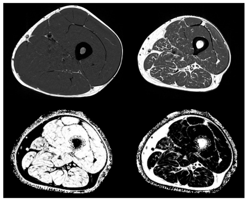
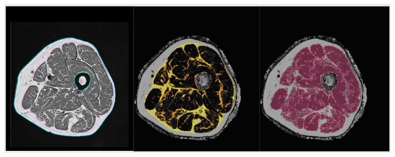
“In clinical routine, the preferred method is semi-quantitative assessment of muscle and adipose tissue area or volume from T1 weighted MR images, which show very good contrast between these two tissues (see Figure 3 top left and top right showing T1 weighted images from a young healthy normal male and from an elderly male with sarcopenia).”
§ Interactions between Muscle and Bone—Where Physics Meets Biology > 4. Muscle and Bone Interactions in Disease—Lessons and Backflashes from Pathology > 4.2. Inflammation and Fatty Degeneration—Lessons from Imaging and Structural Analyses
plain- Asserted: passes — The authors put it forward as true.
- Checkable: passes — It can be checked against evidence.
- Load-bearing: fails — The paper’s conclusions may not depend on it.
- Based: passes — The paper gives a reason why it holds.
Probably not a substantial claim. The paper’s conclusions may not depend on it.
None of the sources cited for this claim have been checked yet.
In the paper
“It impairs mechanotransduction and triggers the production of inhibitors of regeneration in both compartments, namely myostatin, activin, SOST, and DKK-1 under various clinical conditions.”
§ Interactions between Muscle and Bone—Where Physics Meets Biology > 4. Muscle and Bone Interactions in Disease—Lessons and Backflashes from Pathology > 4.2. Inflammation and Fatty Degeneration—Lessons from Imaging and Structural Analyses
What the sources say
- Not checked yetcites [91] Ebert, 2015, Acute phase serum amyloid A induces proinflammatory cytokines and mineralization via toll-like receptor 4 in mesenchymal stem cells, Stem Cell Res. source
- Not checked yetcites [107] Gilbert, 2019, The consequences of ageing, progeroid syndromes and cellular senescence on mechanotransduction and the nucleus, Exp. Cell Res. source
- Not checked yetcites [108] Serrano, 2014, Cellular senescence: from physiology to pathology, Nat. Rev. Mol. Cell Boil. source
- Not checked yetcites [109] Czarkwiani, 2018, Out with the old, in with the new: senescence in development, Curr. Opin. Cell Boil. source
- Not checked yetcites [177] Schell, 2011, Inflammatory phase of bone healing initiates the regenerative healing cascade, Cell Tissue Research
plaindirect- Asserted: passes — The authors put it forward as true.
- Checkable: passes — It can be checked against evidence.
- Load-bearing: passes — The paper’s conclusions depend on it.
- Based: passes — The paper gives a reason why it holds.
It may state two separate claims as one.
None of the sources cited for this claim have been checked yet.
In the paper
“Chronic inflammation can be both a cause and consequence of disuse and atrophy in muscle and bone.”
§ Interactions between Muscle and Bone—Where Physics Meets Biology > 4. Muscle and Bone Interactions in Disease—Lessons and Backflashes from Pathology > 4.2. Inflammation and Fatty Degeneration—Lessons from Imaging and Structural Analyses
What the sources say
- Not checked yetcites [91] Ebert, 2015, Acute phase serum amyloid A induces proinflammatory cytokines and mineralization via toll-like receptor 4 in mesenchymal stem cells, Stem Cell Res. source
- Not checked yetcites [107] Gilbert, 2019, The consequences of ageing, progeroid syndromes and cellular senescence on mechanotransduction and the nucleus, Exp. Cell Res. source
- Not checked yetcites [108] Serrano, 2014, Cellular senescence: from physiology to pathology, Nat. Rev. Mol. Cell Boil. source
- Not checked yetcites [109] Czarkwiani, 2018, Out with the old, in with the new: senescence in development, Curr. Opin. Cell Boil. source
- Not checked yetcites [177] Schell, 2011, Inflammatory phase of bone healing initiates the regenerative healing cascade, Cell Tissue Research
plaindirect- Asserted: passes — The authors put it forward as true.
- Checkable: passes — It can be checked against evidence.
- Load-bearing: passes — The paper’s conclusions depend on it.
- Based: passes — The paper gives a reason why it holds.
It may state two separate claims as one.
A conclusion the paper draws from its own results — it cites no other source for it, so there is none to check. The quote shows it is in the paper.
In the paper
“Both tissues sense mechanical strain, transduce biochemical signals in response, and mutually influence the respective tissue architecture to balance muscle power and fracture resistance.”
§ Interactions between Muscle and Bone—Where Physics Meets Biology > 5. Conclusions and Perspectives
plaindirect- Asserted: passes — The authors put it forward as true.
- Checkable: passes — It can be checked against evidence.
- Load-bearing: passes — The paper’s conclusions depend on it.
- Based: passes — The paper gives a reason why it holds.
It may state two separate claims as one.
A conclusion the paper draws from its own results — it cites no other source for it, so there is none to check. The quote shows it is in the paper.
In the paper
“Epigenetics is strongly involved in aging phenomena, and recent research indicates that both positive exercise and training effects and also exercise resistance and degeneration can be modulated by epigenetic mechanisms.”
§ Interactions between Muscle and Bone—Where Physics Meets Biology > 5. Conclusions and Perspectives
boostedconditional- Asserted: passes — The authors put it forward as true.
- Checkable: passes — It can be checked against evidence.
- Load-bearing: passes — The paper’s conclusions depend on it.
- Based: fails — The paper may give no reason why it holds.
Probably not a substantial claim. The paper may give no reason why it holds. It may state two separate claims as one.
A conclusion the paper draws from its own results — it cites no other source for it, so there is none to check. The quote shows it is in the paper.
In the paper
“Continuous adaptive remodeling appears to be the key to delay aging-associated degeneration; hence, exercise and locomotion in concerted interaction between muscle and bone are core phenomena of health maintenance in higher organisms.”
§ Interactions between Muscle and Bone—Where Physics Meets Biology > 5. Conclusions and Perspectives
hedgedconditional- Asserted: passes — The authors put it forward as true.
- Checkable: passes — It can be checked against evidence.
- Load-bearing: passes — The paper’s conclusions depend on it.
- Based: passes — The paper gives a reason why it holds.
It may state two separate claims as one.
A conclusion the paper draws from its own results — it cites no other source for it, so there is none to check. The quote shows it is in the paper.
In the paper
“Interestingly, the combination of soft tissue parameters discriminated hip fractures as well as a combination of trabecular BMD and cortical thickness, documenting a strong interaction between muscle and bone structure using fatty infiltration and other soft tissue architectural parameters as surrogate parameters.”
§ Interactions between Muscle and Bone—Where Physics Meets Biology > 4. Muscle and Bone Interactions in Disease—Lessons and Backflashes from Pathology > 4.2. Inflammation and Fatty Degeneration—Lessons from Imaging and Structural Analyses
boostedcorrelational- Asserted: passes — The authors put it forward as true.
- Checkable: passes — It can be checked against evidence.
- Load-bearing: passes — The paper’s conclusions depend on it.
- Based: passes — The paper gives a reason why it holds.
It may state two separate claims as one.
A conclusion the paper draws from its own results — it cites no other source for it, so there is none to check. The quote shows it is in the paper.
In the paper
“Physical strain is transformed into a multitude of biochemical signals that causes modeling or remodeling until a new state of homeostasis is reached as long as the physical input remains in a steady state.”
§ Interactions between Muscle and Bone—Where Physics Meets Biology > 5. Conclusions and Perspectives
plaindirect- Asserted: passes — The authors put it forward as true.
- Checkable: passes — It can be checked against evidence.
- Load-bearing: passes — The paper’s conclusions depend on it.
- Based: fails — The paper may give no reason why it holds.
Probably not a substantial claim. The paper may give no reason why it holds.
A conclusion the paper draws from its own results — it cites no other source for it, so there is none to check. The quote shows it is in the paper.
In the paper
“It is physical strain that strongly modulates the biology of cells along their lineage commitment and maturation as well as the hypertrophy versus atrophy of tissues according to physical challenge.”
§ Interactions between Muscle and Bone—Where Physics Meets Biology > 5. Conclusions and Perspectives
boosteddirect- Asserted: passes — The authors put it forward as true.
- Checkable: passes — It can be checked against evidence.
- Load-bearing: passes — The paper’s conclusions depend on it.
- Based: unclear — It is unclear why the paper says it holds.
It may state two separate claims as one.
The paper relies on this without support.
Muscle and bone are derived from common precursors during development.
“Muscle and bone are derived from common precursors during development, indicating a close relationship and interaction.”
The paper relies on this without support.
There are currently no guidelines by which imaging parameter(s) should be quantified to address a given muscle composition question.
“Currently, there are no guidelines by which imaging parameter(s) should be quantified to address a given muscle composition question.”
The paper relies on this without support.
Comparative studies on imaging parameters for muscle composition are still missing.
“Comparative studies are still missing.”
The paper relies on this without support.
A direct comparison of CT muscle density with MRI Dixon FF results has not been published.
“Interestingly, to the best of our knowledge, a direct comparison of CT muscle density with MRI Dixon FF results has not been published so far.”
The paper relies on this without support.
The borders to pathology, which stimulus hurts cells, and which one stimulates adaptive remodeling are not yet exactly known.
“We are beginning to understand the mechanisms of regulation, but we do not yet exactly know where the borders to pathology begin, which stimulus hurts cells, and which one stimulates adaptive remodeling.”
Claims other papers make about this one
Each is checked against this paper’s own text: does it say what the citing paper says it does?
In the citing paper
“The principal products secreted from bone that influence the formation, maintenance and regeneration of muscle are the osteocalcin, a marker protein of osteoblasts, and FGF23, a phosphatonin derived from osteocytes, as well as the inhibitors of the osteogenic wingless (Wnt) signaling pathway, sclerostin [136].”
From Exerkines and Sarcopenia: Unveiling the Mechanism Behind Exercise-Induced Mitochondrial Homeostasis (2025) · cites this paper as [136]
Self-check
Percentages, sums and reported differences re-computed from the paper’s own numbers.
Each contradiction between two passages listed here was confirmed by an independent check.
Rated minor: it would not touch a result — a value no result rests on, a p-value that stays on the same side of the significance level, or a conflict that is clerical, resolved elsewhere in the paper, or outside its own findings.
In the paper
§ Interactions between Muscle and Bone—Where Physics Meets Biology > 2. Principles of Interaction between Muscle and Bone > 2.2. Systemic Mutual Interactions of Skeletal Muscle and Bone
“Seminal work on the muscle cell secretory transcriptome and proteome was published in 2012 by Le Bihan et al. and Terry et al [89,90]”
§ Interactions between Muscle and Bone—Where Physics Meets Biology > References
“90. E Terry E. Zhang X. Hoffmann C. Hughes L.D. A Lewis S. Li J. Wallace M.J. A Riley L. Douglas C.M. A Gutierrez-Monreal M. Transcriptional profiling reveals extraordinary diversity among skeletal muscle tissues eLife 2018 7 e34613”
Citations
Cites
Shedding light on the cell biology of extracellular vesicles
van Niel, G., D'Angelo, G., & Raposo, G. (2018) · Nature Reviews Molecular Cell Biology · doi:10.1038/nrm.2017.125 · Cited by 8,369
Epidemiology of Sarcopenia among the Elderly in New Mexico
Baumgartner, R. N., Koehler, K. M., Gallagher, D., Romero, L., Heymsfield, S. B., Ross, R. R., Garry, P. J., & Lindeman, R. D. (1998) · American Journal of Epidemiology · doi:10.1093/oxfordjournals.aje.a009520 · Cited by 4,004
Bioelectrical impedance analysis?part I: review of principles and methods
KYLE, U. (2004) · Clinical Nutrition · doi:10.1016/j.clnu.2004.06.004 · Cited by 3,200 · cited 1×
“Bioimpedance analysis systems estimate fat and fat-free mass from the electrical impedance measurements of trunk, arms, and legs using age, gender, and ethnic-specific algorithms validated in previous studies, albeit with large prediction errors for individuals [180].”
MethodCellular senescence: from physiology to pathology
Muñoz-Espín, D., & Serrano, M. (2014) · Nature Reviews Molecular Cell Biology · doi:10.1038/nrm3823 · Cited by 2,818
Muscle as an Endocrine Organ: Focus on Muscle-Derived Interleukin-6
Pedersen, B. K., & Febbraio, M. A. (2008) · Physiological Reviews · doi:10.1152/physrev.90100.2007 · Cited by 2,388
Molecular anatomy of a trafficking organelle
Takamori, S., Holt, M., Stenius, K., Lemke, E. A., Grønborg, M., Riedel, D., Urlaub, H., Schenck, S., Brügger, B., Ringler, P., et al. (2006) · Cell · doi:10.1016/j.cell.2006.10.030 · Cited by 2,373
Coupling of angiogenesis and osteogenesis by a specific vessel subtype in bone
Kusumbe, A. P., Ramasamy, S. K., & Adams, R. H. (2014) · Nature · doi:10.1038/nature13145 · Cited by 2,155
WNT signaling in bone homeostasis and disease: From human mutations to treatments
Baron, R., & Kneissel, M. (2013) · Nature Medicine · doi:10.1038/nm.3074 · Cited by 2,093
Fracture healing: mechanisms and interventions
Einhorn, T. A., & Gerstenfeld, L. C. (2014) · Nature Reviews Rheumatology · doi:10.1038/nrrheum.2014.164 · Cited by 1,902
Senescence in Health and Disease
He, S., & Sharpless, N. E. (2017) · Cell · doi:10.1016/j.cell.2017.05.015 · Cited by 1,766
Cited by
Sorted by citations
Novel approaches to target the microenvironment of bone metastasis
Hofbauer, L. C., Bözec, A., Rauner, M., Jakob, F. J., Perner, S., & Pantel, K. (2021) · Nature Reviews Clinical Oncology · doi:10.1038/s41571-021-00499-9 · Cited by 223
Self-citationMolecular mechanisms of exercise contributing to tissue regeneration
Chen, J., Zhou, R. J., Ye, F., & Cheng, L. (2022) · Signal Transduction and Targeted Therapy · doi:10.1038/s41392-022-01233-2 · Cited by 185
Xu, X., Liu, S., Liu, H., Ru, K., Jia, Y., Wu, Z., Liang, S., Khan, Z., Chen, Z., Qian, A., et al. (2021) · International Journal of Molecular Sciences · doi:10.3390/ijms22126429 · Cited by 119
KEMMLER, W. K., Kohl, M., Jakob, F. J., Engelke, K., & von Stengel, S. (2020) · Nutrients · doi:10.3390/nu12082341 · Cited by 88
Self-citationEngelke, K., Chaudry, O., Gast, L. V., Eldib, M., Wang, L., Larédo, J.-D., Schett, G. A., & Nagel, A. M. (2023) · Journal of Orthopaedic Translation · doi:10.1016/j.jot.2023.07.005 · Cited by 82
Self-citationRole of Physical Activity in Bone–Muscle Crosstalk: Biological Aspects and Clinical Implications
Cariati, I., Bonanni, R., Onorato, F., Mastrogregori, A., Rossi, D., Iundusi, R., Gasbarra, E., Tancredi, V., & Tarantino, U. (2021) · Journal of Functional Morphology and Kinesiology · doi:10.3390/jfmk6020055 · Cited by 78
Bone and muscle crosstalk in ageing and disease
Kirk, B., Lombardi, G., & Duque, G. (2025) · Nature Reviews Endocrinology · doi:10.1038/s41574-025-01088-x · Cited by 89
Lloyd‐Lewis, B., Mourikis, P., & Fré, S. (2019) · Current Opinion in Cell Biology · doi:10.1016/j.ceb.2019.06.003 · Cited by 60
Wei, F., Oms, K. F., Kinzel, M., Perotti, L. E., Asiatico, J. T., Omer, M., Hovell, C., Reumers, V., & Coathup, M. J. (2022) · Bone Research · doi:10.1038/s41413-022-00234-9 · Cited by 62
Biomanufacturing in low Earth orbit for regenerative medicine
Sharma, A., Clemens, R. A., Garcia, O., Taylor, D. L., Wagner, N. L., Shepard, K. A., Gupta, A. E., Malany, S., Grodzinsky, A. J., Kearns‐Jonker, M., et al. (2021) · Stem Cell Reports · doi:10.1016/j.stemcr.2021.12.001 · Cited by 57
Li, Y., Wang, X., Pan, C., Yuan, H., Li, X., Chen, Z., & He, H. (2023) · Molecular Medicine · doi:10.1186/s10020-023-00649-y · Cited by 55
A Bidirectional Mendelian Randomization Study of Sarcopenia-Related Traits and Knee Osteoarthritis
Zhang, L., Zhang, C., Zhang, J., Liu, A., Wang, P., & Xu, J. (2023) · Clinical Interventions in Aging · doi:10.2147/cia.s424633 · Cited by 48
Sedentary lifestyle and body composition in type 2 diabetes
Li, D., Yang, Y., Gao, Z.-Y., Zhao, L.-H., Yang, X., Xu, F., Yu, C., Zhang, X., Wang, X., Wang, L., et al. (2022) · Diabetology & Metabolic Syndrome · doi:10.1186/s13098-021-00778-6 · Cited by 54
Bone‐on‐a‐Chip: A Microscale 3D Biomimetic Model to Study Bone Regeneration
Galván-Chacón, V., Zampouka, A., Hesse, B., Bohner, M., Habibović, P., & Barata, D. B. (2021) · Advanced Engineering Materials · doi:10.1002/adem.202101467 · Cited by 48
Tortorella, I., Argentati, C., Emiliani, C., Morena, F., & Martino, S. (2022) · Cells · doi:10.3390/cells11193093 · Cited by 47
Late‐onset Hypogonadism: Bone health
Rochira, V. (2020) · Andrology · doi:10.1111/andr.12827 · Cited by 49
The effect of ageing on fat infiltration of thigh and paraspinal muscles in men
Engelke, K., Ghasemikaram, M., Chaudry, O., Uder, M., Nagel, A. M., Jakob, F. J., & KEMMLER, W. K. (2022) · Aging Clinical and Experimental Research · doi:10.1007/s40520-022-02149-1 · Cited by 44
Self-citationPathophysiological mechanism of acute bone loss after fracture
Zheng, X.-Q., Huang, J., Lin, J., & Song, C. (2022) · Journal of Advanced Research · doi:10.1016/j.jare.2022.08.019 · Cited by 47
Microgravity and Musculoskeletal Health: What Strategies Should Be Used for a Great Challenge?
Bonanni, R., Cariati, I., Marini, M., Tarantino, U., & Tancredi, V. (2023) · Life · doi:10.3390/life13071423 · Cited by 53
Yin, L., Xu, Z., Wang, L., Li, W., Zhao, Y., Su, Y., Sun, W. Y., Liu, Y., Yang, M., Yu, A., et al. (2020) · Frontiers in Endocrinology · doi:10.3389/fendo.2020.00503 · Cited by 37
Self-citationMusculoskeletal health in children and adolescents
Faienza, M. F., Urbano, F., Chiarito, M., Lassandro, G., & Giordano, P. (2023) · Frontiers in Pediatrics · doi:10.3389/fped.2023.1226524 · Cited by 48
Ghasemikaram, M., Chaudry, O., Nagel, A. M., Uder, M., Jakob, F. J., KEMMLER, W. K., Kohl, M., & Engelke, K. (2021) · GeroScience · doi:10.1007/s11357-020-00316-8 · Cited by 37
Self-citationLu, Y., Xu, X., & Li, J. (2023) · Journal of Materials Chemistry B · doi:10.1039/d3tb00251a · Cited by 35
Muscle-derived extracellular vesicles mediate crosstalk between skeletal muscle and other organs
Jia, J., Wang, L., Zhou, Y., Zhang, P., & Chen, X. (2025) · Frontiers in Physiology · doi:10.3389/fphys.2024.1501957 · Cited by 45
The Interconnection Between Muscle and Bone: A Common Clinical Management Pathway
Smith, C., Sim, M., Via, J. D., Levinger, I., & Duque, G. (2023) · Calcified Tissue International · doi:10.1007/s00223-023-01146-4 · Cited by 36
Smith, E. L., Shahien, A. A., Chung, M. C., Stoker, G. E., Niu, R., & Schwarzkopf, R. M. (2020) · The Journal of Arthroplasty · doi:10.1016/j.arth.2020.04.094 · Cited by 30
Role of irisin in effects of chronic exercise on muscle and bone in ovariectomized mice
Kawao, N., Iemura, S., Kawaguchi, M., Mizukami, Y., Takafuji, Y., & Kaji, H. (2021) · Journal of Bone and Mineral Metabolism · doi:10.1007/s00774-020-01201-2 · Cited by 30
Aging, Osteo-Sarcopenia, and Musculoskeletal Mechano-Transduction
Leser, J. M., Harriot, A. D., Buck, H., Ward, C. W., & Stains, J. P. (2021) · Frontiers in Rehabilitation Sciences · doi:10.3389/fresc.2021.782848 · Cited by 29
Proposed Canadian Consensus Guidelines on Osteoarthritis Treatment Based on OA-COAST Stages 1–4
Mosley, C., Edwards, T., Romano, L., Truchetti, G., Dunbar, L., Schiller, T., Gibson, T., Bruce, C., & Troncy, É. (2022) · Frontiers in Veterinary Science · doi:10.3389/fvets.2022.830098 · Cited by 28
Wang, J., Xing, F., Sheng, N., & Xiang, Z. (2022) · Frontiers in Nutrition · doi:10.3389/fnut.2022.860693 · Cited by 28
Time of exercise differentially impacts bone growth in mice
Yu, S., Tang, Q., Lu, X., Chen, G., Xie, M., Yang, J., Yin, Y., Zheng, W., Wang, J., Han, Y., et al. (2024) · Nature Metabolism · doi:10.1038/s42255-024-01057-0 · Cited by 26
Jin, Z., Wang, R., Jin, L., Wan, L., & Li, Y. (2024) · BMC Geriatrics · doi:10.1186/s12877-024-05098-8 · Cited by 27
Argueta‐Figueroa, L., Flores-Mejía, L. A., Ávila-Curiel, B. X., Flores-Ferreyra, B. I., & Rosas, R. T. (2022) · European Journal of Dentistry · doi:10.1055/s-0041-1740220 · Cited by 24
Exerkines and Sarcopenia: Unveiling the Mechanism Behind Exercise-Induced Mitochondrial Homeostasis
Wang, J., Jia, D., Zhang, Z., & Wang, D. (2025) · Metabolites · doi:10.3390/metabo15010059 · Cited by 23
Cellular and Molecular Effects of Microgravity on the Immune System: A Focus on Bioactive Lipids
Fava, M., Dominicis, N. D., Forte, G., Bari, M., Leuti, A., & Maccarrone, M. (2024) · Biomolecules · doi:10.3390/biom14040446 · Cited by 24
Mechanisms and Countermeasures for Muscle Atrophy in Microgravity
Liu, Y., Cao, X., Zhou, Q., Deng, C., Yang, Y., Huang, D., Luo, H., Zhang, S., Li, Y., Xu, J., et al. (2024) · Cells · doi:10.3390/cells13242120 · Cited by 23
Focusing on Mechanoregulation Axis in Fibrosis: Sensing, Transduction and Effecting
Wen, D., Gao, Y., Ho, C., Li, Y., Zhang, Y., Lyu, G., Hu, D., Li, Q., & Zhang, Y. (2022) · Frontiers in Molecular Biosciences · doi:10.3389/fmolb.2022.804680 · Cited by 25
Kast, S., Shojaa, M., Kohl, M., von Stengel, S., Gosch, M., Jakob, F. J., Kerschan‐Schindl, K., Kladny, B., Klöckner, N., Lange, U., et al. (2022) · Osteoporosis International · doi:10.1007/s00198-022-06329-7 · Cited by 23
Self-citationResearch advances in crosstalk between muscle and bone in osteosarcopenia (Review)
Yu, C., Du, Y., Peng, Z. Y., Ma, C., Fang, J., Ma, L., Chen, F., Zhang, C., Geng, R., Zhang, Y., et al. (2023) · Experimental and Therapeutic Medicine · doi:10.3892/etm.2023.11888 · Cited by 24
Physical Activity, Exerkines, and Their Role in Cancer Cachexia
Bilski, J., Szlachcic, A., Ptak‐Belowska, A., & Brzozowski, T. (2025) · International Journal of Molecular Sciences · doi:10.3390/ijms26168011 · Cited by 24
Roth, D. M., Souter, K. M., & Graf, D. (2022) · European Journal of Cell Biology · doi:10.1016/j.ejcb.2022.151258 · Cited by 25
Zhao, X., Sun, J., Xin, S., et al. (2023) · Biomolecules · doi:10.3390/biom13040616 · Cited by 21
Wehrle, E., Günther, D., Mathavan, N., Singh, A., & Müller, R. (2024) · STAR Protocols · doi:10.1016/j.xpro.2024.102986 · Cited by 22
Impact of X-Linked Hypophosphatemia on Muscle Symptoms
Romagnoli, C., Iantomasi, T., & Brandi, M. (2022) · Genes · doi:10.3390/genes13122415 · Cited by 18
Polyamines and Physical Activity in Musculoskeletal Diseases: A Potential Therapeutic Challenge
Galasso, L., Cappella, A., Mulè, A., Castelli, L., Ciorciari, A. M., Stacchiotti, A., & Montaruli, A. (2023) · International Journal of Molecular Sciences · doi:10.3390/ijms24129798 · Cited by 25
Chen, J., Xu, J., Gou, L., et al. (2024) · Archives of Gerontology and Geriatrics · doi:10.1016/j.archger.2024.105486 · Cited by 19
Bone Geometry Is Altered by Follistatin‐Induced Muscle Growth in Young Adult Male Mice
Chan, A. S. M., McGregor, N. E., Poulton, I. J., Hardee, J. P., Cho, H., Martin, T. J., Gregorevic, P., Sims, N. A., & Lynch, G. S. (2021) · JBMR Plus · doi:10.1002/jbm4.10477 · Cited by 19
Osteosarcopenia and Pain: Do We Have a Way Out?
Bonanni, R., Grillo, S. G., Cariati, I., Tranquillo, L., Iundusi, R., Gasbarra, E., Tancredi, V., & Tarantino, U. (2023) · Biomedicines · doi:10.3390/biomedicines11051285 · Cited by 19
Sakamoto, E., Kitase, Y., Fitt, A. J., Zhu, Z., Awad, K. R., Brotto, M. A. P., White, K. E., Welc, S. S., Bergwitz, C., & Bonewald, L. F. (2024) · Cell Reports · doi:10.1016/j.celrep.2024.114397 · Cited by 21
Helicobacter pylori Related Diseases and Osteoporotic Fractures (Narrative Review)
Fisher, L., Fisher, A., & Smith, P. N. (2020) · Journal of Clinical Medicine · doi:10.3390/jcm9103253 · Cited by 18
Impact of 10‐day bed rest on serum levels of irisin and markers of musculoskeletal metabolism
Oranger, A., Storlino, G., Dicarlo, M., Zerlotin, R., Pignataro, P., Sanesi, L., Narici, M. V., Pišot, R., Šimunič, B., Colaianni, G., et al. (2022) · The FASEB Journal · doi:10.1096/fj.202201005rr · Cited by 16
Skeletal muscle-derived exosomes prevent osteoporosis by promoting osteogenesis
Xing, Z., Guo, L., Li, S., Huang, W., Su, J., Chen, X., Li, Y., & Zhang, J. (2024) · Life Sciences · doi:10.1016/j.lfs.2024.123079 · Cited by 20
Wise, P. M., Sahana, J., Neviani, P., Corydon, T. J., Schulz, H., Wehland, M., Infanger, M., & Grimm, D. G. (2022) · International Journal of Molecular Sciences · doi:10.3390/ijms232416095 · Cited by 16
Zhang, Q., Hu, S., Jin, Z., Wang, S., Zhang, B., & Zhao, L. (2024) · Frontiers in Pharmacology · doi:10.3389/fphar.2024.1339148 · Cited by 18
Wang, L., Liu, Y.-D., Li, K., Zhang, W.-S., Yuan, Y., Ma, K.-K., Zhou, F.-Y., Cheng, Z.-T., Geng, J., Su, Y.-B., et al. (2024) · Obesity · doi:10.1002/oby.24040 · Cited by 13
Self-citationHIF-1α mediates osteoclast-induced disuse osteoporosis via cytoophidia in the femur of mice
Bie, M., Tang, Y., Xia, Y., Zhang, Q., Tian, Y., Cheng, C., Li, X., Qi, X., & Kang, F. (2022) · Bone · doi:10.1016/j.bone.2022.116648 · Cited by 15
Novel TNNT1 mutation and mild nemaline myopathy phenotype in an Italian patient
Petrucci, A., Primiano, G., Savarese, M., et al. (2021) · Neuromuscular Disorders · doi:10.1016/j.nmd.2021.03.001 · Cited by 14
Shi, H., Huang, L., Wang, Q., Zhang, N., Lv, C., Lin, C., Shao, L., Xia, D., Zhao, Z., Yang, G., et al. (2025) · International Journal of Surgery · doi:10.1097/js9.0000000000003030 · Cited by 15
Li, Z., Chen, J., Yang, J., Wang, R., & Wang, W. (2024) · BMC Musculoskeletal Disorders · doi:10.1186/s12891-024-07484-0 · Cited by 15
Timofte, D. V., Tudor, R. C., Mocanu, V., & Labusca, L. S. (2024) · Nutrients · doi:10.3390/nu16234231 · Cited by 16
Carbone, J. W., Phillips, S. M., Weaver, C. M., Hughes, J. M., & Pasiakos, S. M. (2024) · Advances in Nutrition · doi:10.1016/j.advnut.2024.100347 · Cited by 15
Guan, X., Zhang, D., Zhang, F., Zong, Y., Wang, H., Shen, Z., & Yin, F. (2023) · Frontiers in Cell and Developmental Biology · doi:10.3389/fcell.2023.1260001 · Cited by 13
Exploring neuronal mechanisms of osteosarcopenia in older adults
Karacan, İ., & Türker, K. S. (2024) · The Journal of Physiology · doi:10.1113/jp285666 · Cited by 13
The “Burden” of Childhood Obesity on Bone Health: A Look at Prevention and Treatment
Farella, I., Chiarito, M., Vitale, R., D’Amato, G., & Faienza, M. F. (2025) · Nutrients · doi:10.3390/nu17030491 · Cited by 16
Miao, T., Li, X., Zhang, W., Yang, F., & Wang, X. (2025) · PeerJ · doi:10.7717/peerj.19616 · Cited by 13
Joint Cartilage in Long-Duration Spaceflight
Ganse, B., Cucchiarini, M., & Madry, H. (2022) · Biomedicines · doi:10.3390/biomedicines10061356 · Cited by 11
Khan, A. A., Rush, E. T., Wakeford, C., et al. (2025) · Advances in Therapy · doi:10.1007/s12325-025-03309-1 · Cited by 10
Deciphering Osteosarcopenia through the hallmarks of aging
Franulic, F., Salech, F., Rivas, D., & Duque, G. (2024) · Mechanisms of Ageing and Development · doi:10.1016/j.mad.2024.111997 · Cited by 14
Kotwal, J. G., Sharma, N. K., Somkunwar, R. K., Bavkar, D. M., Deshmukh, S., Khan, V., & Korade, N. B. (2025) · Iran Journal of Computer Science · doi:10.1007/s42044-025-00271-7 · Cited by 10
Lara-Castillo, N., Masunaga, J., Brotto, L. S., Vallejo, J. A., Javid, K., Wacker, M., Brotto, M., Bonewald, L. F., & Johnson, M. L. (2023) · Bone · doi:10.1016/j.bone.2023.116833 · Cited by 11
Li, J., Wang, Y., Zhang, X., Ping, Z., Su, Y., Bai, L., Wang, Y., Wang, M., & Zhao, J. (2022) · Frontiers in Endocrinology · doi:10.3389/fendo.2022.990487 · Cited by 9
Osteosarcopenia in Finland: prevalence and associated factors
Blomqvist, M., Nuotio, M. S., Sääksjärvi, K., Koskinen, S. V. P., & Stenholm, S. (2024) · Archives of Osteoporosis · doi:10.1007/s11657-024-01439-7 · Cited by 9
SIRT1 and exercise-induced bone metabolism: a regulatory nexus
Liu, L., Zhang, J., Cui, R., Wang, N., Zhang, Y., Liu, L., Zhang, X., & Liu, Q. (2025) · Frontiers in Cell and Developmental Biology · doi:10.3389/fcell.2025.1522821 · Cited by 11
Hind limb muscles influence the architectural properties of long bones in frogs
Vera, M. C., Ferretti, J. L., Cointry, G. R., & Abdala, V. S. L. (2022) · Journal of Anatomy · doi:10.1111/joa.13710 · Cited by 9
Noten, K., & van Amstel, R. N. (2024) · Life · doi:10.3390/life14070799 · Cited by 9
Ambroszkiewicz, J., Gajewska, J., Szamotulska, K., Rowicka, G., Klemarczyk, W., Strucińska, M., & Chełchowska, M. (2024) · Nutrients · doi:10.3390/nu16132009 · Cited by 8
Nguyen, V.-L., Lee, K.-B., & Moon, Y. J. (2025) · International Journal of Molecular Sciences · doi:10.3390/ijms26157060 · Cited by 11
Li, Y., & Xu, Z. (2024) · Geriatric Nursing · doi:10.1016/j.gerinurse.2024.07.001 · Cited by 8
MicroRNAs and their Modulatory Effect on the Hallmarks of Osteosarcopenia
Silva, W. J., Cruz, A., & Duque, G. (2024) · Current Osteoporosis Reports · doi:10.1007/s11914-024-00880-4 · Cited by 9
Müller-Deubert, S. M., Ege, C., Krug, M., Meißner-Weigl, J., Rudert, M., Bischof, O., Jakob, F. J., & Ebert, R. (2020) · Stem Cells International · doi:10.1155/2020/7865484 · Cited by 8
Self-citationVarley, I., Stebbings, G. K., Williams, A. G., et al. (2021) · The Journal of Sports Medicine and Physical Fitness · doi:10.23736/s0022-4707.21.11871-7 · Cited by 8
Macrophages Characterization in an Injured Bone Tissue
Nikovics, K., Durand, M., Castellarin, C., Burger, J., Sicherre, E., Collombet, J., Oger, M., Holy, X., & Favier, A. (2022) · Biomedicines · doi:10.3390/biomedicines10061385 · Cited by 8
Fischer, C., Jakob, F. J., Kohl, M., Kast, S., von Stengel, S., Kerschan‐Schindl, K., Lange, U., Thomasius, F. E., Peters, S., Uder, M., et al. (2023) · Journal of Osteoporosis · doi:10.1155/2023/5570030 · Cited by 10
Self-citationElectrical stimulation paradigms on muscle quality and bone mineral density after spinal cord injury
Gorgey, A. S., Venigalla, S., Deitrich, J. N., et al. (2025) · Osteoporosis International · doi:10.1007/s00198-025-07482-5 · Cited by 7
Wang, L., Lu, S., Feng, J., Ma, K., Duan, F., Wang, R., Cheng, X., Liu, Y., Imai, Y., Chen, H., et al. (2025) · Calcified Tissue International · doi:10.1007/s00223-025-01447-w · Cited by 9
Self-citationAvermectin reduces bone mineralization via the TGF-β signaling pathway in zebrafish
Du, Y., Liu, G., Liu, Z., Mo, J., Zheng, M., Wei, Q., & Xu, Y. (2023) · Comparative Biochemistry and Physiology Part C Toxicology & Pharmacology · doi:10.1016/j.cbpc.2023.109702 · Cited by 9
Ghimire, P. S., Eckart, A. C., Al-Makhzoomy, I. K., & Stavitz, J. (2023) · Sports · doi:10.3390/sports11110215 · Cited by 11
Peres-Ueno, M. J., Capato, L. L., Porto, J. M., Adão, I. F., Maciel, J. G., Herrero, C. F. P. D. S., Nogueira‐Barbosa, M. H., de Paula, F. J. A., Ferrioli, E., & de Abreu, D. C. C. (2022) · Annals of Physical and Rehabilitation Medicine · doi:10.1016/j.rehab.2022.101680 · Cited by 7
Skeletal consequences of heart failure
Bandeira, F. A. F., de Oliveira, L. B., Caldeira, R. B., & Toscano, L. S. (2022) · Women s Health · doi:10.1177/17455057221135501 · Cited by 9
Opportunities for Biomanufacturing in Low Earth Orbit: Current Status and Future DirectionsPreprint
Giulianotti, M. A., Sharma, A., Clemens, R. A., Garcia, O., Taylor, L., Wagner, N. L., Shepard, K. A., Gupta, A., Malany, S., Grodzinsky, A. J., et al. (2021) · Preprints.org · doi:10.20944/preprints202108.0044.v1 · Cited by 7
Fascial Manual Medicine: The Concept of Fascial Continuum
Bordoni, B., & Escher, A. R. (2025) · Cureus · doi:10.7759/cureus.82136 · Cited by 7
Exercise in the Prevention of Age-Related Fragility Fractures (Narrative Review)
Kerschan‐Schindl, K., & Hasenoehrl, T. (2025) · Gerontology · doi:10.1159/000543377 · Cited by 8
Ganoderic Acid A Prevented Osteoporosis by Modulating the PIK3CA /p‐Akt/ TWIST1 Signaling Pathway
Zhao, J., Fan, Y., Li, H., et al. (2025) · Food Science & Nutrition · doi:10.1002/fsn3.70177 · Cited by 6
Li, S., Mei, L., Chen, Q., et al. (2023) · American Journal of Orthodontics and Dentofacial Orthopedics · doi:10.1016/j.ajodo.2023.09.009 · Cited by 6
Banimohamad‐Shotorbani, B., Azizsoltani, A., Khalaj, Z., Rafiei-Baharloo, M., Ghotaslou, A., & Fathi‐karkan, S. (2024) · Microgravity Science and Technology · doi:10.1007/s12217-024-10136-6 · Cited by 7
Ziouti, F., Rummler, M., Steyn, B., Thiele, T., Seliger, A., Duda, G. N., Bogen, B., Willie, B. M., & Jundt, F. (2021) · International Journal of Molecular Sciences · doi:10.3390/ijms22083840 · Cited by 6
Self-citationHart, D. A., Zernicke, R. F., & Shrive, N. G. (2022) · International Journal of Molecular Sciences · doi:10.3390/ijms23179949 · Cited by 6
Li, W., Chen, M., Chen, F., Li, Y., Zhong, Y., Lu, Y., Zhang, K., & Yang, F. (2024) · Trials · doi:10.1186/s13063-024-08498-8 · Cited by 6
Modeling Musculoskeletal Disorders in Zebrafish: Advancements in Muscle and Bone Research
Carbonare, L. D., Braggio, M., Minoia, A., Cominacini, M., Romanelli, M. G., Pessoa, J., Tiso, N., & Valenti, M. T. (2024) · Cells · doi:10.3390/cells14010028 · Cited by 7
The male hormone reset: how GLP-1RAs, lifestyle and testosterone transform obesity-linked problems
Antonič, K. G., & Zitzmann, M. (2025) · The Aging Male · doi:10.1080/13685538.2025.2601423 · Cited by 6
Metadata
- Authors
- Herrmann, M., Engelke, K., Ebert, R., Müller-Deubert, S., Rudert, M., Ziouti, F., Jundt, F., Felsenberg, D., & Jakob, F.
- Affiliations
- University of Würzburg (DE)
- Friedrich-Alexander-Universität Erlangen-Nürnberg (DE)
- Universitätsklinikum Würzburg (DE)
- Privatpraxis für Muskel- und Knochenkrankheiten, 12163 Berlin Germany
- Corresponding author
- Jakob, Franz
- ORCID
- Venue
- Biomolecules
- Publisher
- MDPI AG
- Published
- 2020-03-10
- Volume
- 10
- Issue
- 3
- Pages
- 432
- ISSN
- 2218-273X (ISSN-L)
- PMC
- PMC7175139
- OpenAlex
- W3010713274
- Cited by
- 197
- References
- 217
- Citations by year
- 2026: 23 · 2025: 41 · 2024: 43 · 2023: 32 · 2022: 33 · 2021: 17 · 2020: 6
- FWCI
- 2.11
- Type
- journal article
- Language
- en
- Open access
- gold
- dx.doi.org
- Licence
- Locations
- Biomolecules (publishedVersion, cc-by)
- PubMed Central (publishedVersion, cc-by)
- PubMed (publishedVersion)
- DOAJ (DOAJ: Directory of Open Access Journals) (submittedVersion, cc-by-sa)
- Online Publication Service of Würzburg University (Würzburg University) (submittedVersion, cc-by)
- OPUS Repository (Kooperativer Bibliotheksverbund Berlin-Brandenburg) (submittedVersion, cc-by)
- Funders
- Deutsche Forschungsgemeinschaft (TRR 225, JU426/5-1, 326998133, 01EC1402C, D-361)
- Bundesministerium für Bildung und Forschung (326998133-TRR 225, 01EC1402C, 326998133)
- Topics
- Life Sciences › Biochemistry, Genetics and Molecular Biology › Molecular Biology › Muscle Physiology and Disorders
- Health Sciences › Medicine › Orthopedics and Sports Medicine › Bone health and osteoporosis research
- Health Sciences › Medicine › Physiology (medical) › Spaceflight effects on biology
- Keywords
- muscle-bone interaction, mechanotransduction, osteokines, myokines, exosomes, extracellular matrix, muscle hypertrophy, bone formation, physical activity, microgravity, disuse, sarcopenia, osteoporosis, bone marrow adiposity, tissue stiffness
- MeSH terms
- Bone and Bones, Extracellular Matrix, Humans, Osteoporosis, Exercise, Muscle, Skeletal, Mechanotransduction, Cellular, Sarcopenia
About these checks
Registry, retraction, Cites and statistics results are computed, not judged, with one exception: a printed reference that no index lists is read once by a model before it counts as a citation error. Claims are extracted by a model and kept only when their quote appears verbatim in the text; each cited source is then checked, with a model’s help, for whether it says what the claim relies on. Those verdicts can be wrong — read the claim’s quote and the cited source before relying on one.
A language model drew the claims from the paper on 2026-10-09, under version 3 of the claim definition (model deepseek-v4.1-flash, prompt extract-v3).
A second model labelled each claim on 2026-10-09 (model jev-1.13-20260917, definition revision gold-guideline-v0.2).
Statistics and arithmetic are recomputed, not judged.
Confidence bands are shown only for fields calibrated on our annotated test set. None is calibrated yet, so no band is shown.
What counts as a claim ›How we measure confidence ›How we check this ›
Are you an author, or do you see a problem with this page? Request a review
