

The DOI matches its Crossref record; 169 of the 172 references it cites were found in a registry or an index, and 1 are not checked yet.
Advanced Glycation End Products and Oxidative Stress in Type 2 Diabetes Mellitus
Open accessAbstract
Type 2 diabetes mellitus (T2DM) is a very complex and multifactorial metabolic disease characterized by insulin resistance and β cell failure leading to elevated blood glucose levels. Hyperglycemia is suggested to be the main cause of diabetic complications, which not only decrease life quality and expectancy, but are also becoming a problem regarding the financial burden for health care systems. Therefore, and to counteract the continually increasing prevalence of diabetes, understanding the pathogenesis, the main risk factors, and the underlying molecular mechanisms may establish a basis for prevention and therapy. In this regard, research was performed revealing further evidence that oxidative stress has an important role in hyperglycemia-induced tissue injury as well as in early events relevant for the development of T2DM. The formation of advanced glycation end products (AGEs), a group of modified proteins and/or lipids with damaging potential, is one contributing factor. On the one hand it has been reported that AGEs increase reactive oxygen species formation and impair antioxidant systems, on the other hand the formation of some AGEs is induced per se under oxidative conditions. Thus, AGEs contribute at least partly to chronic stress conditions in diabetes. As AGEs are not only formed endogenously, but also derive from exogenous sources, i.e., food, they have been assumed as risk factors for T2DM. However, the role of AGEs in the pathogenesis of T2DM and diabetic complications—if they are causal or simply an effect—is only partly understood. This review will highlight the involvement of AGEs in the development and progression of T2DM and their role in diabetic complications.
Overview
The claims and statistics of this paper haven’t been checked yet.
Run the check — free, no account needed. It reads the open-access text and checks the sources it cites.
Integrity signals
Registry
Cites
Statistics
Figures
Verification
Claims the paper makes
The claims and statistics of this paper haven’t been checked yet.
Run the check — free, no account needed. It reads the open-access text and checks the sources it cites.
Claims other papers make about this one
Each is checked against this paper’s own text: does it say what the citing paper says it does?
The source backs part of the claim, not all of it — or backs it for a different population, condition or comparison.
In the citing paper
“The formation and accumulation of glycoxidation products occurs during normal, but especially under certain physiological conditions, such as hyperglycemia, hyperlipidemia, and oxidative stress.”
From Protein oxidation - Formation mechanisms, detection and relevance as biomarkers in human diseases (2021) · cites this paper as [152]
Experimental — accuracy not yet measuredThe source backs part of the claim, not all of it — or backs it for a different population, condition or comparison.
In the citing paper
“This indicates their relevance in the progression of metabolic diseases, including neurodegenerative disease, chronic kidney disease (CDK), CVD, and cancer, but in particular, T2D and related complications as reviewed elsewhere [152].”
From Protein oxidation - Formation mechanisms, detection and relevance as biomarkers in human diseases (2021) · cites this paper as [152]
Experimental — accuracy not yet measuredThe source backs part of the claim, not all of it — or backs it for a different population, condition or comparison.
In the citing paper
“a growing body of evidence reveals that levels of glycoxidation products increase as a function of age in several tissues, dependent on the dietary intake and the presence of hyperglycemic conditions [152,153]”
From Protein oxidation - Formation mechanisms, detection and relevance as biomarkers in human diseases (2021) · cites this paper as [152]
Experimental — accuracy not yet measuredIn the citing paper
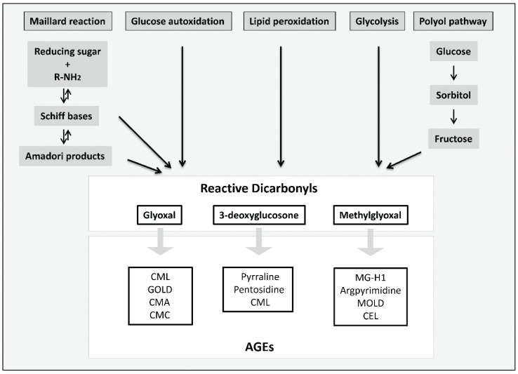
“These molecules can also be generated from glucose autoxidation, lipid peroxidation, and the polyol pathway.[18]”
From The Role of Dietary Advanced Glycation End Products in Metabolic Dysfunction (2021) · cites this paper as [18]
In the citing paper
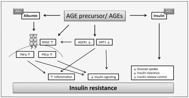
“AGEs can contribute to both pathogenetic features of T2DM by promoting inflammation and oxidative stress in humans. [18]”
From The Role of Dietary Advanced Glycation End Products in Metabolic Dysfunction (2021) · cites this paper as [18]
Self-check
Percentages, sums and reported differences re-computed from the paper’s own numbers.
The self-check needs the paper’s full text.
Citations
Cites
Matthews, D. R., Hosker, J. P., Rudenski, A. S., Naylor, B. A., Treacher, D. F., & Turner, R. C. (1985) · Diabetologia · doi:10.1007/bf00280883 · Cited by 31,502
Toprak, Ö., Kurt, H., Sarı, Y., Şarkış, C., Us, H., & Kırık, A. (2017) · Kidney & Blood Pressure Research · doi:10.1159/000468530 · Cited by 29,772
Global and societal implications of the diabetes epidemic
Zimmet, P., Alberti, K. G. M. M., & Shaw, J. (2001) · Nature · doi:10.1038/414782a · Cited by 5,883
Oxidative stress and diabetic complications
Giacco, F., & Brownlee, M. (2010) · Circulation Research · doi:10.1161/circresaha.110.223545 · Cited by 5,461
The Emerging Risk Factors Collaboration (2010) · The Lancet · doi:10.1016/s0140-6736(10)60484-9 · Cited by 5,125
Gupta, A., & Uribarri, J. (2016) · Hormone Research in Paediatrics · doi:10.1159/000444053 · Cited by 4,023
Role of Oxidative Stress in Development of Complications in Diabetes
Baynes, J. (1991) · Diabetes · doi:10.2337/diab.40.4.405 · Cited by 3,791
Oxidative stress: a concept in redox biology and medicine
Sies, H. (2015) · Redox Biology · doi:10.1016/j.redox.2015.01.002 · Cited by 3,208
Advanced glycation end-products: A review
Singh, R., Barden, A., Mori, T., & Beilin, L. (2001) · Diabetologia · doi:10.1007/s001250051591 · Cited by 2,597
Dehydrated Foods, Chemistry of Browning Reactions in Model Systems
Hodge, J. E. (1953) · Journal of Agricultural and Food Chemistry · doi:10.1021/jf60015a004 · Cited by 1,979
Cited by
Sorted by citations
Items 1,001–1,001 of 1,001
Ansari, P., Khan, J. T., Choudhary, S., Reberio, A. D., Kumar, S., Seidel, V., Abdel-Wahab, Y. H. A., & Flatt, P. R. (2024) · doi:10.20944/preprints202408.1739.v1 · Cited by 89
Metadata
- Authors
- Nowotny, K., Jung, T., Höhn, A., Weber, D., & Grune, T.
- Affiliations
- German Institute of Human Nutrition (DE)
- Corresponding author
- Grune, Tilman
- Venue
- Biomolecules
- Publisher
- MDPI AG
- Published
- 2015-03-16
- Volume
- 5
- Issue
- 1
- Pages
- 194-222
- ISSN
- 2218-273X (ISSN-L)
- PMC
- PMC4384119
- OpenAlex
- W1986014108
- Cited by
- 1,095
- References
- 172
- Citations by year
- 2026: 59 · 2025: 93 · 2024: 106 · 2023: 138 · 2022: 132 · 2021: 118 · 2020: 106 · 2019: 99 · 2018: 99 · 2017: 68 · 2016: 61 · 2015: 14
- FWCI
- 8.00
- Type
- journal article
- Language
- en
- Open access
- gold
- dx.doi.org
- Licence
- Locations
- Biomolecules (publishedVersion, cc-by)
- Europe PMC (PubMed Central) (publishedVersion, cc-by)
- PubMed Central (publishedVersion, cc-by)
- PubMed (publishedVersion)
- Informationszentrum Lebenswissenschaften (ZB MED) (submittedVersion, cc-by)
- DOAJ (DOAJ: Directory of Open Access Journals) (submittedVersion, cc-by-sa)
- Topics
- Life Sciences › Biochemistry, Genetics and Molecular Biology › Clinical Biochemistry › Advanced Glycation End Products research
- Life Sciences › Agricultural and Biological Sciences › Food Science › Potato Plant Research
- Health Sciences › Medicine › Nephrology › Chronic Kidney Disease and Diabetes
- Keywords
- advanced glycation end products, oxidative stress, type 2 diabetes, hyperglycemia, reactive oxygen species, antioxidant defense, diabetes complications, insulin resistance, beta-cell failure
- MeSH terms
- Animals, Diabetes Mellitus, Type 2, Humans, Insulin Resistance, Cell Death, Glycation End Products, Advanced, Oxidative Stress, Insulin-Secreting Cells
About these checks
Registry, retraction, Cites and statistics results are computed, not judged, with one exception: a printed reference that no index lists is read once by a model before it counts as a citation error. Claims are extracted by a model and kept only when their quote appears verbatim in the text; each cited source is then checked, with a model’s help, for whether it says what the claim relies on. Those verdicts can be wrong — read the claim’s quote and the cited source before relying on one.
Statistics and arithmetic are recomputed, not judged.
Confidence bands are shown only for fields calibrated on our annotated test set. None is calibrated yet, so no band is shown.
What counts as a claim ›How we measure confidence ›How we check this ›
Are you an author, or do you see a problem with this page? Request a review
