

The DOI matches its Crossref record; 15 of the 15 references it cites were found in a registry or an index.
Availability of Ferritin-Bound Iron to Enterobacteriaceae
Abstract
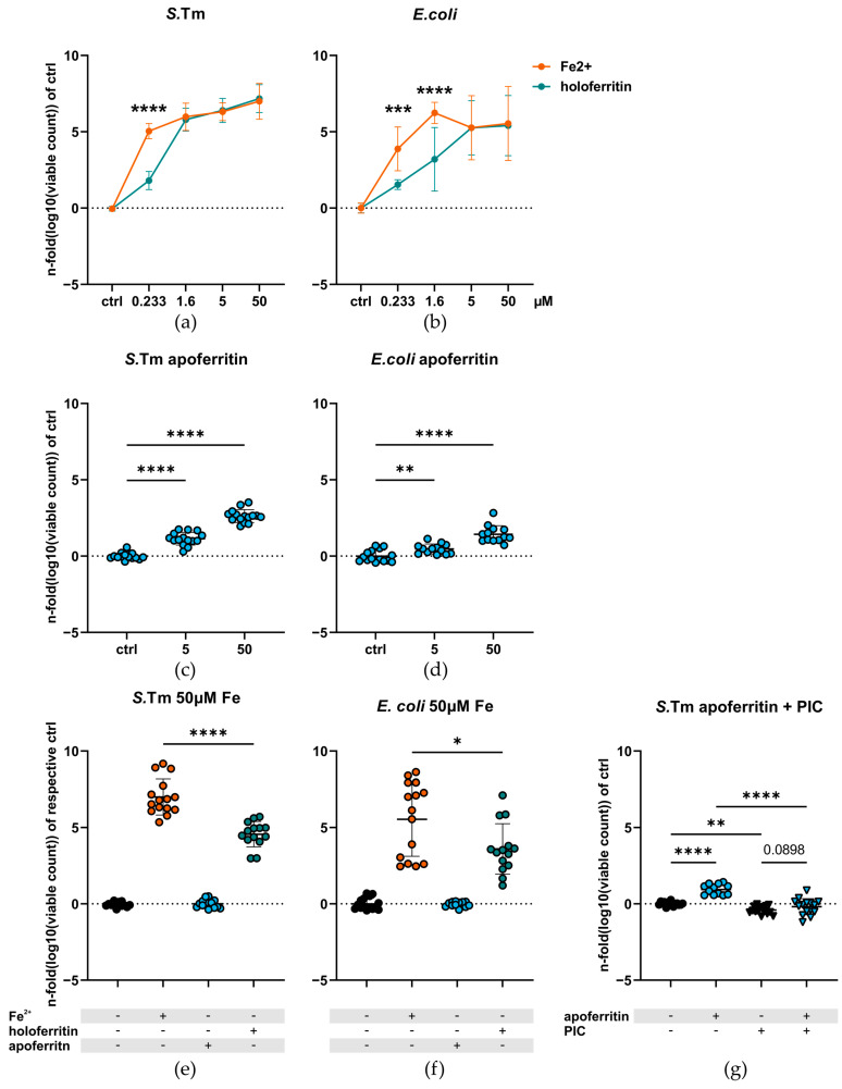
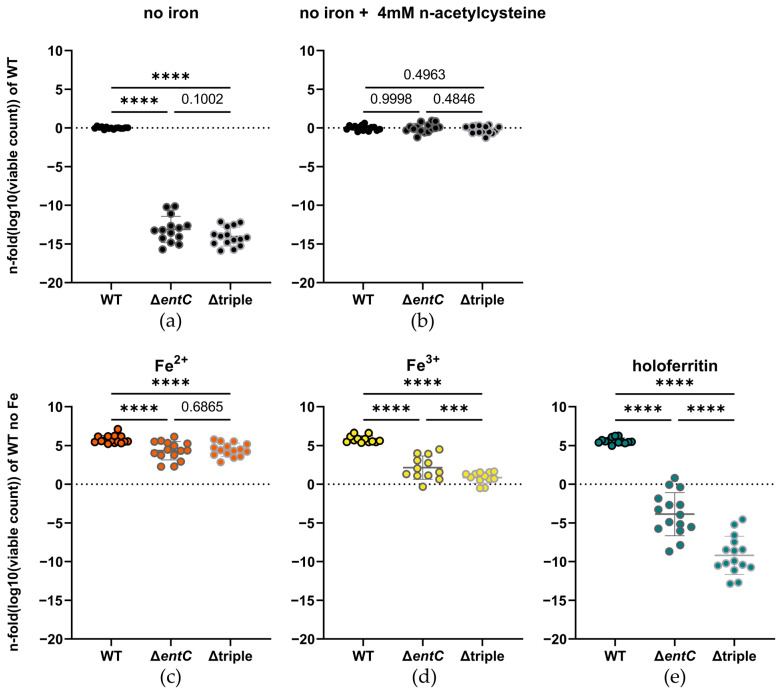
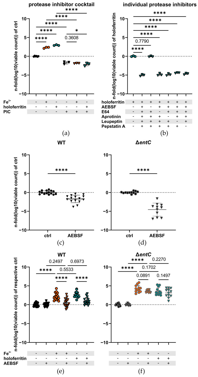
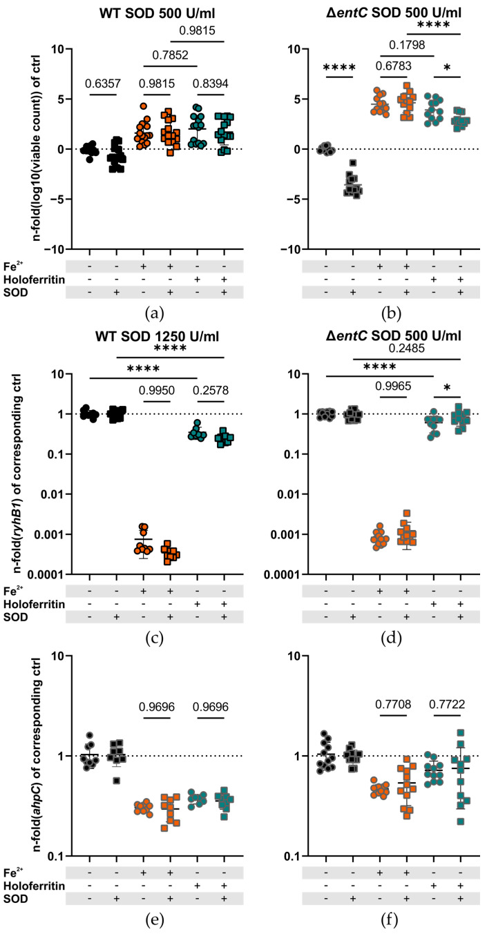
The sequestration of iron in case of infection, termed nutritional immunity, is an established strategy of host defense. However, the interaction between pathogens and the mammalian iron storage protein ferritin is hitherto not completely understood. To better characterize the function of ferritin in Gram-negative infections, we incubated iron-starved cultures of Salmonella Typhimurium and knockout mutant strains defective for major iron uptake pathways or Escherichia coli with horse spleen ferritin or ionic iron as the sole iron source. Additionally, we added bovine superoxide dismutase and protease inhibitors to the growth medium to assess the effect of superoxide and bacterial proteases, respectively, on Salmonella proliferation and reductive iron release. Compared to free ionic iron, ferritin-bound iron was less available to Salmonella, but was still sufficient to significantly enhance the growth of the bacteria. In the absence of various iron acquisition genes, the availability of ferritin iron further decreased. Supplementation with superoxide dismutase significantly reduced the growth of the ΔentC knockout strain with holoferritin as the sole iron source in comparison with ionic ferrous iron. In contrast, this difference was not observed in the wildtype strain, suggesting that superoxide dismutase undermines bacterial iron uptake from ferritin by siderophore-independent mechanisms. Ferritin seems to diminish iron availability for bacteria in comparison to ionic iron, and its iron sequestering effect could possibly be enhanced by host superoxide dismutase activity.
Overview
The claims and statistics of this paper haven’t been checked yet.
Run the check — free, no account needed. It reads the open-access text and checks the sources it cites.
Integrity signals
Registry
Cites
Statistics
Figures
Verification
Claims the paper makes
The claims and statistics of this paper haven’t been checked yet.
Run the check — free, no account needed. It reads the open-access text and checks the sources it cites.
Claims other papers make about this one
Each is checked against this paper’s own text: does it say what the citing paper says it does?
In the citing paper
“Ferritin limits pathogens' capacity to spread infection by binding and sequestering excess iron, preventing them from gaining access to this vital nutrient (Gehrer et al. 2022).”
From Nutritional Immunity in Wound Infection: Unveiling the Role of Dietary Elements in Host–Pathogen Interaction (2025) · cites this paper as Gehrer et al. 2022
Self-check
Percentages, sums and reported differences re-computed from the paper’s own numbers.
The self-check needs the paper’s full text.
Citations
Cites
Nutritional immunity: transition metals at the pathogen–host interface
Hood, M. I., & Skaar, E. P. (2012) · Nature Reviews Microbiology · doi:10.1038/nrmicro2836 · Cited by 1,535
Defining normoxia, physoxia and hypoxia in tumours—implications for treatment response
McKeown, S. R. (2014) · The British Journal of Radiology · doi:10.1259/bjr.20130676 · Cited by 1,050
Non-transferrin bound iron: a key role in iron overload and iron toxicity
Brissot, P., Ropert, M., Le Lan, C., & Loréal, O. (2012) · Biochimica et Biophysica Acta (BBA) - General Subjects · doi:10.1016/j.bbagen.2011.07.014 · Cited by 654 · cited 11×
“Ferritin’s iron storage capability is accompanied by its role as an ROS defense protein.”
BackgroundInfluentialIron in infection and immunity
Nairz, M., & Weiss, G. (2020) · Molecular Aspects of Medicine · doi:10.1016/j.mam.2020.100864 · Cited by 359
Self-citationRuddell, R. G., Hoang‐Le, D., Barwood, J. M., Rutherford, P. S., Piva, T. J., Watters, D. J., Santambrogio, P., Arosio, P., & Ramm, G. A. (2009) · Hepatology · doi:10.1002/hep.22716 · Cited by 298
Distinct mechanisms of ferritin delivery to lysosomes in iron-depleted and iron-replete cells
Asano, T., Komatsu, M., Yamaguchi-Iwai, Y., Ishikawa, F., Mizushima, N., & Iwai, K. (2011) · Molecular and Cellular Biology · doi:10.1128/mcb.01437-10 · Cited by 265
Nairz, M., Schleicher, U., Schroll, A., Sonnweber, T., Theurl, I., Ludwiczek, S., Talasz, H., Brandacher, G., Moser, P. L., Muckenthaler, M. U., et al. (2013) · Journal of Experimental Medicine · doi:10.1084/jem.20121946 · Cited by 204
Self-citationCrouch, M. V., Castor, M., Karlinsey, J. E., Kalhorn, T., & Fang, F. C. (2008) · Molecular Microbiology · doi:10.1111/j.1365-2958.2007.06089.x · Cited by 187
Self-citationCarrano, C. J., & Raymond, K. N. (1979) · Journal of the American Chemical Society · doi:10.1021/ja00512a047 · Cited by 155
Iron Regulatory Proteins Mediate Host Resistance to Salmonella Infection
Nairz, M., Ferring-Appel, D., Casarrubea, D., Sonnweber, T., Viatte, L., Schroll, A., Haschka, D., Fang, F. C., Hentze, M. W., Weiss, G., et al. (2015) · Cell Host & Microbe · doi:10.1016/j.chom.2015.06.017 · Cited by 103
Self-citation
Research works printed but not linked: 78
- Stanaway, 2019, The global burden of non-typhoidal salmonella invasive disease: A systematic analysis for the Global Burden of Disease Study 2017, Lancet Infect. Dis., 19, 1312, 10.1016/S1473-3099(19)30418-9 · DOI · Paper page
- Anderson, 2019, Burden of enterotoxigenic Escherichia coli and shigella non-fatal diarrhoeal infections in 79 low-income and lower middle-income countries: A modelling analysis, Lancet Glob. Health, 7, e321, 10.1016/S2214-109X(18)30483-2 · DOI
- Tan, 2019, Growth and Virulence of Salmonella Typhimurium Mutants Deficient in Iron Uptake, ACS Omega, 4, 13218, 10.1021/acsomega.9b01367 · DOI
- Nairz, 2014, Iron at the interface of immunity and infection, Front. Pharmacol., 5, 152, 10.3389/fphar.2014.00152 · DOI · Paper page
- Healy, 2021, Nutritional immunity: The impact of metals on lung immune cells and the airway microbiome during chronic respiratory disease, Respir. Res., 22, 133, 10.1186/s12931-021-01722-y · DOI · Paper page
- Zhao, 2006, Iron(II) and hydrogen peroxide detoxification by human H-chain ferritin. An EPR spin-trapping study, Biochemistry, 45, 3429, 10.1021/bi052443r · DOI
- Rosário, C., Zandman-Goddard, G., Meyron-Holtz, E.G., D’Cruz, D.P., and Shoenfeld, Y. (2013). The Hyperferritinemic Syndrome: Macrophage activation syndrome, Still’s disease, septic shock and catastrophic antiphospholipid syndrome. BMC Med., 11. · DOI
- Konijn, 1977, Ferritin synthesis in inflammation. I. Pathogenesis of impaired iron release, Br. J. Haematol., 37, 7, 10.1111/j.1365-2141.1977.tb08806.x · DOI
- Hoffmann, 2021, Baseline iron status and presence of anaemia determine the course of systemic Salmonella infection following oral iron supplementation in mice, eBioMedicine, 71, 103568, 10.1016/j.ebiom.2021.103568 · DOI
- Dehner, 2013, Ferritin and ferrihydrite nanoparticles as iron sources for Pseudomonas aeruginosa, J. Biol. Inorg. Chem. JBIC A Publ. Soc. Biol. Inorg. Chem., 18, 371, 10.1007/s00775-013-0981-9 · DOI
- Whitby, 2006, Burkholderia cenocepacia utilizes ferritin as an iron source, J. Med. Microbiol., 55, 661, 10.1099/jmm.0.46199-0 · DOI
- Teawtrakul, 2015, Severe bacterial infections in patients with non-transfusion-dependent thalassemia: Prevalence and clinical risk factors, Int. J. Infect. Dis. IJID Off. Publ. Int. Soc. Infect. Dis., 39, 53
- Adamkiewicz, 1998, Infection due to Yersinia enterocolitica in a series of patients with beta-thalassemia: Incidence and predisposing factors, Clin. Infect. Dis. Off. Publ. Infect. Dis. Soc. Am., 27, 1362, 10.1086/515025 · DOI
- Harrison, 1996, The ferritins: Molecular properties, iron storage function and cellular regulation, Biochim. Et Biophys. Acta BBA Bioenerg., 1275, 161, 10.1016/0005-2728(96)00022-9 · DOI · Paper page
- Mehlenbacher, 2017, Iron Oxidation and Core Formation in Recombinant Heteropolymeric Human Ferritins, Biochemistry, 56, 3900, 10.1021/acs.biochem.7b00024 · DOI
- Narayanan, 2019, On the structure and chemistry of iron oxide cores in human heart and human spleen ferritins using graphene liquid cell electron microscopy, Nanoscale, 11, 16868, 10.1039/C9NR01541H · DOI
- Xu, 1991, Iron oxidation chemistry in ferritin. Increasing Fe/O2 stoichiometry during core formation, J. Biol. Chem., 266, 19965, 10.1016/S0021-9258(18)54877-8 · DOI
- Li, W., Garringer, H.J., Goodwin, C.B., Richine, B., Acton, A., VanDuyn, N., Muhoberac, B.B., Irimia-Dominguez, J., Chan, R.J., and Peacock, M. (2015). Systemic and cerebral iron homeostasis in ferritin knock-out mice. PLoS ONE, 10. · DOI
- Velayudhan, 2007, The role of ferritins in the physiology of Salmonella enterica sv. Typhimurium: A unique role for ferritin B in iron-sulphur cluster repair and virulence, Mol. Microbiol., 63, 1495, 10.1111/j.1365-2958.2007.05600.x · DOI · Paper page
- Harris, 1979, Coordination chemistry of microbial iron transport compounds. 19. Stability constants and electrochemical behavior of ferric enterobactin and model complexes, J. Am. Chem. Soc., 101, 6097, 10.1021/ja00514a037 · DOI
- Aisen, 1978, Stoichiometric and site characteristics of the binding of iron to human transferrin, J. Biol. Chem., 253, 1930, 10.1016/S0021-9258(19)62337-9 · DOI · Paper page
- Buchanan, 1999, Crystal structure of the outer membrane active transporter FepA from Escherichia coli, Nat. Struct. Biol., 6, 56, 10.1038/4931 · DOI
- Sprencel, 2000, Binding of ferric enterobactin by the Escherichia coli periplasmic protein FepB, J. Bacteriol., 182, 5359, 10.1128/JB.182.19.5359-5364.2000 · DOI
- Pierce, 1986, Escherichia coli K-12 envelope proteins specifically required for ferrienterobactin uptake, J. Bacteriol., 166, 930, 10.1128/jb.166.3.930-936.1986 · DOI
- Chenault, 1992, Identification of hydrophobic proteins FepD and FepG of the Escherichia coli ferrienterobactin permease, J. Gen. Microbiol., 138, 2167, 10.1099/00221287-138-10-2167 · DOI
- Brickman, 1992, Overexpression and purification of ferric enterobactin esterase from Escherichia coli. Demonstration of enzymatic hydrolysis of enterobactin and its iron complex, J. Biol. Chem., 267, 12350, 10.1016/S0021-9258(19)49846-3 · DOI
- Zhu, 2005, Functions of the siderophore esterases IroD and IroE in iron-salmochelin utilization, Microbiology, 151, 2363, 10.1099/mic.0.27888-0 · DOI
- Lin, 2005, In vitro characterization of salmochelin and enterobactin trilactone hydrolases IroD, IroE, and Fes, J. Am. Chem. Soc., 127, 11075, 10.1021/ja0522027 · DOI
- Peralta, D.R., Adler, C., Corbalán, N.S., Paz García, E.C., Pomares, M.F., and Vincent, P.A. (2016). Enterobactin as Part of the Oxidative Stress Response Repertoire. PLoS ONE, 11. · DOI
- Achard, 2013, An antioxidant role for catecholate siderophores in Salmonella, Biochem. J., 454, 543, 10.1042/BJ20121771 · DOI
- Kehres, 2002, SitABCD is the alkaline Mn(2+) transporter of Salmonella enterica serovar Typhimurium, J. Bacteriol., 184, 3159, 10.1128/JB.184.12.3159-3166.2002 · DOI · Paper page
- Sabri, 2006, A SitABCD homologue from an avian pathogenic Escherichia coli strain mediates transport of iron and manganese and resistance to hydrogen peroxide, Microbiology, 152, 745, 10.1099/mic.0.28682-0 · DOI
- Janakiraman, 2000, The putative iron transport system SitABCD encoded on SPI1 is required for full virulence of Salmonella typhimurium, Mol. Microbiol., 35, 1146, 10.1046/j.1365-2958.2000.01783.x · DOI
- Lau, 2016, Bacterial ferrous iron transport: The Feo system, FEMS Microbiol. Rev., 40, 273, 10.1093/femsre/fuv049 · DOI · Paper page
- Kim, 2012, The FeoA protein is necessary for the FeoB transporter to import ferrous iron, Biochem. Biophys. Res. Commun., 423, 733, 10.1016/j.bbrc.2012.06.027 · DOI
- Kammler, 1993, Characterization of the ferrous iron uptake system of Escherichia coli, J. Bacteriol., 175, 6212, 10.1128/jb.175.19.6212-6219.1993 · DOI · Paper page
- Kim, 2015, Lon-mediated proteolysis of the FeoC protein prevents Salmonella enterica from accumulating the Fe(II) transporter FeoB under high-oxygen conditions, J. Bacteriol., 197, 92, 10.1128/JB.01826-14 · DOI
- Vartivarian, 1999, Extracellular iron reductases: Identification of a new class of enzymes by siderophore-producing microorganisms, Arch. Biochem. Biophys., 364, 75, 10.1006/abbi.1999.1109 · DOI
- Massey, 1994, Activation of molecular oxygen by flavins and flavoproteins, J. Biol. Chem., 269, 22459, 10.1016/S0021-9258(17)31664-2 · DOI
- Romero, 2018, Same Substrate, Many Reactions: Oxygen Activation in Flavoenzymes, Chem. Rev., 118, 1742, 10.1021/acs.chemrev.7b00650 · DOI
- Turrens, 2003, Mitochondrial formation of reactive oxygen species, J. Physiol., 552, 335, 10.1113/jphysiol.2003.049478 · DOI · Paper page
- Murakami, 2012, Stimulation of the amyloidogenic pathway by cytoplasmic superoxide radicals in an Alzheimer’s disease mouse model, Biosci. Biotechnol. Biochem., 76, 1098, 10.1271/bbb.110934 · DOI
- Weiss, 2013, The complex interplay of iron metabolism, reactive oxygen species, and reactive nitrogen species: Insights into the potential of various iron therapies to induce oxidative and nitrosative stress, Free Radic. Biol. Med., 65, 1174, 10.1016/j.freeradbiomed.2013.09.001 · DOI · Paper page
- Birben, 2012, Oxidative stress and antioxidant defense, World Allergy Organ. J., 5, 9, 10.1097/WOX.0b013e3182439613 · DOI
- Nakashima, 2012, Generation of superoxide anions by a glycation reaction in conventional laboratory media, J. Biosci. Bioeng., 114, 275, 10.1016/j.jbiosc.2012.04.009 · DOI
- Bolann, 1987, Release of iron from ferritin by xanthine oxidase. Role of the superoxide radical, Biochem. J., 243, 55, 10.1042/bj2430055 · DOI
- Boyer, 1987, Superoxide ion as a primary reductant in ascorbate-mediated ferritin iron release, Free Radic. Biol. Med., 3, 389, 10.1016/0891-5849(87)90017-7 · DOI
- Thomas, 1985, Ferritin and superoxide-dependent lipid peroxidation, J. Biol. Chem., 260, 3275, 10.1016/S0021-9258(19)83617-7 · DOI
- Thomas, 1986, Reductive release of iron from ferritin by cation free radicals of paraquat and other bipyridyls, J. Biol. Chem., 261, 13064, 10.1016/S0021-9258(18)69271-3 · DOI
- McNally, 2011, Oxygen catalyzed mobilization of iron from ferritin by iron(III) chelate ligands, Chem. Commun., 47, 731, 10.1039/C0CC03454A · DOI
- Claiborne, 1979, Purification of the o-dianisidine peroxidase from Escherichia coli B. Physicochemical characterization and analysis of its dual catalatic and peroxidatic activities, J. Biol. Chem., 254, 4245, 10.1016/S0021-9258(18)50722-5 · DOI
- Bravo, 1995, Crystal structure of catalase HPII from Escherichia coli, Structure, 3, 491, 10.1016/S0969-2126(01)00182-4 · DOI
- Yost, 1973, An iron-containing superoxide dismutase from Escherichia coli, J. Biol. Chem., 248, 4905, 10.1016/S0021-9258(19)43649-1 · DOI
- Cuesta, 2008, Comparative structural and chemical studies of ferritin cores with gradual removal of their iron contents, J Am Chem Soc, 130, 8062, 10.1021/ja800492z · DOI
- Gottesman, 2002, A small RNA regulates the expression of genes involved in iron metabolism in Escherichia coli, Proc. Natl. Acad. Sci. USA, 99, 4620, 10.1073/pnas.032066599 · DOI · Paper page
- Tsolis, 1996, Contribution of TonB- and Feo-mediated iron uptake to growth of Salmonella typhimurium in the mouse, Infect. Immun., 64, 4549, 10.1128/iai.64.11.4549-4556.1996 · DOI · Paper page
- Boyer, 2002, Acquisition of Mn(II) in addition to Fe(II) is required for full virulence of Salmonella enterica serovar Typhimurium, Infect. Immun., 70, 6032, 10.1128/IAI.70.11.6032-6042.2002 · DOI · Paper page
- Patzer, 2001, Dual repression by Fe(2+)-Fur and Mn(2+)-MntR of the mntH gene, encoding an NRAMP-like Mn(2+) transporter in Escherichia coli, J. Bacteriol., 183, 4806, 10.1128/JB.183.16.4806-4813.2001 · DOI
- Schalk, 2013, Fate of ferrisiderophores after import across bacterial outer membranes: Different iron release strategies are observed in the cytoplasm or periplasm depending on the siderophore pathways, Amino Acids, 44, 1267, 10.1007/s00726-013-1468-2 · DOI · Paper page
- Muselius, 2020, Iron Limitation in Klebsiella pneumoniae Defines New Roles for Lon Protease in Homeostasis and Degradation by Quantitative Proteomics, Front. Microbiol., 11, 546, 10.3389/fmicb.2020.00546 · DOI
- Arends, 2018, An Integrated Proteomic Approach Uncovers Novel Substrates and Functions of the Lon Protease in Escherichia coli, Proteomics, 18, e1800080, 10.1002/pmic.201800080 · DOI
- Budiarti, 2007, Extracellular Protease Activity of Enteropathogenic Escherechia coli on Mucin Substrate, HAYATI J. Biosci., 14, 36, 10.4308/hjb.14.1.36 · DOI
- Grodberg, 1989, Comparison of Escherichia coli K-12 outer membrane protease OmpT and Salmonella typhimurium E protein, J. Bacteriol., 171, 2903, 10.1128/jb.171.5.2903-2905.1989 · DOI
- Pokharel, P., Habouria, H., Bessaiah, H., and Dozois, C.M. (2019). Serine Protease Autotransporters of the Enterobacteriaceae (SPATEs): Out and About and Chopping It Up. Microorganisms, 7. · DOI
- Nguyen, 2017, Neutrophils to the ROScue: Mechanisms of NADPH Oxidase Activation and Bacterial Resistance, Front. Cell. Infect. Microbiol., 7, 373, 10.3389/fcimb.2017.00373 · DOI · Paper page
- Zhang, C., Zhang, X., and Zhao, G. (2020). Ferritin Nanocage: A Versatile Nanocarrier Utilized in the Field of Food, Nutrition, and Medicine. Nanomaterials, 10. · DOI
- Berger, 2006, Lipocalin 2-deficient mice exhibit increased sensitivity to Escherichia coli infection but not to ischemia-reperfusion injury, Proc. Natl. Acad. Sci. USA, 103, 1834, 10.1073/pnas.0510847103 · DOI · Paper page
- Dong, 2008, Nrf2-mediated transcriptional induction of antioxidant response in mouse embryos exposed to ethanol In Vivo: Implications for the prevention of fetal alcohol spectrum disorders, Antioxid. Redox Signal., 10, 2023, 10.1089/ars.2007.2019 · DOI
- Yang, 2019, Salmonella effector SpvB interferes with intracellular iron homeostasis via regulation of transcription factor NRF2, FASEB J. Off. Publ. Fed. Am. Soc. Exp. Biol., 33, 13450
- Grubwieser, 2022, Airway Epithelial Cells Differentially Adapt Their Iron Metabolism to Infection With Klebsiella pneumoniae and Escherichia coli In Vitro, Front. Cell. Infect. Microbiol., 12, 875543, 10.3389/fcimb.2022.875543 · DOI
- Joshi, 2021, NRF2 promotes urothelial cell response to bacterial infection by regulating reactive oxygen species and RAB27B expression, Cell Rep., 37, 109856, 10.1016/j.celrep.2021.109856 · DOI
- Ohshima, 2022, NCOA4 drives ferritin phase separation to facilitate macroferritinophagy and microferritinophagy, J. Cell. Biol., 221, e202203102, 10.1083/jcb.202203102 · DOI
- Wilkinson, 2014, The IRP/IRE system in vivo: Insights from mouse models, Front. Pharmacol., 5, 176, 10.3389/fphar.2014.00176 · DOI · Paper page
- Miao, 2015, A TRP Channel Senses Lysosome Neutralization by Pathogens to Trigger Their Expulsion, Cell, 161, 1306, 10.1016/j.cell.2015.05.009 · DOI
- Liss, 2017, Salmonella enterica Remodels the Host Cell Endosomal System for Efficient Intravacuolar Nutrition, Cell Host Microbe, 21, 390, 10.1016/j.chom.2017.02.005 · DOI
- Kreibich, 2015, Autophagy Proteins Promote Repair of Endosomal Membranes Damaged by the Salmonella Type Three Secretion System 1, Cell Host Microbe, 18, 527, 10.1016/j.chom.2015.10.015 · DOI
- Chen, 2009, Superoxide is the major reactive oxygen species regulating autophagy, Cell Death Differ., 16, 1040, 10.1038/cdd.2009.49 · DOI
- Suzuki, 2000, Quantitative analysis of small-subunit rRNA genes in mixed microbial populations via 5′-nuclease assays, Appl. Environ. Microbiol., 66, 4605, 10.1128/AEM.66.11.4605-4614.2000 · DOI
Cited by
Sorted by citations
Chen, C., Hu, X., He, D., He, X., & Shen, L. (2025) · Food Science & Nutrition · doi:10.1002/fsn3.70854 · Cited by 6
Metadata
- Authors
- Gehrer, C. M., Hoffmann, A., Hilbe, R., Grubwieser, P., Mitterstiller, A.-M., Talasz, H., Fang, F. C., Meyron-Holtz, E. G., Atkinson, S. H., Weiss, G., & Nairz, M.
- Affiliations
- Department of Internal Medicine II, Infectious Diseases, Immunology, Rheumatology, Medical University of Innsbruck, 6020 Innsbruck, Austria
- Biocenter, Institute of Medical Biochemistry, Medical Universitiy of Innsbruck, 6020 Innsbruck, Austria
- Department of Laboratory Medicine and Pathology, University of Washington School of Medicine, Seattle, WA 98195-7110, USA
- Laboratory of Molecular Nutrition, Faculty of Biotechnology and Food Engineering, Technion-Israel Institute of Technology, Haifa 32000, Israel
- Kenya Medical Research Institute (KEMRI), Centre for Geographic Medicine Research Coast, KEMRI-Wellcome Trust Research Programme, Kilifi 80108, Kenya
- ORCID
- Venue
- International Journal of Molecular Sciences
- Publisher
- MDPI AG
- Published
- 2022-10-28
- Volume
- 23
- Issue
- 21
- Pages
- 13087
- ISSN
- 1422-0067
- PMC
- PMC9657528
- Cited by
- 10
- References
- 15
- Language
- en
- Funders
- Austrian Science Fund (FWF) (P 33062-B)
About these checks
Registry, retraction, Cites and statistics results are computed, not judged, with one exception: a printed reference that no index lists is read once by a model before it counts as a citation error. Claims are extracted by a model and kept only when their quote appears verbatim in the text; each cited source is then checked, with a model’s help, for whether it says what the claim relies on. Those verdicts can be wrong — read the claim’s quote and the cited source before relying on one.
Statistics and arithmetic are recomputed, not judged.
Confidence bands are shown only for fields calibrated on our annotated test set. None is calibrated yet, so no band is shown.
What counts as a claim ›How we measure confidence ›How we check this ›
Are you an author, or do you see a problem with this page? Request a review
