

The DOI matches its Crossref record; 210 of the 212 references it cites were found in a registry or an index.
Glutamine: Metabolism and Immune Function, Supplementation and Clinical Translation
Open accessPublished version of a preprint
Abstract
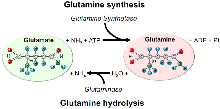
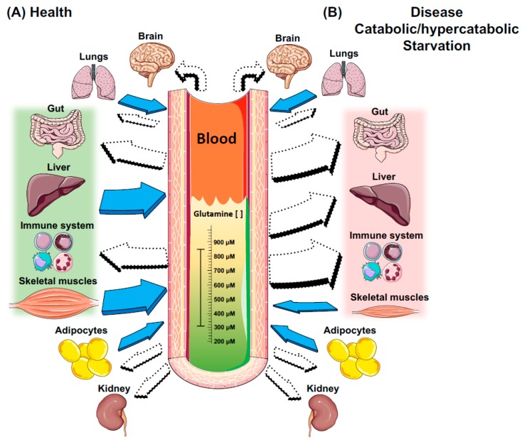
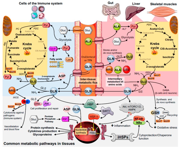
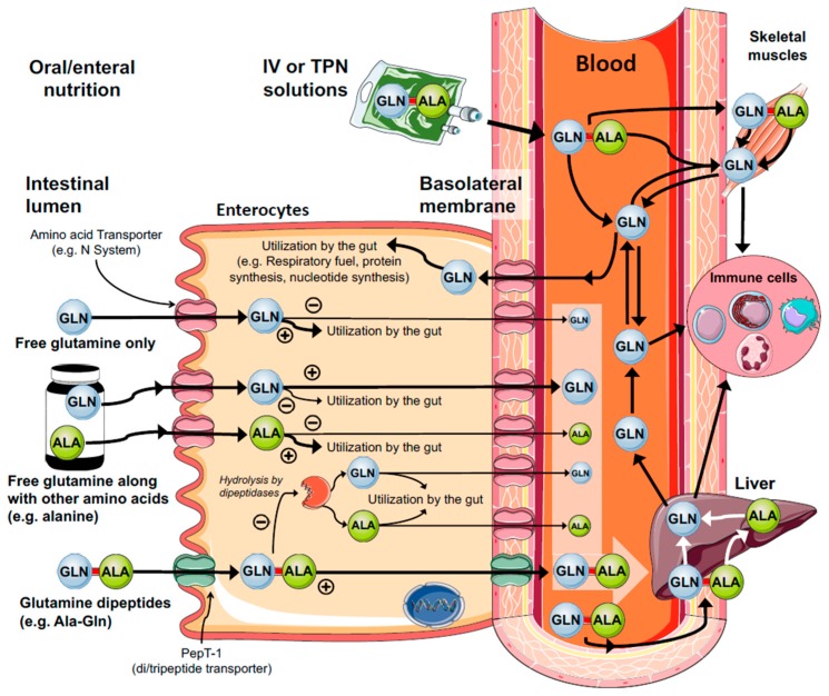
Glutamine is the most abundant and versatile amino acid in the body. In health and disease, the rate of glutamine consumption by immune cells is similar or greater than glucose. For instance, in vitro and in vivo studies have determined that glutamine is an essential nutrient for lymphocyte proliferation and cytokine production, macrophage phagocytic plus secretory activities, and neutrophil bacterial killing. Glutamine release to the circulation and availability is mainly controlled by key metabolic organs, such as the gut, liver, and skeletal muscles. During catabolic/hypercatabolic situations glutamine can become essential for metabolic function, but its availability may be compromised due to the impairment of homeostasis in the inter-tissue metabolism of amino acids. For this reason, glutamine is currently part of clinical nutrition supplementation protocols and/or recommended for immune suppressed individuals. However, in a wide range of catabolic/hypercatabolic situations (e.g., ill/critically ill, post-trauma, sepsis, exhausted athletes), it is currently difficult to determine whether glutamine supplementation (oral/enteral or parenteral) should be recommended based on the amino acid plasma/bloodstream concentration (also known as glutaminemia). Although the beneficial immune-based effects of glutamine supplementation are already established, many questions and evidence for positive in vivo outcomes still remain to be presented. Therefore, this paper provides an integrated review of how glutamine metabolism in key organs is important to cells of the immune system. We also discuss glutamine metabolism and action, and important issues related to the effects of glutamine supplementation in catabolic situations.
Overview
The claims and statistics of this paper haven’t been checked yet.
Run the check — free, no account needed. It reads the open-access text and checks the sources it cites.
Integrity signals
Registry
Cites
Statistics
Figures
Verification
Claims the paper makes
The claims and statistics of this paper haven’t been checked yet.
Run the check — free, no account needed. It reads the open-access text and checks the sources it cites.
Claims other papers make about this one
Each is checked against this paper’s own text: does it say what the citing paper says it does?
In the citing paper
“L-glutamine is the most abundant amino acid in the body, with an essential role in lymphocyte proliferation and cytokine production [175].”
From International Society of Sports Nutrition Position Stand: nutritional considerations for single-stage ultra-marathon training and racing (2019) · cites this paper as [175]
In the citing paper
“In catabolic and hypercatabolic situations, L-glutamine can be essential to help maintain normal metabolic function and is, therefore, included in clinical nutritional supplementation protocols and recommended for immune-suppressed individuals [175].”
From International Society of Sports Nutrition Position Stand: nutritional considerations for single-stage ultra-marathon training and racing (2019) · cites this paper as [175]
Self-check
Percentages, sums and reported differences re-computed from the paper’s own numbers.
The self-check needs the paper’s full text.
Citations
Cites
Exploring the full spectrum of macrophage activation
Mosser, D. M., & Edwards, J. P. (2008) · Nature Reviews Immunology · doi:10.1038/nri2448 · Cited by 9,144
Meister, A., & Anderson, M. E. (1983) · Annual Review of Biochemistry · doi:10.1146/annurev.bi.52.070183.003431 · Cited by 5,694
Monocyte and macrophage heterogeneity
Gordon, S., & Taylor, P. R. (2005) · Nature Reviews Immunology · doi:10.1038/nri1733 · Cited by 5,190
The metabolism of tumors in the body
Warburg, O., Wind, F., & Negelein, E. (1927) · Journal of General Physiology · doi:10.1085/jgp.8.6.519 · Cited by 4,416
Alternative activation of macrophages: mechanism and functions
Gordon, S., & Martinez, F. O. (2010) · Immunity · doi:10.1016/j.immuni.2010.05.007 · Cited by 4,197
Succinate is an inflammatory signal that induces IL-1β through HIF-1α
Tannahill, G. M., Curtis, A. M., Adamik, J., Palsson-McDermott, E. M., McGettrick, A. F., Goel, G., Frezza, C., Bernard, N. J., Kelly, B., Foley, N. H., et al. (2013) · Nature · doi:10.1038/nature11986 · Cited by 4,176 · cited 2×
“, succinate, α-ketoglutarate, citrate, and itaconate) that regulates LPS macrophage activation [119].”
BackgroundMacrophage activation and polarization
Martinez, F. O. (2008) · Frontiers in Bioscience · doi:10.2741/2692 · Cited by 3,160
Posttranscriptional Control of T Cell Effector Function by Aerobic Glycolysis
Chang, C.-H., Curtis, J. D., Maggi, L. B., Faubert, B., Villarino, A. V., O’Sullivan, D., Huang, S. C.-C., van der Windt, G. J. W., Blagih, J., Qiu, J., et al. (2013) · Cell · doi:10.1016/j.cell.2013.05.016 · Cited by 2,204
From Krebs to clinic: glutamine metabolism to cancer therapy
Altman, B. J., Stine, Z. E., & Dang, C. V. (2016) · Nature Reviews Cancer · doi:10.1038/nrc.2016.71 · Cited by 2,119
Michalek, R. D., Gerriets, V. A., Jacobs, S. R., Macintyre, A. N., MacIver, N. J., Mason, E. F., Sullivan, S. A., Nichols, A. G., & Rathmell, J. C. (2011) · The Journal of Immunology · doi:10.4049/jimmunol.1003613 · Cited by 2,091
Research works printed but not linked: 1
- Rogero, 2007, Ffect of glutamine supplementation and in vivo infection with mycobacterium bovis (bacillus calmette-guerin) in the function of peritoneal macrophages in early weaned mice, Ann. Nutr. Metab., 51, 173
Cited by
Sorted by citations
Glutamine: Metabolism and Immune Function, Supplementation and Clinical Translation
Cruzat, V., Macedo Rogero, M., Noel Keane, K., Curi, R., & Newsholme, P. (2018) · doi:10.20944/preprints201809.0459.v1 · Cited by 1,261
Self-citationLactate in the Regulation of Tumor Microenvironment and Therapeutic Approaches
de la Cruz-López, K. G., Castro-Muñoz, L. J., Reyes-Hernández, D. O., García-Carrancá, A., & Manzo-Merino, J. (2019) · Frontiers in Oncology · doi:10.3389/fonc.2019.01143 · Cited by 940
Role of oxidative stress in depression
Bhatt, S., Nagappa, A. N., & Patil, C. R. (2020) · Drug Discovery Today · doi:10.1016/j.drudis.2020.05.001 · Cited by 854
Iddir, M., Alex Brito, O., Dingeo, G., Sosa, S., Samouda, H., Frano, M. R. L., & Bohn, T. (2020) · Nutrients · doi:10.3390/nu12061562 · Cited by 825
Diabetic vascular diseases: molecular mechanisms and therapeutic strategies
Li, Y., Liu, Y., Liu, S., Gao, M., Wang, W., Chen, K., Huang, L., & Liu, Y. (2023) · Signal Transduction and Targeted Therapy · doi:10.1038/s41392-023-01400-z · Cited by 799
Metadata
- Authors
- Cruzat, V., Macedo Rogero, M., Noel Keane, K., Curi, R., & Newsholme, P.
- Affiliations
- School of Pharmacy and Biomedical Sciences, Curtin Health Innovation Research Institute, Biosciences, Curtin University, Perth 6102, Australia
- Department of Nutrition, Faculty of Public Health, University of São Paulo, Avenida Doutor Arnaldo 715, São Paulo 01246-904, Brazil
- Universidade Cruzeiro do Sul (BR)
- Curtin University (AU)
- Corresponding author
- Newsholme, Philip
- Venue
- Nutrients
- Publisher
- MDPI AG
- Published
- 2018-10-23
- Volume
- 10
- Issue
- 11
- Pages
- 1564
- ISSN
- 2072-6643 (ISSN-L)
- PMC
- PMC6266414
- OpenAlex
- W2897213700
- Cited by
- 1,261
- References
- 212
- Citations by year
- 2026: 155 · 2025: 244 · 2024: 204 · 2023: 203 · 2022: 180 · 2021: 145 · 2020: 94 · 2019: 29 · 2018: 2
- FWCI
- 8.66
- Type
- journal article
- Language
- en
- Open access
- gold
- dx.doi.org
- Licence
- Locations
- Nutrients (publishedVersion, cc-by)
- Preprints.org (acceptedVersion, cc-by)
- Europe PMC (PubMed Central) (publishedVersion, other-oa)
- PubMed Central (publishedVersion, cc-by)
- PubMed (publishedVersion)
- DOAJ (DOAJ: Directory of Open Access Journals) (submittedVersion, cc-by-sa)
- eSpace (Curtin University) (submittedVersion, cc-by)
- Figshare (submittedVersion, cc-by)
- Funders
- Curtin University of Technology
- Topics
- Health Sciences › Nursing › Nutrition and Dietetics › Clinical Nutrition and Gastroenterology
- Life Sciences › Biochemistry, Genetics and Molecular Biology › Biochemistry › Amino Acid Enzymes and Metabolism
- Life Sciences › Agricultural and Biological Sciences › Animal Science and Zoology › Animal Nutrition and Physiology
- Keywords
- glutamine, glutamine metabolism, immune function, glutamine supplementation, catabolic state, hypercatabolism, lymphocyte proliferation, cytokine production, macrophage function, neutrophil function, critically ill patients, sepsis, trauma (physical injury), athletes, immunosuppression
- MeSH terms
- Enteral Nutrition, Glutamine, Humans, Parenteral Nutrition, Dietary Supplements
About these checks
Registry, retraction, Cites and statistics results are computed, not judged, with one exception: a printed reference that no index lists is read once by a model before it counts as a citation error. Claims are extracted by a model and kept only when their quote appears verbatim in the text; each cited source is then checked, with a model’s help, for whether it says what the claim relies on. Those verdicts can be wrong — read the claim’s quote and the cited source before relying on one.
Statistics and arithmetic are recomputed, not judged.
Confidence bands are shown only for fields calibrated on our annotated test set. None is calibrated yet, so no band is shown.
What counts as a claim ›How we measure confidence ›How we check this ›
Are you an author, or do you see a problem with this page? Request a review
