

The DOI matches its Crossref record; 106 of the 107 references it cites were found in a registry or an index.
Potential toxicity of superparamagnetic iron oxide nanoparticles (SPION)
Open accessAbstract
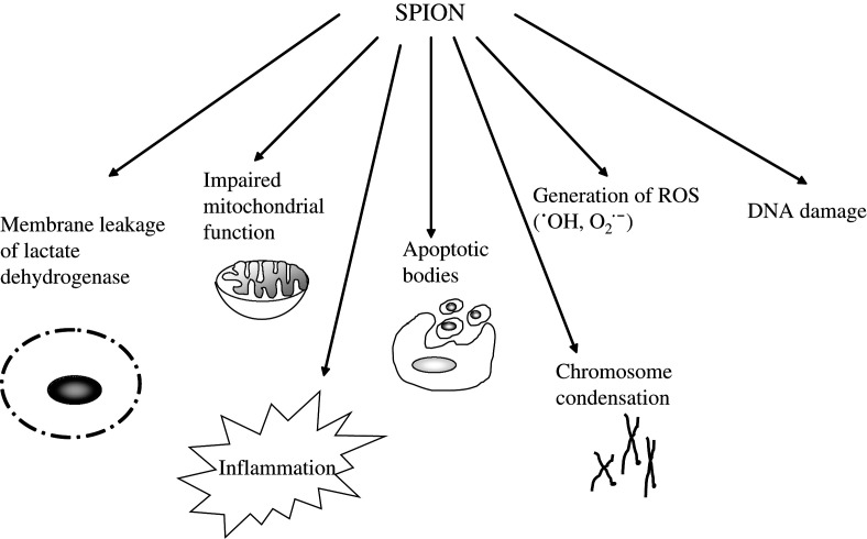
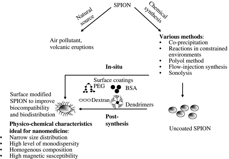
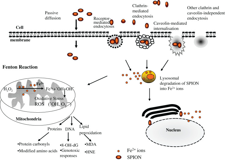
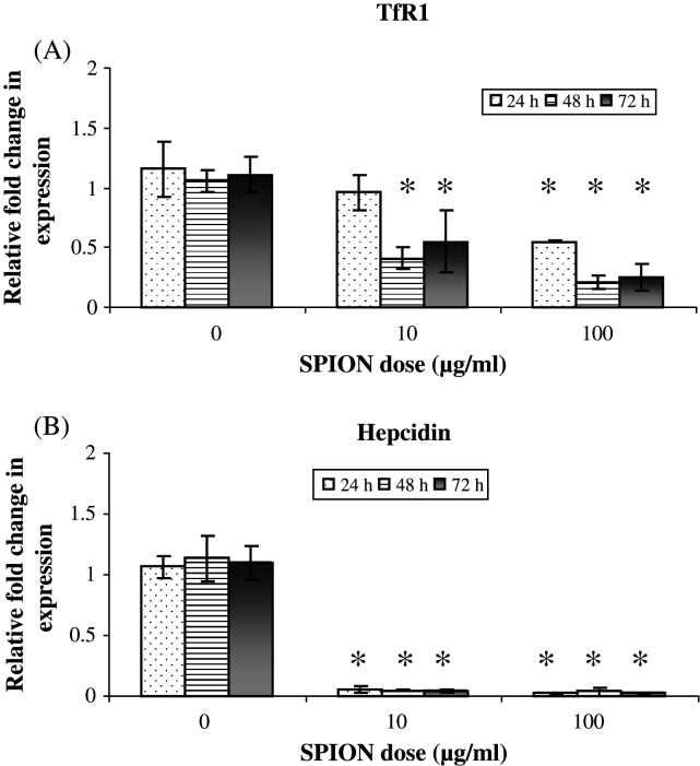
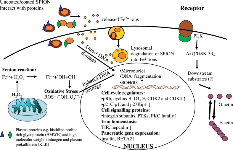
Superparamagnetic iron oxide nanoparticles (SPION) are being widely used for various biomedical applications, for example, magnetic resonance imaging, targeted delivery of drugs or genes, and in hyperthermia. Although, the potential benefits of SPION are considerable, there is a distinct need to identify any potential cellular damage associated with these nanoparticles. Besides focussing on cytotoxicity, the most commonly used determinant of toxicity as a result of exposure to SPION, this review also mentions the importance of studying the subtle cellular alterations in the form of DNA damage and oxidative stress. We review current studies and discuss how SPION, with or without different surface coating, may cause cellular perturbations including modulation of actin cytoskeleton, alteration in gene expression profiles, disturbance in iron homeostasis and altered cellular responses such as activation of signalling pathways and impairment of cell cycle regulation. The importance of protein-SPION interaction and various safety considerations relating to SPION exposure are also addressed.
Overview
The claims and statistics of this paper haven’t been checked yet.
Run the check — free, no account needed. It reads the open-access text and checks the sources it cites.
Integrity signals
Registry
Cites
Statistics
Figures
Verification
Claims the paper makes
The claims and statistics of this paper haven’t been checked yet.
Run the check — free, no account needed. It reads the open-access text and checks the sources it cites.
Claims other papers make about this one
Each is checked against this paper’s own text: does it say what the citing paper says it does?
The source backs part of the claim, not all of it — or backs it for a different population, condition or comparison.
In the citing paper
“Metallic NPs are valuable for their theragnostic capabilities but face significant barriers to translation due to potential long-term toxicity and bioaccumulation, which limit clinical progress [102, 103].”
From Targeted Nanoparticles for Drug Delivery Across the Blood–Brain Barrier in Early and Late Stages of Alzheimer's Disease: A Review (2026) · cites this paper as [102]
Experimental — accuracy not yet measuredIn the citing paper
“In vitro, upon prolonged incubation, surface-coated SPION are typically nontoxic at iron concentration up to and 10 μg/ml, while slight cytotoxicity is seen at 100 μg/ml.”
From Iron oxide nanoparticles: Diagnostic, therapeutic and theranostic applications (2019) · cites this paper as [120]
In the citing paper
“In general, SPION toxicity depends on a variety of factors, including - besides dosing and exposure time - also the nature of the coating material, the hydrodynamic diameter of the particle and SPION-protein interactions [119–122].”
From Iron oxide nanoparticles: Diagnostic, therapeutic and theranostic applications (2019) · cites this paper as [120]
Self-check
Percentages, sums and reported differences re-computed from the paper’s own numbers.
The self-check needs the paper’s full text.
Citations
Cites
Free radicals in biology and medicine
Gutteridge, J. M. C. (1991) · General Pharmacology The Vascular System · doi:10.1016/0306-3623(91)90478-o · Cited by 17,178
Free radicals and antioxidants in normal physiological functions and human disease
Valko, M., Leibfritz, D., Moncol, J., Cronin, M. T. D., Mazur, M., & Telser, J. (2007) · The International Journal of Biochemistry & Cell Biology · doi:10.1016/j.biocel.2006.07.001 · Cited by 14,269
Toxic potential of materials at the nanolevel
Nel, A., Xia, T., Mädler, L., & Li, N. (2006) · Science · doi:10.1126/science.1114397 · Cited by 9,324
Free Radicals in Biology and Medicine
Halliwell, B., & Gutteridge, J. M. C. (2015) · doi:10.1093/acprof:oso/9780198717478.001.0001 · Cited by 8,990
Synthesis and surface engineering of iron oxide nanoparticles for biomedical applications
Gupta, A. K., & Gupta, M. S. (2004) · Biomaterials · doi:10.1016/j.biomaterials.2004.10.012 · Cited by 6,906
Laurent, S., Forge, D., Port, M., Roch, A., Robic, C., Vander Elst, L., & Muller, R. N. (2008) · Chemical Reviews · doi:10.1021/cr068445e · Cited by 6,852
Hepcidin Regulates Cellular Iron Efflux by Binding to Ferroportin and Inducing Its Internalization
Nemeth, E., Tuttle, M. S., Powelson, J., Vaughn, M. B., Donovan, A., Ward, D. M., Ganz, T., & Kaplan, J. (2004) · Science · doi:10.1126/science.1104742 · Cited by 4,803
Biodegradable nanoparticles for drug and gene delivery to cells and tissue
Panyam, J., & Labhasetwar, V. D. (2003) · Advanced Drug Delivery Reviews · doi:10.1016/s0169-409x(02)00228-4 · Cited by 3,417
Fischer, D., Li, Y., Ahlemeyer, B., Krieglstein, J., & Kissel, T. (2003) · Biomaterials · doi:10.1016/s0142-9612(02)00445-3 · Cited by 2,329
In vitro toxicity of nanoparticles in BRL 3A rat liver cells
Hussain, S. M., Hess, K. L., Gearhart, J. M., Geiss, K. T., & Schlager, J. J. (2005) · Toxicology in Vitro · doi:10.1016/j.tiv.2005.06.034 · Cited by 2,025
Protein-nanoparticle interactions
Lynch, I., & Dawson, K. A. (2008) · Nano Today · doi:10.1016/s1748-0132(08)70014-8 · Cited by 1,783
Nanocarriers’ entry into the cell: relevance to drug delivery
Hillaireau, H., & Couvreur, P. (2009) · Cellular and Molecular Life Sciences · doi:10.1007/s00018-009-0053-z · Cited by 1,620
Safe handling of nanotechnology
Maynard, A., Aitken, R. J., Butz, T., Colvin, V. L., Donaldson, K., Oberdörster, G., Philbert, M. A., Ryan, J. F., Seaton, A., Stone, V., et al. (2006) · Nature · doi:10.1038/444267a · Cited by 1,502
Karlsson, H. L., Cronholm, P., Gustafsson, J., & Möller, L. (2008) · Chemical Research in Toxicology · doi:10.1021/tx800064j · Cited by 1,480
Medical application of functionalized magnetic nanoparticles
Itô, A., Shinkai, M., Honda, H., & Kobayashi, T. (2005) · Journal of Bioscience and Bioengineering · doi:10.1263/jbb.100.1 · Cited by 1,469
Synthesis, Properties, and Applications of Iron Nanoparticles
Huber, D. L. (2005) · Small · doi:10.1002/smll.200500006 · Cited by 1,437
Static and dynamic magnetic properties of spherical magnetite nanoparticles
Goya, G. F., Berquó, T. S., Fonseca, F. C. C., & Morales, M. P. (2003) · Journal of Applied Physics · doi:10.1063/1.1599959 · Cited by 1,393
Brunner, T. J., Wick, P., Manser, P., Spohn, P., Grass, R. N., Limbach, L. K., Bruinink, A., & Stark, W. J. (2006) · Environmental Science & Technology · doi:10.1021/es052069i · Cited by 1,385
Wang, Y.-X. J., Hussain, S. M., & Krestin, G. P. (2001) · European Radiology · doi:10.1007/s003300100908 · Cited by 1,381
Carracedo, A., Ma, L., Teruya-Feldstein, J., Rojo, F., Salmena, L., Alimonti, A., Egia, A., Sasaki, A. T., Thomas, G., Kozma, S. C., et al. (2008) · Journal of Clinical Investigation · doi:10.1172/jci34739 · Cited by 1,381
Dysmetabolic Hyperferritinemia: All Iron Overload Is Not Hemochromatosis
Makker, J. S., Hanif, A., Bajantri, B., & Chilimuri, S. S. (2015) · Case Reports in Gastroenterology · doi:10.1159/000373883 · Cited by 1,353
The potential risks of nanomaterials: a review carried out for ECETOC.
Borm, P. J. A., Robbins, D. J., Haubold, S., Kuhlbusch, T. A. J., Fissan, H. J., Donaldson, K., Schins, R. P. F., Stone, V., Kreyling, W. G., Lademann, J. M., et al. (2006) · Particle and Fibre Toxicology · doi:10.1186/1743-8977-3-11 · Cited by 1,344
Nicolas, G., Bennoun, M., Devaux, I., Beaumont, C., Grandchamp, B., Kahn, A., & Vaulont, S. (2001) · Proceedings of the National Academy of Sciences · doi:10.1073/pnas.151179498 · Cited by 1,233
NanoGenotoxicology: The DNA damaging potential of engineered nanomaterials
Singh, N., Manshian, B. B., Jenkins, G. J. S., Griffiths, S. M., Williams, P. M., Maffeïs, T. G. G., Wright, C. J., & Doak, S. H. (2009) · Biomaterials · doi:10.1016/j.biomaterials.2009.04.009 · Cited by 1,212
Self-citationDesai, N. P., Trieu, V. N., Yao, Z., Louie, L. G., Ci, S., Yang, A. J., Tao, C., De, T., Beals, B., Dykes, D. J., et al. (2006) · Clinical Cancer Research · doi:10.1158/1078-0432.ccr-05-1634 · Cited by 1,183
Magnetic nanoparticles for theragnostics
Shubayev, V. I., Pisanic, T. R., & Jin, S. (2009) · Advanced Drug Delivery Reviews · doi:10.1016/j.addr.2009.03.007 · Cited by 1,052
Magnetodendrimers allow endosomal magnetic labeling and in vivo tracking of stem cells
Bulte, J. W. M., Douglas, T., Witwer, B. P., Zhang, S., Strable, E., Lewis, B. K., Zywicke, H. A., Miller, B., van Gelderen, P., Moskowitz, B. M., et al. (2001) · Nature Biotechnology · doi:10.1038/nbt1201-1141 · Cited by 1,027
Toxicology of nanoparticles: A historical perspective
Oberdörster, G., Stone, V., & Donaldson, K. (2007) · Nanotoxicology · doi:10.1080/17435390701314761 · Cited by 989
Size-dependent toxicity of metal oxide particles—A comparison between nano- and micrometer size
Karlsson, H. L., Gustafsson, J., Cronholm, P., & Möller, L. (2009) · Toxicology Letters · doi:10.1016/j.toxlet.2009.03.014 · Cited by 949
Multifunctional magnetic nanoparticles for targeted imaging and therapy
MCCARTHY, J., & WEISSLEDER, R. (2008) · Advanced Drug Delivery Reviews · doi:10.1016/j.addr.2008.03.014 · Cited by 892
FIEVET, F., LAGIER, J., BLIN, B., BEAUDOIN, B., & FIGLARZ, M. (1989) · Solid State Ionics · doi:10.1016/0167-2738(89)90222-1 · Cited by 840
Toxicity of Metal Oxide Nanoparticles in Mammalian Cells
Jeng, H. A., & Swanson, J. R. (2006) · Journal of Environmental Science and Health Part A · doi:10.1080/10934520600966177 · Cited by 786
Biodistribution, Clearance, and Biocompatibility of Iron Oxide Magnetic Nanoparticles in Rats
Jain, T. K., Reddy, M. K., Morales, M. A., Leslie‐Pelecky, D. L., & Labhasetwar, V. D. (2008) · Molecular Pharmaceutics · doi:10.1021/mp7001285 · Cited by 709
Wilhelm, C., Billotey, C., Roger, J., Pons, J.-N., Bacri, J.-C., & Gazeau, F. (2002) · Biomaterials · doi:10.1016/s0142-9612(02)00440-4 · Cited by 675
Toxicity and Tissue Distribution of Magnetic Nanoparticles in Mice
Kim, J. S., Yoon, T., Yu, K. N., Kim, B. G., Park, S. J., Kim, H. W., Lee, K. H., Park, S. B., Lee, J.-K., & Cho, M. (2005) · Toxicological Sciences · doi:10.1093/toxsci/kfj027 · Cited by 643
The lectin‐complement pathway – its role in innate immunity and evolution
Fujita, T., Matsushita, M., & Endo, Y. (2004) · Immunological Reviews · doi:10.1111/j.0105-2896.2004.0123.x · Cited by 606
Nanotoxicity of iron oxide nanoparticle internalization in growing neurons
Pisanic, T. R., Blackwell, J. D., Shubayev, V. I., Fiñones, R. R., & Jin, S. (2007) · Biomaterials · doi:10.1016/j.biomaterials.2007.01.043 · Cited by 592
Colman, R. W., & Schmaier, A. H. (1997) · Blood · doi:10.1182/blood.v90.10.3819 · Cited by 582
Interaction of Nanoparticles with Cells
Mailänder, V., & Landfester, K. (2009) · Biomacromolecules · doi:10.1021/bm900266r · Cited by 576
Magnetoferritin: in Vitro Synthesis of a Novel Magnetic Protein
Meldrum, F. C., Heywood, B. R., & Mann, S. (1992) · Science · doi:10.1126/science.1636086 · Cited by 572
Dextran and albumin derivatised iron oxide nanoparticles: influence on fibroblasts in vitro
Berry, C. C., Wells, S., Charles, S. W., & Curtis, A. S. G. (2003) · Biomaterials · doi:10.1016/s0142-9612(03)00237-0 · Cited by 524
Biomimetic amplification of nanoparticle homing to tumors
Simberg, D., Duza, T., Park, J. H., Essler, M., Pilch, J., Zhang, L., Derfus, A. M., Yang, M., Hoffman, R. M., Bhatia, S. N., et al. (2007) · Proceedings of the National Academy of Sciences · doi:10.1073/pnas.0610298104 · Cited by 473
Iron-induced carcinogenesis: The role of redox regulation
Toyokuni, S. (1996) · Free Radical Biology and Medicine · doi:10.1016/0891-5849(95)02111-6 · Cited by 471
Penetration of Metallic Nanoparticles in Human Full-Thickness Skin
Baroli, B. M., Ennas, M. G., Loffredo, F., Isola, M., Pinna, R., & López-Quintela, M. A. (2007) · Journal of Investigative Dermatology · doi:10.1038/sj.jid.5700733 · Cited by 464
Apopa, P. L., Qian, Y., Shao, R., Guo, N. L., Schwegler-Berry, D., Pacurari, M., Porter, D., Shi, X., Vallyathan, V., Castranova, V., et al. (2009) · Particle and Fibre Toxicology · doi:10.1186/1743-8977-6-1 · Cited by 445
Body Iron Stores and the Risk of Cancer
Stevens, R. G., Jones, D. Y., Micozzi, M. S., & Taylor, P. R. (1988) · New England Journal of Medicine · doi:10.1056/nejm198810203191603 · Cited by 430
Gupta, A. K., & Curtis, A. S. G. (2004) · Journal of Materials Science Materials in Medicine · doi:10.1023/b:jmsm.0000021126.32934.20 · Cited by 402
Magnetic Nanoparticles and Their Applications in Medicine
Duguet, É., Vasseur, S., Mornet, S., & Devoisselle, J. (2006) · Nanomedicine · doi:10.2217/17435889.1.2.157 · Cited by 396
Cell toxicity of superparamagnetic iron oxide nanoparticles
Mahmoudi, M., Simchi, A., Milani, A. S., & Stroeve, P. (2009) · Journal of Colloid and Interface Science · doi:10.1016/j.jcis.2009.04.046 · Cited by 376
Ferritin as a source of iron for oxidative damage
Reif, D. W. (1992) · Free Radical Biology and Medicine · doi:10.1016/0891-5849(92)90091-t · Cited by 357
Arbab, A. S., Wilson, L. B., Ashari, P., Jordan, E. K., Lewis, B. K., & Frank, J. A. (2005) · NMR in Biomedicine · doi:10.1002/nbm.970 · Cited by 347
Huang, D., Hsiao, J., Chen, Y., Chien, L., Yao, M., Chen, Y., Ko, B., Hsu, S.-C., Tai, L., Cheng, H., et al. (2009) · Biomaterials · doi:10.1016/j.biomaterials.2009.03.032 · Cited by 337
Maschio, A. D., Zanetti, A., Corada, M., Rival, Y., Ruco, L., Lampugnani, M. G., & Dejana, E. (1996) · The Journal of Cell Biology · doi:10.1083/jcb.135.2.497 · Cited by 332
Jones, A. L., Hulett, M. D., & Parish, C. R. (2005) · Immunology and Cell Biology · doi:10.1111/j.1440-1711.2005.01320.x · Cited by 319
Cell response to dextran-derivatised iron oxide nanoparticles post internalisation
Berry, C. C., Wells, S., Charles, S. W., Aitchison, G., & Curtis, A. S. G. (2004) · Biomaterials · doi:10.1016/j.biomaterials.2003.12.046 · Cited by 313
Starke-Reed, P. E., & Farber, J. L. (1985) · Journal of Biological Chemistry · doi:10.1016/s0021-9258(17)39218-9 · Cited by 298
Mahmoudi, M., Simchi, A., Imani, M., Shokrgozar, M. A., Milani, A. S., Häfeli, U. O., & Stroeve, P. (2010) · Colloids and Surfaces B Biointerfaces · doi:10.1016/j.colsurfb.2009.08.044 · Cited by 294 · cited 2×
“(62) on a mouse fibroblast cell line showed that the uncoated particles induce greater toxicity than the biocompatible polyvinyl alcohol (PVA)-coated particles.”
BackgroundAnzai, Y., Piccoli, C. W., Outwater, E. K., Stanford, W. L., Bluemke, D. A., Nurenberg, P., Saini, S., Maravilla, K. R., Feldman, D. E., Schmiedl, U. P., et al. (2003) · Radiology · doi:10.1148/radiol.2283020872 · Cited by 289
Cell Uptake and in Vitro Toxicity of Magnetic Nanoparticles Suitable for Drug Delivery
Häfeli, U. O., Riffle, J. S., Harris-Shekhawat, L., Carmichael-Baranauskas, A. Y., Mark, F., Dailey, J. P., & Bardenstein, D. S. (2009) · Molecular Pharmaceutics · doi:10.1021/mp900083m · Cited by 278
Ankamwar, B., Lai, T. C., Huang, J. H., Liu, R. S., Hsiao, M., Chen, C. H., & Hwu, Y. K. (2010) · Nanotechnology · doi:10.1088/0957-4484/21/7/075102 · Cited by 267
Soenen, S. J., Nuytten, N., Meyer, S. F. D., Smedt, S. C. D., & Cuyper, M. D. (2010) · Small · doi:10.1002/smll.200902084 · Cited by 263
Veranth, J. M., Kaser, E. G., Veranth, M. M., Koch, M., & Yost, G. S. (2007) · Particle and Fibre Toxicology · doi:10.1186/1743-8977-4-2 · Cited by 251
Thoeny, H. C., Triantafyllou, M., Birkhaeuser, F. D., Froehlich, J. M., Vogel, D. W. T., Binser, T., Fleischmann, A., Vermathen, P., & Studer, U. E. (2009) · European Urology · doi:10.1016/j.eururo.2008.12.034 · Cited by 238
Novel flow injection synthesis of iron oxide nanoparticles with narrow size distribution
Salazar‐Alvarez, G., Muhammed, M. A., & Zagorodni, A. A. (2006) · Chemical Engineering Science · doi:10.1016/j.ces.2006.02.032 · Cited by 230
Gao, J., & Richardson, D. R. (2001) · Blood · doi:10.1182/blood.v98.3.842 · Cited by 224
Jung, C. W. (1995) · Magnetic Resonance Imaging · doi:10.1016/0730-725x(95)00023-a · Cited by 224
Iron-regulatory proteins, iron-responsive elements and ferritin mRNA translation
Thomson, A. M., Rogers, J. T., & Leedman, P. J. (1999) · The International Journal of Biochemistry & Cell Biology · doi:10.1016/s1357-2725(99)00080-1 · Cited by 223
Gold nanoparticles delivery in mammalian live cells: a critical review
Lévy, R., Shaheen, U., Cesbron, Y., & Sée, V. (2010) · Nano Reviews · doi:10.3402/nano.v1i0.4889 · Cited by 223
Distribution of SPARC in normal and neoplastic human tissue.
Porter, P. L., Sage, E. H., Lane, T. F., Funk, S. E., & Gown, A. M. (1995) · Journal of Histochemistry & Cytochemistry · doi:10.1177/43.8.7622842 · Cited by 209
Auffan, M., Decome, L., Rose, J., Orsière, T. G., Méo, M. D., Briois, V., Chanéac, C., Olivi, L., Bergé‐Lefranc, J., Botta, A., et al. (2006) · Environmental Science & Technology · doi:10.1021/es060691k · Cited by 207
Hiura, T. S., Li, N., Kaplan, R., Horwitz, M. A., Seagrave, J., & Nel, A. E. (2000) · The Journal of Immunology · doi:10.4049/jimmunol.165.5.2703 · Cited by 205
Stroh, A., Zimmer, C., Gutzeit, C., Jakstadt, M., Marschinke, F., Jung, T., Pilgrimm, H., & Grune, T. (2004) · Free Radical Biology and Medicine · doi:10.1016/j.freeradbiomed.2004.01.016 · Cited by 203
Influence of polysaccharide coating on the interactions of nanoparticles with biological systems
Lemarchand, C., Gref, R., Passirani, C., Garcion, E., Petri, B., Müller, R. H., Costantini, D., & Couvreur, P. (2005) · Biomaterials · doi:10.1016/j.biomaterials.2005.04.041 · Cited by 197
Particulate Matter Induces Alveolar Epithelial Cell DNA Damage and Apoptosis
Upadhyay, D., Panduri, V., Ghio, A. J., & Kamp, D. W. (2003) · American Journal of Respiratory Cell and Molecular Biology · doi:10.1165/rcmb.2002-0269oc · Cited by 191
Body Distribution of Inhaled Fluorescent Magnetic Nanoparticles in the Mice
Kwon, J., Hwang, S., Jin, H., Kim, D., Minai‐Tehrani, A., Yoon, H., Choi, M., Yoon, T., Han, D., Kang, Y., et al. (2008) · Journal of Occupational Health · doi:10.1539/joh.50.1 · Cited by 185
Liu, G., Men, P., Harris, P. L. R., Rolston, R. K., Perry, G. L., & Smith, M. A. (2006) · Neuroscience Letters · doi:10.1016/j.neulet.2006.07.020 · Cited by 177
Hepcidin regulation: ironing out the details
Domenico, I. D., Ward, D. M., & Kaplan, J. (2007) · Journal of Clinical Investigation · doi:10.1172/jci32701 · Cited by 171
Iron and carcinogenesis: from Fenton reaction to target genes
Toyokuni, S. (2002) · Redox Report · doi:10.1179/135100002125000596 · Cited by 166
Simberg, D., Park, J., Karmali, P. P., Zhang, W.-M., Merkulov, S. M., McCrae, K. R., Bhatia, S. N., Sailor, M. J., & Ruoslahti, E. I. (2009) · Biomaterials · doi:10.1016/j.biomaterials.2009.03.056 · Cited by 164
Chen, Y.-C., Hsiao, J., Liu, H., Lai, I.-Y., Yao, M., Hsu, S.-C., Ko, B., Chen, Y., Yang, C., & Huang, D. (2010) · Toxicology and Applied Pharmacology · doi:10.1016/j.taap.2010.03.011 · Cited by 163
Cytotoxicity and cellular uptake of iron nanowires
Song, M.-M., Song, W., Bi, H., Wang, J., Wu, W., Sun, J., & Yu, M. (2009) · Biomaterials · doi:10.1016/j.biomaterials.2009.11.034 · Cited by 158
Mechanisms Related to the Genotoxicity of Particles in the Subway and from Other Sources
Karlsson, H. L., Holgersson, Å., & Möller, L. (2008) · Chemical Research in Toxicology · doi:10.1021/tx7003568 · Cited by 152
Cellular Uptake and Trafficking of a Prototypical Magnetic Iron Oxide Label In Vitro
Schulze, E., Ferrucci, J. T., Poss, K., LAPOINTE, L., Bogdanova, A., & Weissleder, R. (1995) · Investigative Radiology · doi:10.1097/00004424-199510000-00006 · Cited by 142
Iron supplementation generates hydroxyl radical in vivo. An ESR spin-trapping investigation.
Kadiiska, M. B., Burkitt, M. J., Xiang, Q.-H., & Mason, R. P. (1995) · Journal of Clinical Investigation · doi:10.1172/jci118205 · Cited by 140
Berry, C. C., Charles, S. W., Wells, S., Dalby, M. J., & Curtis, A. S. G. (2003) · International Journal of Pharmaceutics · doi:10.1016/j.ijpharm.2003.09.042 · Cited by 138
Sodum, R. S., & Chung, F. (1988) · PubMed · Cited by 130
Effect of Fe3O4magnetic nanoparticles on lysozyme amyloid aggregation
Bellova, A., Bystrenová, E., Koneracká, M., Kopčanský, P., Valle, F., Tomašovičová, N., Timko, M., Bágeľová, J., Biscarini, F., & Gažová, Z. (2010) · Nanotechnology · doi:10.1088/0957-4484/21/6/065103 · Cited by 130
Mahmoudi, M., Simchi, A., Imani, M., Milani, A. S., & Stroeve, P. (2009) · Nanotechnology · doi:10.1088/0957-4484/20/22/225104 · Cited by 129
RhoA inactivation enhances endothelial barrier function
Carbajal, J. M., & Schaeffer, R. C. (1999) · American Journal of Physiology-Cell Physiology · doi:10.1152/ajpcell.1999.277.5.c955 · Cited by 124
Human serum albumin coated iron oxide nanoparticles for efficient celllabeling
Xie, J., Wang, J., Niu, G., Huang, J., Chen, K., Li, X., & Chen, X. S. (2009) · Chemical Communications · doi:10.1039/b917195a · Cited by 118
Sadeghiani, N., Barbosa, L. S., Silva, L. P., Azevedo, R. B., Morais, P. C., & Lacava, Z. G. M. (2004) · Journal of Magnetism and Magnetic Materials · doi:10.1016/j.jmmm.2004.11.131 · Cited by 116
Soenen, S. J., Illyés, E., Vercauteren, D., Braeckmans, K., Májer, Z., Smedt, S. C. D., & Cuyper, M. D. (2009) · Biomaterials · doi:10.1016/j.biomaterials.2009.08.050 · Cited by 115
Imaging circulating cells and lymphoid tissues with iron oxide nanoparticles
Elias, A., & Tsourkas, A. (2009) · Hematology · doi:10.1182/asheducation-2009.1.720 · Cited by 104
Morris, J. B., Olzinski, A. R., Bernard, R. E., Aravindhan, K., Mirabile, R. C., Boyce, R. W., Willette, R. N., & Jucker, B. M. (2007) · Arteriosclerosis Thrombosis and Vascular Biology · doi:10.1161/atvbaha.107.151175 · Cited by 91
Hautot, D., Pankhurst, Q. A., Morris, C. M., Curtis, A. R. J., Burn, J., & Dobson, J. (2006) · Biochimica et Biophysica Acta (BBA) - Molecular Basis of Disease · doi:10.1016/j.bbadis.2006.09.011 · Cited by 69
Induction of 8-hydroxydeoxyguanosine in Ah-responsive mouse liver by iron and Aroclor 1254
Faux, S. P., Francis, J. E., Smith, A. G., & Chipman, J. K. (1992) · Carcinogenesis · doi:10.1093/carcin/13.2.247 · Cited by 68
Seligman, P. A., Kovár̆, J., & Gelfand, E. W. (2008) · Pathobiology · doi:10.1159/000163692 · Cited by 68
Nanomagnetism shows in vivo potential
Gould, P. (2006) · Nano Today · doi:10.1016/s1748-0132(06)70115-3 · Cited by 63
Small Molecule Inhibitors of Lysozyme Amyloid Aggregation
Vieira, M. N. N., Figueroa‐Villar, J. D., Meirelles, M. N. L., Ferreira, S. T., & Felice, F. G. D. (2006) · Cell Biochemistry and Biophysics · doi:10.1385/cbb:44:3:549 · Cited by 61
Elevated hepatic iron: A confounding factor in chronic hepatitis C
Isom, H. C., McDevitt, E. I., & Moon, M. S. (2009) · Biochimica et Biophysica Acta (BBA) - General Subjects · doi:10.1016/j.bbagen.2009.04.009 · Cited by 54
Soenen, S. J., Baert, J. H., & Cuyper, M. D. (2007) · ChemBioChem · doi:10.1002/cbic.200700327 · Cited by 49
Iron augments stage-I and stage-II tumor promotion in murine skin
Bhasin, G., Kauser, H., & Athar, M. (2002) · Cancer Letters · doi:10.1016/s0304-3835(02)00116-7 · Cited by 48
Regulation of hepcidin in HepG2 and RINm5F cells
Fein, E., Merle, U., Ehehalt, R., Herrmann, T. R. W., & Kulaksiz, H. (2007) · Peptides · doi:10.1016/j.peptides.2007.01.016 · Cited by 41
Barisani, D., & Conte, D. (2002) · Blood Cells Molecules and Diseases · doi:10.1006/bcmd.2002.0588 · Cited by 29
Kim, H. S., Choi, Y., Song, I. C., & Moon, W. K. (2009) · NMR in Biomedicine · doi:10.1002/nbm.1398 · Cited by 27
Conference Scene - Nanotoxicology: Health and Environmental Impacts
Singh, N. (2009) · Nanomedicine · doi:10.2217/nnm.09.20 · Cited by 24
Self-citation
Research works printed but not linked: 3
- Gould P, 2006, Nano Today, 1, 34
- 10.1016/j.biomaterials.2004.10.012 · DOI
- Sodum RS, 1988, Cancer Res, 48, 320
Cited by
Sorted by citations
Reddy, L. H., Arias, J. L., Nicolas, J., & Couvreur, P. (2012) · Chemical Reviews · doi:10.1021/cr300068p · Cited by 2,010
Chemistry of MRI Contrast Agents: Current Challenges and New Frontiers
Wahsner, J., Gale, E. M., Rodríguez‐Rodríguez, A., & Caravan, P. D. (2018) · Chemical Reviews · doi:10.1021/acs.chemrev.8b00363 · Cited by 1,647
Nanochemistry and Nanomedicine for Nanoparticle-based Diagnostics and Therapy
Chen, G., Roy, I., Yang, C., & Prasad, P. N. (2016) · Chemical Reviews · doi:10.1021/acs.chemrev.5b00148 · Cited by 1,528
Diverse Applications of Nanomedicine
Pelaz, B., Alexiou, C., Alvarez-Puebla, R. A., Alves, F., Andrews, A. M., Ashraf, S., Balogh, L. P., Ballerini, L., Bestetti, A., Brendel, C., et al. (2017) · ACS Nano · doi:10.1021/acsnano.6b06040 · Cited by 1,350
Iron oxide nanoparticles: Diagnostic, therapeutic and theranostic applications
Dadfar, S. M., Roemhild, K., Drude, N. I., von Stillfried, S., Knüchel, R., Kiessling, F., & Lammers, T. (2019) · Advanced Drug Delivery Reviews · doi:10.1016/j.addr.2019.01.005 · Cited by 1,181
Metadata
- Authors
- Singh, N., Jenkins, G. J. S., Asadi, R., & Doak, S. H.
- Affiliations
- Swansea University (GB)
- Cardiff University (GB)
- Corresponding author
- Singh, Neenu
- Venue
- Nano Reviews
- Publisher
- Informa UK Limited
- Published
- 2010-01-01
- Volume
- 1
- Issue
- 1
- Pages
- 5358
- ISSN
- 2000-5121 (ISSN-L)
- PMC
- PMC3215220
- OpenAlex
- W2045080998
- Cited by
- 1,112
- References
- 107
- Citations by year
- 2026: 30 · 2025: 63 · 2024: 69 · 2023: 70 · 2022: 87 · 2021: 109 · 2020: 91 · 2019: 78 · 2018: 92 · 2017: 83 · 2016: 83 · 2015: 92 · 2014: 57 · 2013: 60 · 2012: 33
- FWCI
- 16.82
- Type
- journal article
- Language
- en
- Open access
- hybrid
- doi.org
- Licence
- cc-by-nc
- Locations
- Nano Reviews (publishedVersion, cc-by-nc)
- Europe PMC (PubMed Central) (publishedVersion, cc-by-nc)
- PubMed Central (publishedVersion, cc-by-nc)
- PubMed (publishedVersion)
- DOAJ (DOAJ: Directory of Open Access Journals) (submittedVersion)
- Topics
- Physical Sciences › Materials Science › Materials Chemistry › Nanoparticles: synthesis and applications
- Physical Sciences › Engineering › Biomedical Engineering › Characterization and Applications of Magnetic Nanoparticles
- Physical Sciences › Materials Science › Biomaterials › Nanoparticle-Based Drug Delivery
- Keywords
- superparamagnetic iron oxide nanoparticles, nanoparticle toxicity, cytotoxicity, DNA damage, oxidative stress, actin cytoskeleton, gene expression profiling, iron homeostasis, signaling pathways, cell cycle regulation, nanoparticle-protein interactions, surface coating, MRI contrast agents, drug delivery, hyperthermia
About these checks
Registry, retraction, Cites and statistics results are computed, not judged, with one exception: a printed reference that no index lists is read once by a model before it counts as a citation error. Claims are extracted by a model and kept only when their quote appears verbatim in the text; each cited source is then checked, with a model’s help, for whether it says what the claim relies on. Those verdicts can be wrong — read the claim’s quote and the cited source before relying on one.
Statistics and arithmetic are recomputed, not judged.
Confidence bands are shown only for fields calibrated on our annotated test set. None is calibrated yet, so no band is shown.
What counts as a claim ›How we measure confidence ›How we check this ›
Are you an author, or do you see a problem with this page? Request a review
