Cited by 69

The DOI matches its Crossref record; 7 of the 7 references it cites were found in a registry or an index.

Neuronal autophagy aggravates microglial inflammatory injury by downregulating CX3CL1/fractalkine after ischemic stroke

Deng, Yi-Hao, He, Hong-Yun, Ren, Lu, Guo, Tao·2019·Neural Regeneration Research

Overview

The claims and statistics of this paper haven’t been checked yet.

Run the check — free, no account needed. It reads the open-access text and checks the sources it cites.

A few free checks a day · papers already verified open instantly · results are public

Integrity signals

Registry

Crossref · 10.4103/1673-5374.244793

Cites

7 of 7 references verified0 citation errors · 0 not checkable

Statistics

Figures

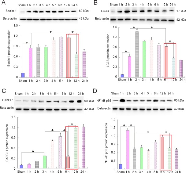
Figure 1 Correlation between autophagy and CX3CL1/fractalkine expression or NF-κB p65 activation after ischemic stroke (western blot assay). Autophagic signaling in the penumbra was remarkably activated by ischemia within 6 hours(h) after cerebral ischemia (A, B); however, the autophagy level was obviously declined at 12 h after insult. At this time point, the NF-κB p65 expression level (D) was also decreased, but CX3CL1 expression (C) was contrarily increased. There is a trend that autophagy level was negatively correlated with CX3CL1 expression, but positively correlated with activation of inflammatory signaling (as shown in the red squares). Relative protein expression was expressed as the optical density ratio to β-actin. Data are presented as the mean ± SEM (n = 6; one-way analysis of variance followed by Dunett’s test). *P < 0.01. NF-κB: Nuclear factor kappa B; LC3B: light chain 3B.
Figure 1 Correlation between autophagy and CX3CL1/fractalkine expression or NF-κB p65 activation after ischemic stroke (western blot assay). Autophagic signaling in the penumbra was remarkably activated by ischemia within 6 hours(h) after cerebral ischemia (A, B); however, the autophagy level was obviously declined at 12 h after insult. At this time point, the NF-κB p65 expression level (D) was also decreased, but CX3CL1 expression (C) was contrarily increased. There is a trend that autophagy level was negatively correlated with CX3CL1 expression, but positively correlated with activation of inflammatory signaling (as shown in the red squares). Relative protein expression was expressed as the optical density ratio to β-actin. Data are presented as the mean ± SEM (n = 6; one-way analysis of variance followed by Dunett’s test). *P < 0.01. NF-κB: Nuclear factor kappa B; LC3B: light chain 3B.CC BY-NC-SA · PubMed Central
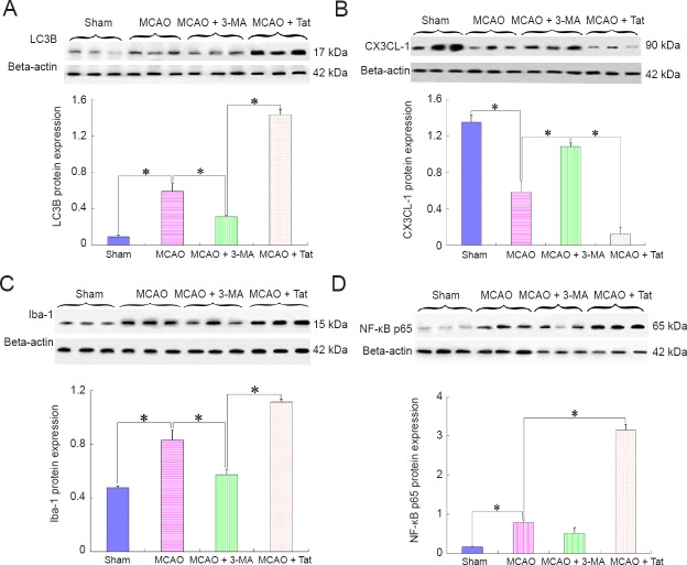
Figure 2 Effect of altered autophagy on CX3CL1 expression and inflammatory responses after ischemic stroke (western blot assay). Autophagy inducer Tat-Beclin 1 and autophagy inhibitor 3-MA were additionally administered in MCAO rats, separately. CX3CL1, Iba-1, and NF-κB p65 expression were detected 12 hours after insult. The results indicated that autophagy inhibition (A) dramatically enhanced CX3CL1 expression (B); meanwhile, both Iba-1 (C) and NF-κB p65 (D) expression levels were markedly declined. Compared with the MCAO group, CX3CL1 expression was further reduced after treatment with an autophagy inducer in the MCAO + Tat group; whereas, Iba-1 and NF-κB p65 were increasingly expressed. Relative protein expression was expressed as the optical density ratio to β-actin. Data are presented as the mean ± SEM (n = 6; one-way analysis of variance followed by Dunett’s test). *P < 0.01. MCAO: Middle cerebral artery occlusion; 3-MA: 3-methyladenine; Tat: Tat-Beclin 1; NF-κB: nuclear factor kappa B.
Figure 2 Effect of altered autophagy on CX3CL1 expression and inflammatory responses after ischemic stroke (western blot assay). Autophagy inducer Tat-Beclin 1 and autophagy inhibitor 3-MA were additionally administered in MCAO rats, separately. CX3CL1, Iba-1, and NF-κB p65 expression were detected 12 hours after insult. The results indicated that autophagy inhibition (A) dramatically enhanced CX3CL1 expression (B); meanwhile, both Iba-1 (C) and NF-κB p65 (D) expression levels were markedly declined. Compared with the MCAO group, CX3CL1 expression was further reduced after treatment with an autophagy inducer in the MCAO + Tat group; whereas, Iba-1 and NF-κB p65 were increasingly expressed. Relative protein expression was expressed as the optical density ratio to β-actin. Data are presented as the mean ± SEM (n = 6; one-way analysis of variance followed by Dunett’s test). *P < 0.01. MCAO: Middle cerebral artery occlusion; 3-MA: 3-methyladenine; Tat: Tat-Beclin 1; NF-κB: nuclear factor kappa B.CC BY-NC-SA · PubMed Central
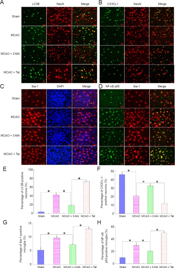
Figure 3 Neuronal autophagy reduces CX3CL1 expression on neurons and activates microglial inflammatory responses after cerebral stroke. Immunofluorescence was performed to determine cellular localization of CX3CL1 and its effect on microglial inflammatory responses. The results showed that enhanced neuronal autophagy [A, E (MCAO + Tat group)] reduced CX3CL1 expression on neurons (B, F), and further boosted microglial activity (C, G). In contrast, inhibition of neuron autophagy improved the percentage of CX3CL1-positive neurons, and reduced the ratio of NF-κB p65-positive microglia (D, H). In A and B, green staining (DyLight 488) indicates LC3B and CX3CL1-positive cells, while red staining (Alexa Fluor® 594) indicates NeuN-positive cells and yellow (merged) indicated the LC3B and CX3CL1-positive neurons. In C, red indicates Iba-1-positive cells; blue indicates total cells; and the merged picture shows the percentage of activated microglia. In D, green staining (DyLight 488) indicates NF-κB p65-positive cells, while red staining (Alexa Fluor® 594) indicates Iba-1-positive cells and yellow (merged) indicates the ratio of NF-κB p65-positive microglia. Arrows indicate positive neurons or microglia. Scale bars: 10 μm. Data are presented as the mean ± SEM (n = 6; one-way analysis of variance followed by Dunett’s test). *P < 0.01. MCAO: Middle cerebral artery occlusion; 3-MA: 3-methyladenine; Tat: Tat-Beclin 1; NF-κB: nuclear factor kappa B.
Figure 3 Neuronal autophagy reduces CX3CL1 expression on neurons and activates microglial inflammatory responses after cerebral stroke. Immunofluorescence was performed to determine cellular localization of CX3CL1 and its effect on microglial inflammatory responses. The results showed that enhanced neuronal autophagy [A, E (MCAO + Tat group)] reduced CX3CL1 expression on neurons (B, F), and further boosted microglial activity (C, G). In contrast, inhibition of neuron autophagy improved the percentage of CX3CL1-positive neurons, and reduced the ratio of NF-κB p65-positive microglia (D, H). In A and B, green staining (DyLight 488) indicates LC3B and CX3CL1-positive cells, while red staining (Alexa Fluor® 594) indicates NeuN-positive cells and yellow (merged) indicated the LC3B and CX3CL1-positive neurons. In C, red indicates Iba-1-positive cells; blue indicates total cells; and the merged picture shows the percentage of activated microglia. In D, green staining (DyLight 488) indicates NF-κB p65-positive cells, while red staining (Alexa Fluor® 594) indicates Iba-1-positive cells and yellow (merged) indicates the ratio of NF-κB p65-positive microglia. Arrows indicate positive neurons or microglia. Scale bars: 10 μm. Data are presented as the mean ± SEM (n = 6; one-way analysis of variance followed by Dunett’s test). *P < 0.01. MCAO: Middle cerebral artery occlusion; 3-MA: 3-methyladenine; Tat: Tat-Beclin 1; NF-κB: nuclear factor kappa B.CC BY-NC-SA · PubMed Central
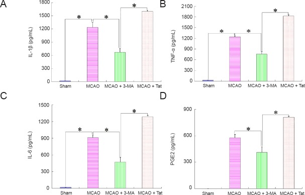
Figure 4 Effect of neuronal autophagy on production of inflammatory molecules after ischemic stroke (enzyme-linked immunosorbent assay). Tissues in the penumbra area were obtained 12 hours after insult to detect the production of inflammatory markers. (A–D) IL-1β, TNF-α, IL-6, and PGE2 production, respectively. Concentrations of inflammatory molecules in brain tissues from the MCAO group were markedly higher than those from the sham or MCAO + 3-MA groups, but notably lower than those from the MCAO + Tat group. Data are presented as the mean ± SEM (n = 6; one-way analysis of variance followed by Dunett’s test). *P < 0.01. IL: Interleukin; TNF-α: tumor necrosis factor alpha; PGE2: prostaglandin E2; MCAO: middle cerebral artery occlusion; 3-MA: 3-methyladenine; Tat: Tat-Beclin 1.
Figure 4 Effect of neuronal autophagy on production of inflammatory molecules after ischemic stroke (enzyme-linked immunosorbent assay). Tissues in the penumbra area were obtained 12 hours after insult to detect the production of inflammatory markers. (A–D) IL-1β, TNF-α, IL-6, and PGE2 production, respectively. Concentrations of inflammatory molecules in brain tissues from the MCAO group were markedly higher than those from the sham or MCAO + 3-MA groups, but notably lower than those from the MCAO + Tat group. Data are presented as the mean ± SEM (n = 6; one-way analysis of variance followed by Dunett’s test). *P < 0.01. IL: Interleukin; TNF-α: tumor necrosis factor alpha; PGE2: prostaglandin E2; MCAO: middle cerebral artery occlusion; 3-MA: 3-methyladenine; Tat: Tat-Beclin 1.CC BY-NC-SA · PubMed Central
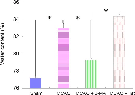
Figure 5 Effect of neuronal autophagy on brain edema in ischemic stroke rats. To observe the effect of autophagy on ischemia-induced inflammatory edema, brain water content was assessed. The ratio of water content in brain tissues from MCAO + 3-MA group was obviously lower than that from MCAO + Tat or MCAO groups, and was similar to that of the sham group. Data are presented as the mean ± SEM (n = 6; one-way analysis of variance followed by Dunett’s test). *P < 0.01. MCAO: Middle cerebral artery occlusion; 3-MA: 3-methyladenine; Tat: Tat-Beclin 1.
Figure 5 Effect of neuronal autophagy on brain edema in ischemic stroke rats. To observe the effect of autophagy on ischemia-induced inflammatory edema, brain water content was assessed. The ratio of water content in brain tissues from MCAO + 3-MA group was obviously lower than that from MCAO + Tat or MCAO groups, and was similar to that of the sham group. Data are presented as the mean ± SEM (n = 6; one-way analysis of variance followed by Dunett’s test). *P < 0.01. MCAO: Middle cerebral artery occlusion; 3-MA: 3-methyladenine; Tat: Tat-Beclin 1.CC BY-NC-SA · PubMed Central
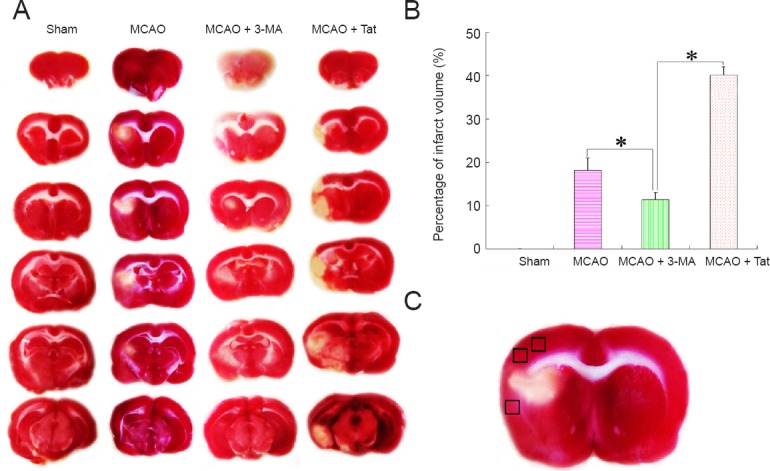
Figure 6 Effect of neuronal autophagy on cerebral infarct volume in rats with ischemic stroke. (A) To observe the effect of microglial inflammation on cerebral infarct volume after ischemic stroke, TTC staining was performed to assess infarct size. (B) Cerebral infarct volume was obviously reduced after treatment with the autophagy inhibitor of 3-MA compared with MCAO rats, and was similar to that observed in sham animals. In contrast, infarct volume was further expanded after administration with the autophagy inducer Tat. Data are presented as the mean ± SEM (n = 6; one-way analysis of variance followed by Dunett’s test). *P < 0.01. (C) Black squares indicate the penumbral area (derived from the third graph in the MCAO group from A). MCAO: Middle cerebral artery occlusion; 3-MA: 3-methyladenine; Tat: Tat-Beclin 1; TTC: 2,3,5-triphenyltetrazolium chloride.
Figure 6 Effect of neuronal autophagy on cerebral infarct volume in rats with ischemic stroke. (A) To observe the effect of microglial inflammation on cerebral infarct volume after ischemic stroke, TTC staining was performed to assess infarct size. (B) Cerebral infarct volume was obviously reduced after treatment with the autophagy inhibitor of 3-MA compared with MCAO rats, and was similar to that observed in sham animals. In contrast, infarct volume was further expanded after administration with the autophagy inducer Tat. Data are presented as the mean ± SEM (n = 6; one-way analysis of variance followed by Dunett’s test). *P < 0.01. (C) Black squares indicate the penumbral area (derived from the third graph in the MCAO group from A). MCAO: Middle cerebral artery occlusion; 3-MA: 3-methyladenine; Tat: Tat-Beclin 1; TTC: 2,3,5-triphenyltetrazolium chloride.CC BY-NC-SA · PubMed Central

Verification

Claims the paper makes

The claims and statistics of this paper haven’t been checked yet.

Run the check — free, no account needed. It reads the open-access text and checks the sources it cites.

A few free checks a day · papers already verified open instantly · results are public

Claims other papers make about this one

Each is checked against this paper’s own text: does it say what the citing paper says it does?

Self-check

Percentages, sums and reported differences re-computed from the paper’s own numbers.

The self-check needs the paper’s full text.

Citations

Cites

Show all 7 references

Research works printed but not linked: 25

  1. Button RW, Luo S, Rubinsztein DC (2015) Autophagic activity in neuronal cell death. Neurosci Bull 31:382-394. · DOI
  2. Chen W, Sun Y, Liu K, Sun X (2014) Autophagy: a double-edged sword for neuronal survival after cerebral ischemia. Neural Regen Res 9:1210-1216. · DOI
  3. Deng YH, He HY, Yang LQ, Zhang PY (2016) Dynamic changes in neuronal autophagy and apoptosis in the ischemic penumbra following permanent ischemic stroke. Neural Regen Res 11:1108-1114. · DOI
  4. Descloux C, Ginet V, Clarke PG, Puyal J, Truttmann AC (2015) Neuronal death after perinatal cerebral hypoxia-ischemia: Focus on autophagy-mediated cell death. Int J Dev Neurosci 45:75-85. · DOI
  5. Fonarow GC, Smith EE, Saver JL, Reeves MJ, Bhatt DL, Grau-Sepulveda MV, Olson DM, Hernandez AF, Peterson ED, Schwamm LH (2011) Timeliness of tissue-type plasminogen activator therapy in acute ischemic stroke: patient characteristics, hospital factors, and outcomes associated with door-to-needle times within 60 minutes. Circulation 123:750-758. · DOI
  6. Gusel’nikova VV, Korzhevskiy DE (2015) NeuN as a neuronal nuclear antigen and neuron differentiation marker. Acta Naturae 7:42-47. · DOI · Paper page
  7. Hara T, Nakamura K, Matsui M, Yamamoto A, Nakahara Y, Suzuki-Migishima R, Yokoyama M, Mishima K, Saito I, Okano H, Mizushima N (2006) Suppression of basal autophagy in neural cells causes neurodegenerative disease in mice. Nature 441:885-889. · DOI · Paper page
  8. Hongyun H, Tao G, Pengyue Z, Liqiang Y, Yihao D (2017) Puerarin provides a neuroprotection against transient cerebral ischemia by attenuating autophagy at the ischemic penumbra in neurons but not in astrocytes. Neurosci Lett 643:45-51. · DOI
  9. Iadecola C, Anrather J (2011) The immunology of stroke: from mechanisms to translation. Nat Med 17:796-808. · DOI · Paper page
  10. Jiang P, Mizushima N (2014) Autophagy and human diseases. Cell Res 24:69-79. · DOI
  11. Kaur J, Debnath J (2015) Autophagy at the crossroads of catabolism and anabolism. Nat Rev Mol Cell Biol 16:461-472. · DOI
  12. Kohman RA, Rhodes JS (2013) Neurogenesis, inflammation and behavior. Brain Behav Immun 27:22-32. · DOI
  13. Kono H, Rock KL (2008) How dying cells alert the immune system to danger. Nat Rev Immunol 8:279-289. · DOI · Paper page
  14. Lo EH (2008) A new penumbra: transitioning from injury into repair after stroke. Nat Med 14:497-500. · DOI · Paper page
  15. Martinez B, Peplow PV (2017) Immunomodulators and microRNAs as neurorestorative therapy for ischemic stroke. Neural Regen Res 12:865-874. · DOI
  16. Puyal J, Ginet V, Clarke PG (2013) Multiple interacting cell death mechanisms in the mediation of excitotoxicity and ischemic brain damage: a challenge for neuroprotection. Prog Neurobiol 105:24-48. · DOI
  17. Rana DS, Anand I, Batra A, Sethi PK, Bhargava S (2018) Serum levels of high-sensitivity C-reactive protein in acute ischemic stroke and its subtypes: a prospective case-control study. Asia Pac J Clin Trials Nerv Syst Dis 3:128-135. · DOI
  18. Saitoh T, Akira S (2010) Regulation of innate immune responses by autophagy-related proteins. J Cell Biol 189:925-935. · DOI
  19. Sheridan GK, Murphy KJ (2013) Neuron-glia crosstalk in health and disease: fractalkine and CX3CR1 take centre stage. Open Biol 3:130181. · DOI
  20. Spite M, Serhan CN (2010) Novel lipid mediators promote resolution of acute inflammation: impact of aspirin and statins. Circ Res 107:1170-1184. · DOI
  21. Su P, Zhang J, Wang D, Zhao F, Cao Z, Aschner M, Luo W (2016) The role of autophagy in modulation of neuroinflammation in microglia. Neuroscience 319:155-167. · DOI
  22. Tremblay MÈ, Stevens B, Sierra A, Wake H, Bessis A, Nimmerjahn A (2011) The role of microglia in the healthy brain. J Neurosci 31:16064-16069. · DOI · Paper page
  23. Walker FR, Beynon SB, Jones KA, Zhao Z, Kongsui R, Cairns M, Nilsson M (2014) Dynamic structural remodelling of microglia in health and disease: a review of the models, the signals and the mechanisms. Brain Behav Immun 37:1-14. · DOI
  24. Wang JF, Mei ZG, Fu Y, Yang SB, Zhang SZ, Huang WF, Xiong L, Zhou HJ, Tao W, Feng ZT (2018) Puerarin protects rat brain against schemia/reperfusion injury by suppressing autophagy via the AMPK-mTOR-ULK1 signaling pathway. Neural Regen Res 13:989-998. · DOI
  25. Yang Y, Chen S, Zhang Y, Lin X, Song Y, Xue Z, Qian H, Wang S, Wan G, Zheng X, Zhang L (2017) Induction of autophagy by spermidine is neuroprotective via inhibition of caspase 3-mediated Beclin 1 cleavage. Cell Death Dis 8:e2738. · DOI

Cited by

Sorted by citations

See all 69

Metadata

Authors
Deng, Y.-H., He, H.-Y., Ren, L., & Guo, T.
Venue
Neural Regeneration Research
Published
2019
Volume
14
Issue
2
Pages
280
ISSN
1673-5374
Cited by
69
References
7
Language
en

About these checks

Registry, retraction, Cites and statistics results are computed, not judged, with one exception: a printed reference that no index lists is read once by a model before it counts as a citation error. Claims are extracted by a model and kept only when their quote appears verbatim in the text; each cited source is then checked, with a model’s help, for whether it says what the claim relies on. Those verdicts can be wrong — read the claim’s quote and the cited source before relying on one.

Statistics and arithmetic are recomputed, not judged.

Confidence bands are shown only for fields calibrated on our annotated test set. None is calibrated yet, so no band is shown.

What counts as a claim ›How we measure confidence ›How we check this ›

Are you an author, or do you see a problem with this page? Request a review

How to cite

Cite CoChat for the checks on this page, and the paper for its own findings.

This page
CoChat. (2026). Citation check: Neuronal autophagy aggravates microglial inflammatory injury by downregulating CX3CL1/fractalkine after ischemic stroke. https://app.cochat.ai/papers/10.4103/1673-5374.244793
The paper
Deng, Y.-H., He, H.-Y., Ren, L., & Guo, T. (2019). Neuronal autophagy aggravates microglial inflammatory injury by downregulating CX3CL1/fractalkine after ischemic stroke. Neural Regeneration Research. https://doi.org/10.4103/1673-5374.244793