

The DOI matches its Crossref record; 52 of the 53 references it cites were found in a registry or an index.
Deep Learning for Classification of Colorectal Polyps on Whole-slide Images
Open accessAbstract
CONTEXT: Histopathological characterization of colorectal polyps is critical for determining the risk of colorectal cancer and future rates of surveillance for patients. However, this characterization is a challenging task and suffers from significant inter- and intra-observer variability. AIMS: We built an automatic image analysis method that can accurately classify different types of colorectal polyps on whole-slide images to help pathologists with this characterization and diagnosis. SETTING AND DESIGN: Our method is based on deep-learning techniques, which rely on numerous levels of abstraction for data representation and have shown state-of-the-art results for various image analysis tasks. SUBJECTS AND METHODS: Our method covers five common types of polyps (i.e., hyperplastic, sessile serrated, traditional serrated, tubular, and tubulovillous/villous) that are included in the US Multisociety Task Force guidelines for colorectal cancer risk assessment and surveillance. We developed multiple deep-learning approaches by leveraging a dataset of 2074 crop images, which were annotated by multiple domain expert pathologists as reference standards. STATISTICAL ANALYSIS: We evaluated our method on an independent test set of 239 whole-slide images and measured standard machine-learning evaluation metrics of accuracy, precision, recall, and F1 score and their 95% confidence intervals. RESULTS: Our evaluation shows that our method with residual network architecture achieves the best performance for classification of colorectal polyps on whole-slide images (overall accuracy: 93.0%, 95% confidence interval: 89.0%-95.9%). CONCLUSIONS: Our method can reduce the cognitive burden on pathologists and improve their efficacy in histopathological characterization of colorectal polyps and in subsequent risk assessment and follow-up recommendations.
Overview
The claims and statistics of this paper haven’t been checked yet.
Run the check — free, no account needed. It reads the open-access text and checks the sources it cites.
Integrity signals
Registry
Cites
Statistics
Figures
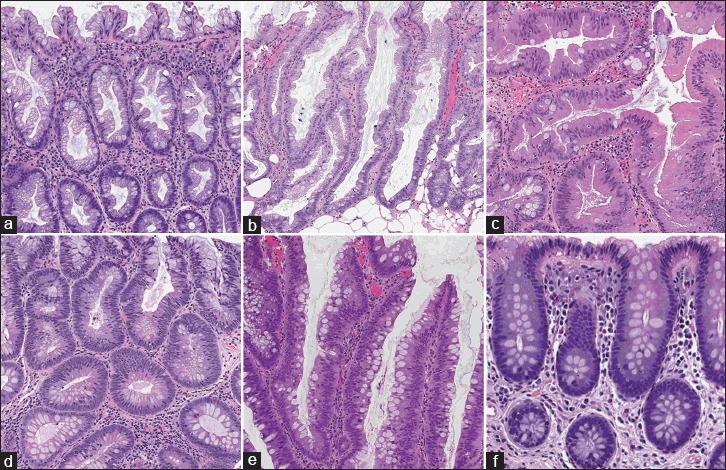
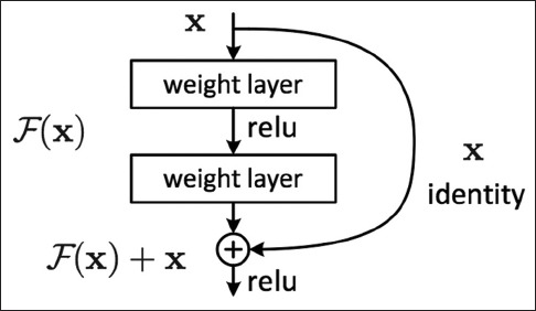
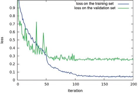
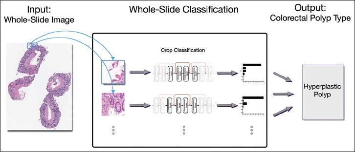
Verification
Claims the paper makes
The claims and statistics of this paper haven’t been checked yet.
Run the check — free, no account needed. It reads the open-access text and checks the sources it cites.
Claims other papers make about this one
Each is checked against this paper’s own text: does it say what the citing paper says it does?
In the citing paper
“Korbar and colleagues (72) developed a deep learning model to classify colorectal polyps into one of five common types based on H&E whole slide images.”
From Artificial Intelligence in Oncology: Current Landscape, Challenges, and Future Directions (2024) · cites this paper as [72]
Self-check
Percentages, sums and reported differences re-computed from the paper’s own numbers.
The self-check needs the paper’s full text.
Citations
Cites
Deep Residual Learning for Image Recognition
He, K., Zhang, X., Ren, S., & Sun, J. (2016) · 2016 IEEE Conference on Computer Vision and Pattern Recognition (CVPR) · doi:10.1109/cvpr.2016.90 · Cited by 235,474
Very Deep Convolutional Networks for Large-Scale Image Recognition
Simonyan, K., & Zisserman, A. (2014) · arXiv · doi:10.48550/arxiv.1409.1556 · Cited by 114,148
ImageNet classification with deep convolutional neural networks
Krizhevsky, A., Sutskever, I., & Hinton, G. E. (2017) · Communications of the ACM · doi:10.1145/3065386 · Cited by 112,155
LeCun, Y., Bengio, Y., & Hinton, G. (2015) · Nature · doi:10.1038/nature14539 · Cited by 84,126
Going deeper with convolutions
Szegedy, C., Liu, W., Jia, Y., Sermanet, P., Reed, S., Anguelov, D., Erhan, D., Vanhoucke, V., & Rabinovich, A. (2015) · 2015 IEEE Conference on Computer Vision and Pattern Recognition (CVPR) · doi:10.1109/cvpr.2015.7298594 · Cited by 48,562
Microsoft COCO: Common Objects in Context
Lin, T.-Y., Maire, M., Belongie, S., Hays, J., Perona, P., Ramanan, D., Dollár, P., & Zitnick, C. L. (2014) · Lecture Notes in Computer Science · doi:10.1007/978-3-319-10602-1_48 · Cited by 45,460
ImageNet Large Scale Visual Recognition Challenge
Russakovsky, O., Deng, J., Su, H., Krause, J., Satheesh, S., Ma, S., Huang, Z., Karpathy, A., Khosla, A., Bernstein, M., et al. (2015) · International Journal of Computer Vision · doi:10.1007/s11263-015-0816-y · Cited by 39,380
Delving Deep into Rectifiers: Surpassing Human-Level Performance on ImageNet Classification
He, K., Zhang, X., Ren, S., & Sun, J. (2015) · 2015 IEEE International Conference on Computer Vision (ICCV) · doi:10.1109/iccv.2015.123 · Cited by 19,741
Proceedings of IEEE Conference on Computer Vision and Pattern Recognition
(1993) · IEEE Computer Society Conference on Computer Vision and Pattern Recognition/Proceedings - IEEE Computer Society Conference on Computer Vision and Pattern Recognition/Proceedings · doi:10.1109/cvpr.1993.341010 · Cited by 14,475
Proceedings of IEEE International Conference on Computer Vision
(1995) · IEEE/CVF International Conference on Computer Vision (ICCV) · doi:10.1109/iccv.1995.466933 · Cited by 13,117
Advances in Neural Information Processing Systems 25
Zhang, Y., Sutton, C. A., Storkey, A., & Zoubin Ghahramani, A. (2012) · Cited by 12,653
(1968) · American Association of Industrial Nurses Journal · doi:10.1177/216507996801601208 · Cited by 8,811
Evaluation: from precision, recall and F-measure to ROC, informedness, markedness and correlation
Powers, D. M. W. (2020) · arXiv · doi:10.48550/arxiv.2010.16061 · Cited by 5,440
Deep Inside Convolutional Networks: Visualising Image Classification Models and Saliency Maps
Simonyan, K., Vedaldi, A., & Zisserman, A. (2013) · arXiv · doi:10.48550/arxiv.1312.6034 · Cited by 5,370
THE USE OF CONFIDENCE OR FIDUCIAL LIMITS ILLUSTRATED IN THE CASE OF THE BINOMIAL
Clopper, C. J., & Pearson, E. S. (1934) · Biometrika · doi:10.1093/biomet/26.4.404 · Cited by 5,327
(1989) · neural information processing systems · Cited by 3,736
Advances in Neural Information Processing Systems 37
Touretzky, D. S. (2024) · doi:10.52202/079017 · Cited by 2,874
Histopathological Image Analysis: A Review
Gurcan, M. N., Boucheron, L. E., Can, A., Madabhushi, A., Rajpoot, N. M., & Yener, B. (2009) · IEEE Reviews in Biomedical Engineering · doi:10.1109/rbme.2009.2034865 · Cited by 2,052
Lieberman, D. A., Rex, D. K., Winawer, S. J., Giardiello, F. M., Johnson, D. A., & Levin, T. R. (2012) · Gastroenterology · doi:10.1053/j.gastro.2012.06.001 · Cited by 1,889
Deep learning for digital pathology image analysis: A comprehensive tutorial with selected use cases
Janowczyk, A. R., & Madabhushi, A. (2016) · Journal of Pathology Informatics · doi:10.4103/2153-3539.186902 · Cited by 1,387
Sirinukunwattana, K., Raza, S. E. A., Tsang, Y.-W., Snead, D. R. J., Cree, I. A., & Rajpoot, N. M. (2016) · IEEE Transactions on Medical Imaging · doi:10.1109/tmi.2016.2525803 · Cited by 1,224
Image analysis and machine learning in digital pathology: challenges and opportunities
Madabhushi, A., & Lee, G. (2016) · Medical Image Analysis · doi:10.1016/j.media.2016.06.037 · Cited by 1,093
Role of the Serrated Pathway in Colorectal Cancer Pathogenesis
Leggett, B., & Whitehall, V. (2010) · Gastroenterology · doi:10.1053/j.gastro.2009.12.066 · Cited by 1,015
Update on the serrated pathway to colorectal carcinoma
Snover, D. C. (2010) · Human Pathology · doi:10.1016/j.humpath.2010.06.002 · Cited by 642
Irshad, H., Veillard, A., Roux, L., & Racoceanu, D. (2014) · IEEE Reviews in Biomedical Engineering · doi:10.1109/rbme.2013.2295804 · Cited by 641
Assessment of algorithms for mitosis detection in breast cancer histopathology images
Veta, M., van Diest, P. J., Willems, S. M., Wang, H., Madabhushi, A., Cruz-Roa, Á., Gonzalez, F. A., Larsen, A., Vestergaard, J. S., Dahl, A. B., et al. (2014) · Medical Image Analysis · doi:10.1016/j.media.2014.11.010 · Cited by 480
Cruz-Roa, A. A., Arevalo Ovalle, J. E., Madabhushi, A., & González Osorio, F. A. (2013) · Lecture Notes in Computer Science · doi:10.1007/978-3-642-40763-5_50 · Cited by 403 · cited 1×
“mage processing techniques (Xie et al., 2015; Sirinukunwattana et al., 2016; Janowczyk and Madabhushi, 2016). As examples, deep-learning models have been developed to detect metastatic breast cancer (Cruz-Roa et al., 2013), to nd mitotically active cells (Ertosun and Rubin, 2015), to identify basal-cell carcinoma (Mal…”
MethodLearning long‐range vision for autonomous off‐road driving
Hadsell, R. T., Sermanet, P., Ben, J., Erkan, A., Scoffier, M., Kavukcuoglu, K., Müller, U., & LeCun, Y. (2009) · Journal of Field Robotics · doi:10.1002/rob.20276 · Cited by 314
A Stochastic Polygons Model for Glandular Structures in Colon Histology Images
Sirinukunwattana, K., Snead, D. R. J., & Rajpoot, N. (2015) · IEEE Transactions on Medical Imaging · doi:10.1109/tmi.2015.2433900 · Cited by 257
Ertosun, M. G., & Rubin, D. L. (2015) · PubMed · Cited by 255
AUTOMATED GRADING OF PROSTATE CANCER USING ARCHITECTURAL AND TEXTURAL IMAGE FEATURES
Doyle, S. T., Hwang, M. E., Shah, K., Madabhushi, A., Feldman, M., & Tomaszeweski, J. E. (2007) · IEEE International Symposium on Biomedical Imaging (ISBI) · doi:10.1109/isbi.2007.357094 · Cited by 235
Classification of mitotic figures with convolutional neural networks and seeded blob features
Malon, C. D., & Cosatto, E. (2013) · Journal of Pathology Informatics · doi:10.4103/2153-3539.112694 · Cited by 199
Clinical impact and frequency of anatomic pathology errors in cancer diagnoses
Raab, S. S., Grzybicki, D. M., Janosky, J. E., Zarbo, R. J., Meier, F. A., Jensen, C. S., & Geyer, S. J. (2005) · Cancer · doi:10.1002/cncr.21431 · Cited by 188
Sertel, O., Kong, J., Çatalyürek, Ü. V., Lozanski, G., Saltz, J. H., & Gürcan, M. N. (2008) · Journal of Signal Processing Systems · doi:10.1007/s11265-008-0201-y · Cited by 184
Abdeljawad, K. H., Vemulapalli, K. C., Kahi, C. J., Cummings, O. W., Snover, D. C., & Rex, D. K. (2014) · Gastrointestinal Endoscopy · doi:10.1016/j.gie.2014.04.064 · Cited by 178
Scene Parsing with Multiscale Feature Learning, Purity Trees, and Optimal Covers
Farabet, C., Couprie, C., Najman, L. A., & LeCun, Y. (2012) · HAL · doi:10.48550/arxiv.1202.2160 · Cited by 176
Assisted Primary Screening Using the Automated ThinPrep Imaging System
Biscotti, C. V., Dawson, A. E., Dziura, B. R., Galup, L., Darragh, T. M., Rahemtulla, A., & Wills-Frank, L. A. (2005) · American Journal of Clinical Pathology · doi:10.1309/agb1mj9h5n43megx · Cited by 172
Observer agreement in the diagnosis of serrated polyps of the large bowel
Wong, N. A. C. S., Hunt, L. P., Novelli, M. R., Shepherd, N. A., & Warren, B. F. (2009) · Histopathology · doi:10.1111/j.1365-2559.2009.03329.x · Cited by 125
Kallenbach‐Thieltges, A., Großerueschkamp, F., Mosig, A., Diem, M., Tannapfel, A., & Gerwert, K. B. (2012) · Journal of Biophotonics · doi:10.1002/jbio.201200132 · Cited by 116
Cascaded ensemble of convolutional neural networks and handcrafted features for mitosis detection
Wang, H., Roa, Á. A. C., Basavanhally, A. N., Gilmore, H. L., Shih, N. N. C., Feldman, M., Tomaszewski, J., Gonzalez, F. A., & Madabhushi, A. (2014) · Proceedings of SPIE, the International Society for Optical Engineering/Proceedings of SPIE · doi:10.1117/12.2043902 · Cited by 108
Image analysis and morphometry in the diagnosis of breast cancer
Gil, J., Wu, H., & Wang, B. Y. (2002) · Microscopy Research and Technique · doi:10.1002/jemt.10182 · Cited by 96
Deep Voting: A Robust Approach Toward Nucleus Localization in Microscopy Images
Xie, Y., Kong, X., Xing, F., Liu, F., Su, H., & Yang, L. (2015) · Lecture notes in computer science · doi:10.1007/978-3-319-24574-4_45 · Cited by 80
Individuals With Sessile Serrated Polyps Express an Aggressive Colorectal Phenotype
Vu, H. T., López, R., Bennett, A. E., & Burke, C. A. (2011) · Diseases of the Colon & Rectum · doi:10.1097/dcr.0b013e318228f8a9 · Cited by 70
SVM Optimization for Hyperspectral Colon Tissue Cell Classification
Rajpoot, K., & Rajpoot, N. (2004) · Lecture notes in computer science · doi:10.1007/978-3-540-30136-3_101 · Cited by 59
Manjunath, B. S., & Boucheron, L. E. (2008) · Cited by 54
2007 4th IEEE International Symposium on Biomedical Imaging: From Nano to Macro
(2007) · IEEE International Symposium on Biomedical Imaging (ISBI) · doi:10.1109/isbi.2007.356765 · Cited by 40
Error reduction in surgical pathology.
Nakhleh, R. E. (2006) · PubMed · doi:10.5858/2006-130-630-erisp · Cited by 33
How Does the Serrated Polyp Pathway Alter CRC Screening and Surveillance?
Kahi, C. J. (2015) · Digestive Diseases and Sciences · doi:10.1007/s10620-014-3449-z · Cited by 19
Mitosis detection in breast cancer histological images with mathematical morphology
Aptoula, E., Courty, N., & Lefèvre, S. (2013) · Signal Processing and Communications Applications Conference (SIU) · doi:10.1109/siu.2013.6531502 · Cited by 12
Image analysis can be used to detect spatial changes in the histopathology of pancreatic tumours
Sims, A. J., Bennett, M. K., & Murray, A. (2003) · Physics in Medicine and Biology · doi:10.1088/0031-9155/48/13/401 · Cited by 11
Malkin, H. M. (1998) · Annals of Diagnostic Pathology · doi:10.1016/s1092-9134(98)80036-1 · Cited by 8
Research works printed but not linked: 1
- Bengio, 2009, Vol. 2, 1
Cited by
Sorted by citations
Applications of machine learning in drug discovery and development
Vamathevan, J., Clark, D., Czodrowski, P., Dunham, I., Ferran, E., Lee, G., Li, B., Madabhushi, A., Shah, P., Spitzer, M., et al. (2019) · Nature Reviews Drug Discovery · doi:10.1038/s41573-019-0024-5 · Cited by 3,228
Machine Learning in Drug Discovery: A Review
Dara, S., Dhamercherla, S., Jadav, S. S., Babu, C. M., & Ahsan, M. J. (2021) · Artificial Intelligence Review · doi:10.1007/s10462-021-10058-4 · Cited by 893
Artificial Intelligence in Cancer Research and Precision Medicine
Bhinder, B., Gilvary, C., Madhukar, N. S., & Elemento, O. (2021) · Cancer Discovery · doi:10.1158/2159-8290.cd-21-0090 · Cited by 781
Deep learning in cancer pathology: a new generation of clinical biomarkers
Echle, A., Rindtorff, N. T., Brinker, T. J., Luedde, T., Pearson, A. T., & Kather, J. N. (2021) · British Journal of Cancer · doi:10.1038/s41416-020-01122-x · Cited by 667
Deep learning based tissue analysis predicts outcome in colorectal cancer
Bychkov, D., Linder, N., Turkki, R., Nordling, S., Kovanen, P. E., Verrill, C., Walliander, M., Lundin, M., Haglund, C., & Lundin, J. (2018) · Scientific Reports · doi:10.1038/s41598-018-21758-3 · Cited by 644
Metadata
- Authors
- Korbar, B., Olofson, A. M., Miraflor, A. P., Nicka, C. M., Suriawinata, M. A., Torresani, L., Suriawinata, A. A., & Hassanpour, S.
- Affiliations
- Dartmouth College (US)
- Corresponding author
- Hassanpour, Saeed
- Venue
- Journal of Pathology Informatics
- Publisher
- Elsevier BV
- Published
- 2017-01
- Volume
- 8
- Issue
- 1
- Pages
- 30
- ISSN
- 2153-3539 (ISSN-L)
- PMC
- PMC5545773
- OpenAlex
- W2594760301
- Cited by
- 349
- References
- 53
- Citations by year
- 2026: 16 · 2025: 30 · 2024: 48 · 2023: 41 · 2022: 49 · 2021: 65 · 2020: 51 · 2019: 33 · 2018: 13 · 2017: 3
- FWCI
- 16.31
- Type
- journal article
- Language
- en
- Open access
- gold
- doi.org
- Licence
- Locations
- Journal of Pathology Informatics (publishedVersion, cc-by-nc-sa)
- Europe PMC (PubMed Central) (publishedVersion, cc-by-nc-sa)
- PubMed Central (publishedVersion, cc-by-nc-sa)
- PubMed (publishedVersion)
- arXiv (Cornell University) (submittedVersion)
- DOAJ (DOAJ: Directory of Open Access Journals) (submittedVersion, cc-by-sa)
- arXiv (Cornell University)
- Funders
- Dartmouth College
- Topics
- Physical Sciences › Computer Science › Artificial Intelligence › AI in cancer detection
- Health Sciences › Medicine › Oncology › Colorectal Cancer Screening and Detection
- Physical Sciences › Computer Science › Computer Vision and Pattern Recognition › Advanced Neural Network Applications
- Keywords
- colorectal polyps, whole slide images, histological classification, colorectal cancer risk assessment, hyperplastic polyps, sessile serrated polyps, traditional serrated adenoma, tubular adenoma, villous adenoma, ResNet, computer-aided diagnosis, observer variability
About these checks
Registry, retraction, Cites and statistics results are computed, not judged, with one exception: a printed reference that no index lists is read once by a model before it counts as a citation error. Claims are extracted by a model and kept only when their quote appears verbatim in the text; each cited source is then checked, with a model’s help, for whether it says what the claim relies on. Those verdicts can be wrong — read the claim’s quote and the cited source before relying on one.
Statistics and arithmetic are recomputed, not judged.
Confidence bands are shown only for fields calibrated on our annotated test set. None is calibrated yet, so no band is shown.
What counts as a claim ›How we measure confidence ›How we check this ›
Are you an author, or do you see a problem with this page? Request a review
