

The DOI matches its Crossref record; 126 of the 126 references it cites were found in a registry or an index.
Effects of blue light on the circadian system and eye physiology
Open accessAbstract
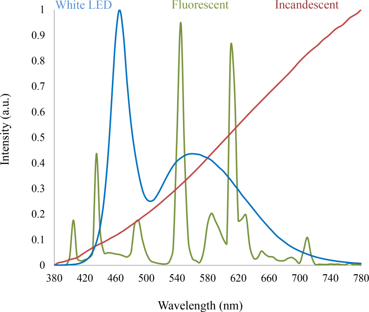
Light-emitting diodes (LEDs) have been used to provide illumination in industrial and commercial environments. LEDs are also used in TVs, computers, smart phones, and tablets. Although the light emitted by most LEDs appears white, LEDs have peak emission in the blue light range (400-490 nm). The accumulating experimental evidence has indicated that exposure to blue light can affect many physiologic functions, and it can be used to treat circadian and sleep dysfunctions. However, blue light can also induce photoreceptor damage. Thus, it is important to consider the spectral output of LED-based light sources to minimize the danger that may be associated with blue light exposure. In this review, we summarize the current knowledge of the effects of blue light on the regulation of physiologic functions and the possible effects of blue light exposure on ocular health.
Overview
The claims and statistics of this paper haven’t been checked yet.
Run the check — free, no account needed. It reads the open-access text and checks the sources it cites.
Integrity signals
Registry
Cites
Statistics
Figures
Verification
Claims the paper makes
The claims and statistics of this paper haven’t been checked yet.
Run the check — free, no account needed. It reads the open-access text and checks the sources it cites.
Claims other papers make about this one
Each is checked against this paper’s own text: does it say what the citing paper says it does?
The source backs part of the claim, not all of it — or backs it for a different population, condition or comparison.
In the citing paper
“The severity of the blue‐light induced damage depends on the time of the day and is thus related with the circadian rhythm 52.”
From The inner clock-Blue light sets the human rhythm (2019) · cites this paper as [52]
Experimental — accuracy not yet measuredThe source backs part of the claim, not all of it — or backs it for a different population, condition or comparison.
In the citing paper
“While short wavelengths in the range of 460 nm are very efficient in phase shifting of circadian system, intensive blue light in the range between 400 and 440 nm (see Figure 4) is damaging to the retina via a photochemical reaction called photoreversal of bleaching 52.”
From The inner clock-Blue light sets the human rhythm (2019) · cites this paper as [52]
Experimental — accuracy not yet measuredThe source backs part of the claim, not all of it — or backs it for a different population, condition or comparison.
In the citing paper
“However, the detrimental effect of blue light on the retina should be taken with caution. Most of the damaging effects through blue light are demonstrated in nocturnal animals only, or in isolated animal cell cultures directly exposed to blue light 52, 53, 54.”
From The inner clock-Blue light sets the human rhythm (2019) · cites this paper as [52]
Experimental — accuracy not yet measuredThe source backs part of the claim, not all of it — or backs it for a different population, condition or comparison.
In the citing paper
“Accumulation of ROS and lipofuscin may be due to both acute and cumulative exposure to blue light 52.”
From The inner clock-Blue light sets the human rhythm (2019) · cites this paper as [52]
Experimental — accuracy not yet measuredThe cited source says what the claim attributes to it.
In the citing paper
“The amount of lipofuscin in the retina increases with age and is related to age‐related macular degeneration 52.”
From The inner clock-Blue light sets the human rhythm (2019) · cites this paper as [52]
Self-check
Percentages, sums and reported differences re-computed from the paper’s own numbers.
The self-check needs the paper’s full text.
Citations
Cites
Repertoires of Autophagy in the Pathogenesis of Ocular Diseases
Li, Y., Jiang, Q. L., Cao, G.-F., Yao, J., & Yan, B. (2015) · Cellular Physiology and Biochemistry · doi:10.1159/000373980 · Cited by 16,351
Phototransduction by retinal ganglion cells that set the circadian clock
Berson, D. M., Dunn, F. A., & Takao, M. (2002) · Science · doi:10.1126/science.1067262 · Cited by 3,629
Hattar, S., Liao, H.-W., Takao, M., Berson, D. M., & Yau, K.-W. (2002) · Science · doi:10.1126/science.1069609 · Cited by 2,734
Pimputkar, S., Speck, J. S., DenBaars, S. P., & Nakamura, S. (2009) · Nature Photonics · doi:10.1038/nphoton.2009.32 · Cited by 2,196
Action Spectrum for Melatonin Regulation in Humans: Evidence for a Novel Circadian Photoreceptor
Brainard, G. C., Hanifin, J. P., Greeson, J. M., Byrne, B., Glickman, G., Gerner, E., & Rollag, M. D. (2001) · The Journal of Neuroscience · doi:10.1523/jneurosci.21-16-06405.2001 · Cited by 1,930
Chang, A.-M., Aeschbach, D., Duffy, J. F., & Czeisler, C. A. (2015) · Proceedings of the National Academy of Sciences · doi:10.1073/pnas.1418490112 · Cited by 1,430
Thapan, K., Arendt, J., & Skene, D. J. (2001) · The Journal of Physiology · doi:10.1111/j.1469-7793.2001.t01-1-00261.x · Cited by 1,320
A Novel Human Opsin in the Inner Retina
Provencio, I., Rodriguez, I. R., Jiang, G., Hayes, W. P., Moreira, E. F., & Rollag, M. D. (2000) · The Journal of Neuroscience · doi:10.1523/jneurosci.20-02-00600.2000 · Cited by 1,290
Measuring and using light in the melanopsin age
Lucas, R. J., Peirson, S. N., Berson, D. M., Brown, T. M., Cooper, H. M., Czeisler, C. A., Figueiro, M. G., Gamlin, P. D., Lockley, S. W., O’Hagan, J. B., et al. (2014) · Trends in Neurosciences · doi:10.1016/j.tins.2013.10.004 · Cited by 1,278
A retinohypothalamic projection in the rat
Moore, R. Y., & Lenn, N. J. (1972) · The Journal of Comparative Neurology · doi:10.1002/cne.901460102 · Cited by 1,263
Cited by
Sorted by citations
The new world atlas of artificial night sky brightness
Falchi, F., Cinzano, P., Duriscoe, D. M., Kyba, C. C. M., Elvidge, C. D., Baugh, K., Portnov, B. A., Rybnikova, N. A., & Furgoni, R. (2016) · Science Advances · doi:10.1126/sciadv.1600377 · Cited by 1,695
Digital eye strain: prevalence, measurement and amelioration
Sheppard, A. L., & Wolffsohn, J. S. (2018) · BMJ Open Ophthalmology · doi:10.1136/bmjophth-2018-000146 · Cited by 822
Wang, Z., Yuan, F., Li, X., Li, Y., Zhong, H., Fan, L., & Yang, S. (2017) · Advanced Materials · doi:10.1002/adma.201702910 · Cited by 786
Managing the health of the eSport athlete: an integrated health management model
DiFrancisco‐Donoghue, J., Balentine, J., Schmidt, G., & Zwibel, H. (2019) · BMJ Open Sport & Exercise Medicine · doi:10.1136/bmjsem-2018-000467 · Cited by 353
Touitou, Y., Touitou, D., & Reinberg, A. (2016) · Journal of Physiology-Paris · doi:10.1016/j.jphysparis.2017.05.001 · Cited by 297
Metadata
- Authors
- Tosini, G., Ferguson, I., & Tsubota, K.
- Affiliations
- Morehouse School of Medicine (US)
- Missouri University of Science and Technology (US)
- Keio University (JP)
- Corresponding author
- Tosini, Gianluca
- Venue
- Molecular Vision
- Publisher
- Molecular Vision
- Published
- 2016-01
- Volume
- 22
- Pages
- 61-72
- ISSN
- 1090-0535 (ISSN-L)
- PMC
- PMC4734149
- OpenAlex
- W2294594166
- Cited by
- 517
- References
- 126
- Citations by year
- 2026: 52 · 2025: 51 · 2024: 43 · 2023: 51 · 2022: 54 · 2021: 54 · 2020: 103 · 2019: 42 · 2018: 37 · 2017: 27 · 2016: 3
- FWCI
- 18.19
- Type
- journal article
- Language
- en
- Open access
- diamond
- doi.org
- Locations
- Molecular vision (publishedVersion, cc-by)
- Europe PMC (PubMed Central) (publishedVersion, cc-by-nc-nd)
- PubMed Central (publishedVersion, cc-by-nc-nd)
- PubMed (publishedVersion)
- PubMed
- Funders
- National Aeronautics and Space Administration (NCC958)
- National Space Biomedical Research Institute (NCC 9‐58)
- National Institutes of Health (NCC9-58)
- National Eye Institute (R01EY022216)
- Topics
- Life Sciences › Neuroscience › Endocrine and Autonomic Systems › Circadian rhythm and melatonin
- Health Sciences › Medicine › Ophthalmology › Ocular and Laser Science Research
- Life Sciences › Agricultural and Biological Sciences › Plant Science › Light effects on plants
- Keywords
- blue light, light-emitting diodes, circadian rhythm, circadian system, sleep disorders, photoreceptor damage, eye health, eye physiology, blue light exposure, light spectrum
- MeSH terms
- Animals, Circadian Rhythm, Humans, Light, Ocular Physiological Phenomena, Photic Stimulation, Photoreceptor Cells, Vertebrate
About these checks
Registry, retraction, Cites and statistics results are computed, not judged, with one exception: a printed reference that no index lists is read once by a model before it counts as a citation error. Claims are extracted by a model and kept only when their quote appears verbatim in the text; each cited source is then checked, with a model’s help, for whether it says what the claim relies on. Those verdicts can be wrong — read the claim’s quote and the cited source before relying on one.
Statistics and arithmetic are recomputed, not judged.
Confidence bands are shown only for fields calibrated on our annotated test set. None is calibrated yet, so no band is shown.
What counts as a claim ›How we measure confidence ›How we check this ›
Are you an author, or do you see a problem with this page? Request a review
