

The DOI matches its Crossref record; 5 of the 5 references it cites were found in a registry or an index.
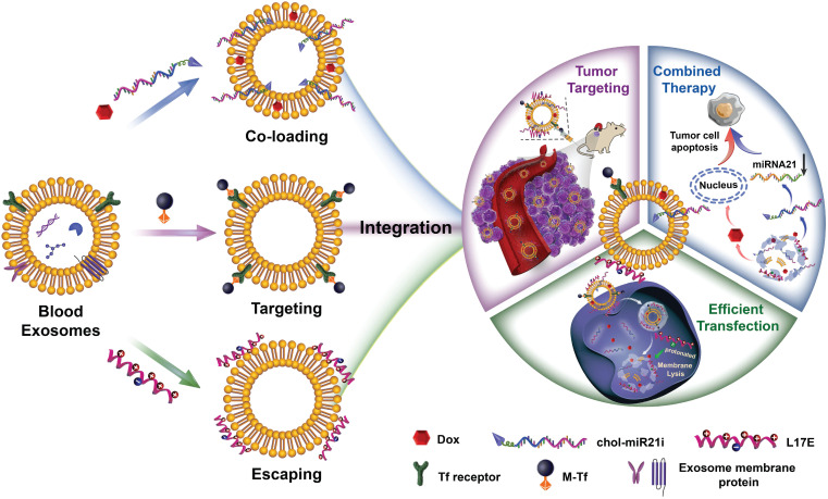
Engineering blood exosomes for tumor-targeting efficient gene/chemo combination therapy
Overview
The claims and statistics of this paper haven’t been checked yet.
Run the check — free, no account needed. It reads the open-access text and checks the sources it cites.
Integrity signals
Registry
Cites
Statistics
Figures
Verification
Claims the paper makes
The claims and statistics of this paper haven’t been checked yet.
Run the check — free, no account needed. It reads the open-access text and checks the sources it cites.
Claims other papers make about this one
Each is checked against this paper’s own text: does it say what the citing paper says it does?
In the citing paper
“For example, exosomes carrying Doxorubicin (Dox) and miR-21 inhibitors have been found to significantly reduce tumor volume [88].”
From Emerging role of exosomes in cancer therapy: progress and challenges (2025) · cites this paper as [88]
Self-check
Percentages, sums and reported differences re-computed from the paper’s own numbers.
The self-check needs the paper’s full text.
Citations
Cites
Principles of nanoparticle design for overcoming biological barriers to drug delivery
Blanco, E., Shen, H., & Ferrari, M. (2015) · Nature Biotechnology · doi:10.1038/nbt.3330 · Cited by 7,002
Exosomes facilitate therapeutic targeting of oncogenic KRAS in pancreatic cancer
Kamerkar, S., LeBleu, V. S., Sugimoto, H., Yang, S., Ruivo, C. F., Melo, S. A., Lee, J. J., & Kalluri, R. (2017) · Nature · doi:10.1038/nature22341 · Cited by 2,639
Overcoming cellular barriers for RNA therapeutics
Dowdy, S. F. (2017) · Nature Biotechnology · doi:10.1038/nbt.3802 · Cited by 1,139
Advances in therapeutic applications of extracellular vesicles
Wiklander, O. P. B., Brennan, M. Á., Lötvall, J., Breakefield, X. O., & EL Andaloussi, S. (2019) · Science Translational Medicine · doi:10.1126/scitranslmed.aav8521 · Cited by 1,039
Responsive Exosome Nano-bioconjugates for Synergistic Cancer Therapy
Nie, W., Wu, G., Zhang, J., Huang, L., Ding, J., Jiang, A., Zhang, Y., Liu, Y., Li, J., Pu, K., et al. (2020) · Angewandte Chemie International Edition · doi:10.1002/anie.201912524 · Cited by 376
Research works printed but not linked: 49
- [1]Shi J Kantoff PW Wooster R Farokhzad OC Cancer nanomedicine: progress, challenges and opportunities Nat Rev Cancer 2017 17 20 37 27834398 PMC5575742
- [2]Xu X Ho W Zhang X Bertrand N Farokhzad O Cancer nanomedicine: from targeted delivery to combination therapy Trends Mol Med 2015 21 223 32 25656384 PMC4385479
- [3]Hu Q Sun W Wang C Gu Z Recent advances of cocktail chemotherapy by combination drug delivery systems Adv Drug Deliv Rev 2016 98 19 34 26546751 PMC4998845
- [4]Sun Q Zhou Z Qiu N Shen Y Rational Design of Cancer Nanomedicine: Nanoproperty Integration and Synchronization Adv Mater 2017 29 1606628 28234430
- [5]van der Meel R Sulheim E Shi Y Kiessling F Mulder WJM Lammers T Smart cancer nanomedicine Nat Nanotechnol 2019 14 1007 17 31695150 PMC7227032
- [6]Huang W Chen L Kang L Jin M Sun P Xin X Nanomedicine-based combination anticancer therapy between nucleic acids and small-molecular drugs Adv Drug Deliv Rev 2017 115 82 97 28624478
- [7]Ding Y Su S Zhang R Shao L Zhang Y Wang B Precision combination therapy for triple negative breast cancer via biomimetic polydopamine polymer core-shell nanostructures Biomaterials 2017 113 243 52 27829203
- [8]Li C Yang XQ Zhang MZ Song YY Cheng K An J In vivo Imaging-Guided Nanoplatform for Tumor Targeting Delivery and Combined Chemo-, Gene- and Photothermal Therapy Theranostics 2018 8 5662 75 30555572 PMC6276300
- [9]Prencipe G Tabakman SM Welsher K Liu Z Goodwin AP Zhang L PEG branched polymer for functionalization of nanomaterials with ultralong blood circulation J Am Chem Soc 2009 131 4783 7 19173646 PMC2827329
- [11]Allen TM Ligand-targeted therapeutics in anticancer therapy Nat Rev Cancer 2002 2 750 63 12360278
- [12]Vaughan HJ Green JJ Tzeng SY Cancer-Targeting Nanoparticles for Combinatorial Nucleic Acid Delivery Adv Mater. 2019: e1901081 PMC6923623 31222852
- [13]Samal SK Dash M Van Vlierberghe S Kaplan DL Chiellini E van Blitterswijk C Cationic polymers and their therapeutic potential Chem Soc Rev 2012 41 7147 94 22885409
- [14]Yu H Guo C Feng B Liu J Chen X Wang D Triple-Layered pH-Responsive Micelleplexes Loaded with siRNA and Cisplatin Prodrug for NF-Kappa B Targeted Treatment of Metastatic Breast Cancer Theranostics 2016 6 14 27 26722370 PMC4679351
- [15]Mura S Nicolas J Couvreur P Stimuli-responsive nanocarriers for drug delivery Nat Mater 2013 12 991 1003 24150417
- [17]Wang S Huang P Chen X Stimuli-Responsive Programmed Specific Targeting in Nanomedicine ACS Nano 2016 10 2991 4 26881288 PMC5223089
- [18]El-Sawy HS Al-Abd AM Ahmed TA El-Say KM Torchilin VP Stimuli-Responsive Nano-Architecture Drug-Delivery Systems to Solid Tumor Micromilieu: Past, Present, and Future Perspectives ACS Nano 2018 12 10636 64 30335963
- [19]Wang C Li Z Cao D Zhao YL Gaines JW Bozdemir OA Stimulated release of size-selected cargos in succession from mesoporous silica nanoparticles Angew Chem Int Ed Engl 2012 51 5460 5 22505218
- [20]Sun Q Sun X Ma X Zhou Z Jin E Zhang B Integration of nanoassembly functions for an effective delivery cascade for cancer drugs Adv Mater 2014 26 7615 21 25328159
- [21]Chen B Dai W He B Zhang H Wang X Wang Y Current Multistage Drug Delivery Systems Based on the Tumor Microenvironment Theranostics 2017 7 538 58 28255348 PMC5327631
- [22]Saw PE Yao H Lin C Tao W Farokhzad OC Xu X Stimuli-Responsive Polymer-Prodrug Hybrid Nanoplatform for Multistage siRNA Delivery and Combination Cancer Therapy Nano Lett 2019 19 5967 74 31381852
- [23]Mathieu M Martin-Jaular L Lavieu G Théry C Specificities of secretion and uptake of exosomes and other extracellular vesicles for cell-to-cell communication Nat Cell Biol 2019 21 9 17 30602770
- [24]Syn NL Wang L Chow EK Lim CT Goh BC Exosomes in Cancer Nanomedicine and Immunotherapy: Prospects and Challenges Trends Biotechnol 2017 35 665 76 28365132
- [25]Yang B Chen Y Shi J Exosome Biochemistry and Advanced Nanotechnology for Next-Generation Theranostic Platforms Adv Mater 2019 31 e1802896 30126052
- [26]Lener T Gimona M Aigner L Borger V Buzas E Camussi G Applying extracellular vesicles based therapeutics in clinical trials - an ISEV position paper J Extracell Vesicles 2015 4 30087 26725829 PMC4698466
- [28]Pegtel DM Gould SJ Exosomes Annu Rev Biochem 2019 88 487 514 31220978
- [29]S ELA Mäger I Breakefield XO Wood MJ Extracellular vesicles: biology and emerging therapeutic opportunities Nat Rev Drug Discov 2013 12 347 57 23584393
- [30]Valadi H Ekström K Bossios A Sjöstrand M Lee JJ Lötvall JO Exosome-mediated transfer of mRNAs and microRNAs is a novel mechanism of genetic exchange between cells Nat Cell Biol 2007 9 654 9 17486113
- [31]Alvarez-Erviti L Seow Y Yin H Betts C Lakhal S Wood MJ Delivery of siRNA to the mouse brain by systemic injection of targeted exosomes Nat Biotechnol 2011 29 341 5 21423189
- [33]Yong T Zhang X Bie N Zhang H Zhang X Li F Tumor exosome-based nanoparticles are efficient drug carriers for chemotherapy Nat Commun 2019 10 3838 31444335 PMC6707218
- [34]Cheng Q Shi X Han M Smbatyan G Lenz HJ Zhang Y Reprogramming Exosomes as Nanoscale Controllers of Cellular Immunity J Am Chem Soc 2018 140 16413 7 30452238 PMC6469991
- [35]Sancho-Albero M Rubio-Ruiz B Perez-Lopez AM Sebastian V Martin-Duque P Arruebo M Cancer-derived exosomes loaded with ultrathin palladium nanosheets for targeted bioorthogonal catalysis Nat Catal 2019 2 864 72 31620674 PMC6795537
- [37]Gong C Tian J Wang Z Gao Y Wu X Ding X Functional exosome-mediated co-delivery of doxorubicin and hydrophobically modified microRNA 159 for triple-negative breast cancer therapy J Nanobiotechnology 2019 17 93 31481080 PMC6721253
- [38]Blanc L De Gassart A Géminard C Bette-Bobillo P Vidal M Exosome release by reticulocytes-an integral part of the red blood cell differentiation system Blood Cells Mol Dis 2005 35 21 6 15946868
- [39]Qi H Liu C Long L Ren Y Zhang S Chang X Blood Exosomes Endowed with Magnetic and Targeting Properties for Cancer Therapy ACS Nano 2016 10 3323 33 26938862
- [40]Akishiba M Takeuchi T Kawaguchi Y Sakamoto K Yu HH Nakase I Cytosolic antibody delivery by lipid-sensitive endosomolytic peptide Nat Chem 2017 9 751 61 28754944
- [41]Pi F Binzel DW Lee TJ Li Z Sun M Rychahou P Nanoparticle orientation to control RNA loading and ligand display on extracellular vesicles for cancer regression Nat Nanotechnol 2018 13 82 9 29230043 PMC5762263
- [42]Wan Y Wang L Zhu C Zheng Q Wang G Tong J Aptamer-Conjugated Extracellular Nanovesicles for Targeted Drug Delivery Cancer Res 2018 78 798 808 29217761 PMC5811376
- [43]Yerneni SS Lathwal S Shrestha P Shirwan H Matyjaszewski K Weiss L Rapid On-Demand Extracellular Vesicle Augmentation with Versatile Oligonucleotide Tethers ACS Nano 2019 13 10555 65 31436946 PMC6800810
- [44]Armstrong JP Holme MN Stevens MM Re-Engineering Extracellular Vesicles as Smart Nanoscale Therapeutics ACS Nano 2017 11 69 83 28068069 PMC5604727
- [45]Kooijmans SAA Stremersch S Braeckmans K de Smedt SC Hendrix A Wood MJA Electroporation-induced siRNA precipitation obscures the efficiency of siRNA loading into extracellular vesicles J Control Release 2013 172 229 38 23994516
- [47]Rogers S Wells R Rechsteiner M Amino acid sequences common to rapidly degraded proteins: the PEST hypothesis Science 1986 234 364 8 2876518
- [48]Selby LI Cortez-Jugo CM Such GK Johnston APR Nanoescapology: progress toward understanding the endosomal escape of polymeric nanoparticles Wiley Interdiscip Rev Nanomed Nanobiotechnol 2017 9 e1452 28160452
- [49]Lewinski N Colvin V Drezek R Cytotoxicity of nanoparticles Small 2008 4 26 49 18165959
- [50]Deng H Lin L Wang S Yu G Zhou Z Liu Y X-ray-Controlled Bilayer Permeability of Bionic Nanocapsules Stabilized by Nucleobase Pairing Interactions for Pulsatile Drug Delivery Adv Mater 2019 31 e1903443 31379091
- [51]Zhu S Wu H Wu F Nie D Sheng S Mo YY MicroRNA-21 targets tumor suppressor genes in invasion and metastasis Cell Res 2008 18 350 9 18270520
- [52]Hong L Han Y Zhang Y Zhang H Zhao Q Wu K MicroRNA-21: a therapeutic target for reversing drug resistance in cancer Expert Opin Ther Targets 2013 17 1073 80 23865553
- [53]Zhou X Ren Y Moore L Mei M You Y Xu P Downregulation of miR-21 inhibits EGFR pathway and suppresses the growth of human glioblastoma cells independent of PTEN status Lab Invest 2010 90 144 55 20048743
- [54]Han L Liu C Qi H Zhou J Wen J Wu D Systemic Delivery of Monoclonal Antibodies to the Central Nervous System for Brain Tumor Therapy Adv Mater 2019 31 e1805697 30773720
- [55]Usman WM Pham TC Kwok YY Vu LT Ma V Peng B Efficient RNA drug delivery using red blood cell extracellular vesicles Nat Commun 2018 9 2359 29907766 PMC6004015
Cited by
Sorted by citations
Engineered Extracellular Vesicles for Cancer Therapy
Zhang, X., Zhang, H., Gu, J., Zhang, J., Shi, H., Qian, H., Wang, D., Xu, W., Pan, J., & Santos, H. A. (2021) · Advanced Materials · doi:10.1002/adma.202005709 · Cited by 428
Recent advances in exosome-mediated nucleic acid delivery for cancer therapy
Zhang, Y., Liu, Q., Zhang, X., Huang, H., Tang, S., Chai, Y., Xu, Z., Li, M., Chen, X., Liu, J., et al. (2022) · Journal of Nanobiotechnology · doi:10.1186/s12951-022-01472-z · Cited by 306
Emerging role of exosomes in cancer therapy: progress and challenges
Li, J., Wang, J., & Chen, Z. (2025) · Molecular Cancer · doi:10.1186/s12943-024-02215-4 · Cited by 297
Ghosh, S., & Ghosh, S. (2022) · Frontiers in Pharmacology · doi:10.3389/fphar.2022.878058 · Cited by 13
Metadata
- Authors
- Zhan, Q., Yi, K., Qi, H., Li, S., Li, X., Wang, Q., Wang, Y., Liu, C., Qiu, M., Yuan, X., Zhao, J., Hou, X., & Kang, C.
- Venue
- Theranostics
- Publisher
- Ivyspring International Publisher
- Published
- 2020
- Volume
- 10
- Issue
- 17
- Pages
- 7889-7905
- ISSN
- 1838-7640
- PMC
- PMC7359100
- Cited by
- 168
- References
- 5
- Language
- en
About these checks
Registry, retraction, Cites and statistics results are computed, not judged, with one exception: a printed reference that no index lists is read once by a model before it counts as a citation error. Claims are extracted by a model and kept only when their quote appears verbatim in the text; each cited source is then checked, with a model’s help, for whether it says what the claim relies on. Those verdicts can be wrong — read the claim’s quote and the cited source before relying on one.
Statistics and arithmetic are recomputed, not judged.
Confidence bands are shown only for fields calibrated on our annotated test set. None is calibrated yet, so no band is shown.
What counts as a claim ›How we measure confidence ›How we check this ›
Are you an author, or do you see a problem with this page? Request a review
How to cite
Cite CoChat for the checks on this page, and the paper for its own findings.
- This page
- CoChat. (2026). Citation check: Engineering blood exosomes for tumor-targeting efficient gene/chemo combination therapy. https://app.cochat.ai/papers/10.7150/thno.45028
- The paper
- Zhan, Q., Yi, K., Qi, H., Li, S., Li, X., Wang, Q., Wang, Y., Liu, C., Qiu, M., Yuan, X., Zhao, J., Hou, X., & Kang, C. (2020). Engineering blood exosomes for tumor-targeting efficient gene/chemo combination therapy. Theranostics. https://doi.org/10.7150/thno.45028
