

The DOI matches its Crossref record; 63 of the 63 references it cites were found in a registry or an index.
Ultrastructural analysis of adult mouse neocortex comparing aldehyde perfusion with cryo fixation
Open accessAbstract
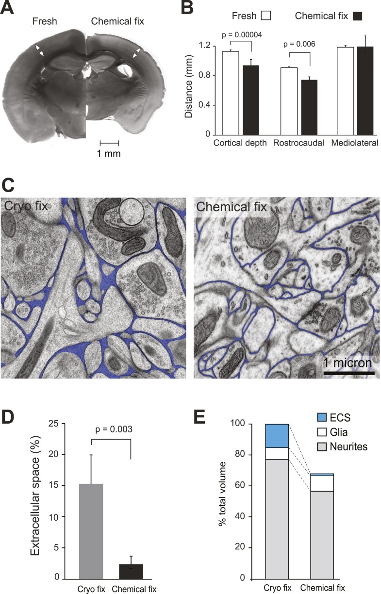
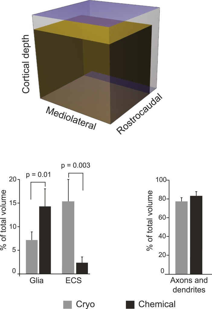
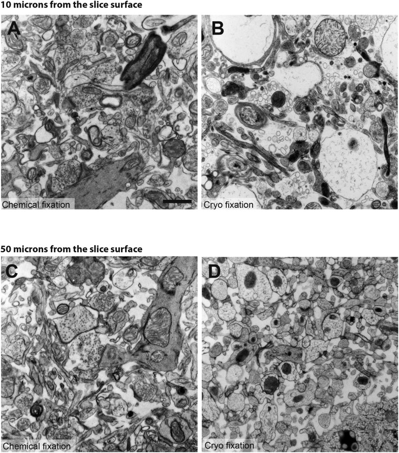
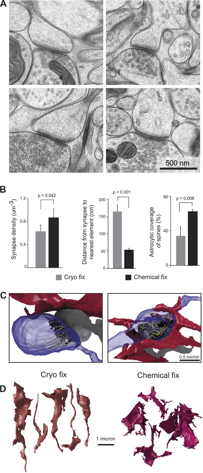
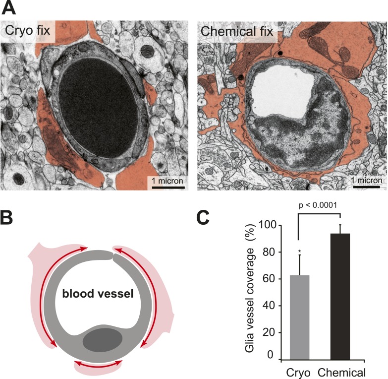
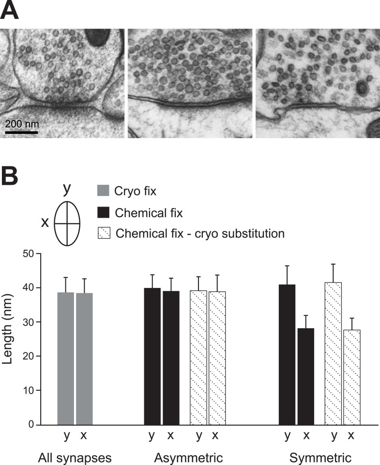
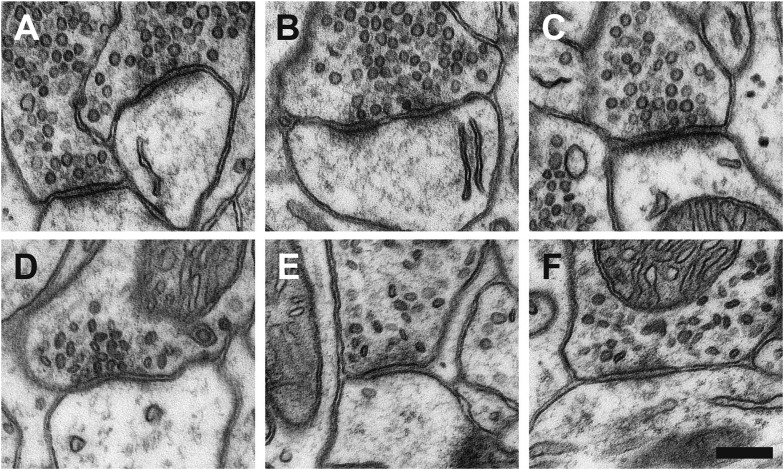
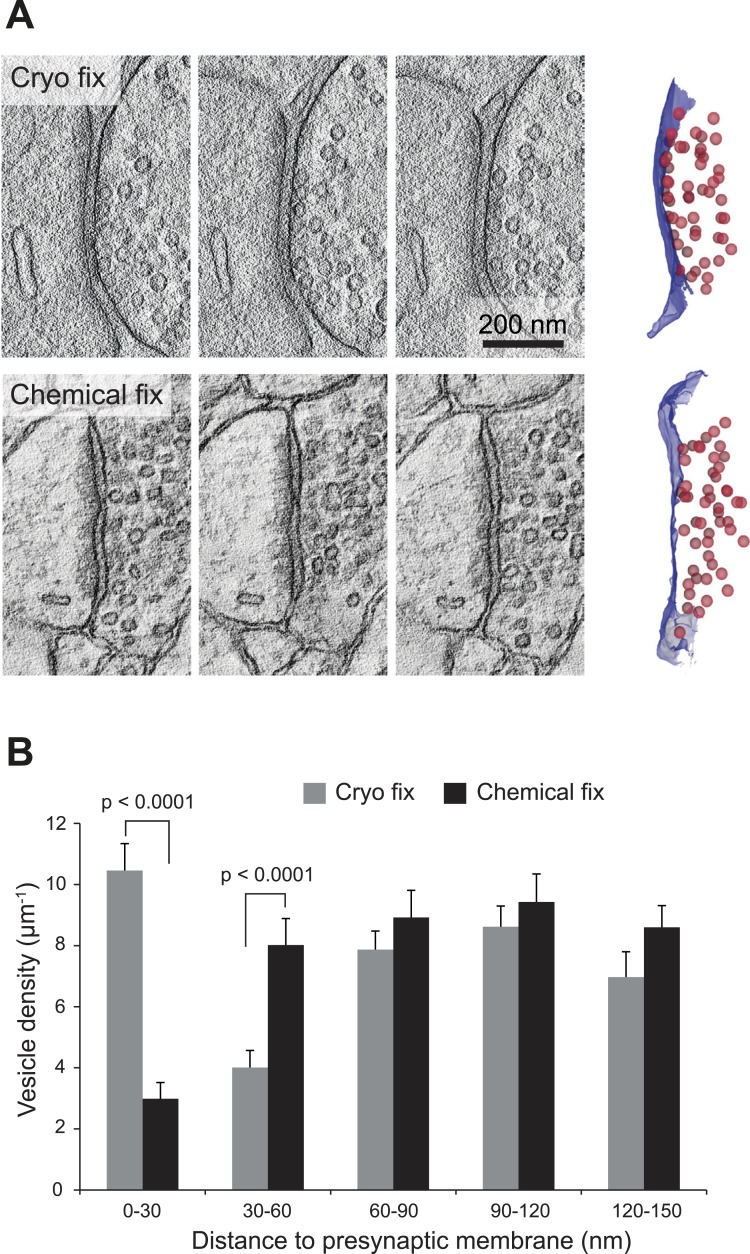
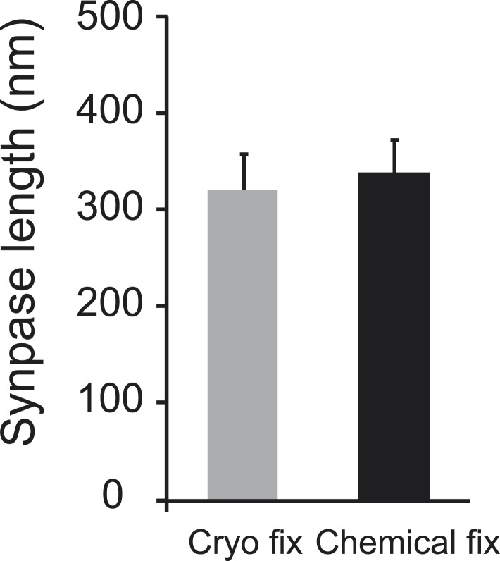
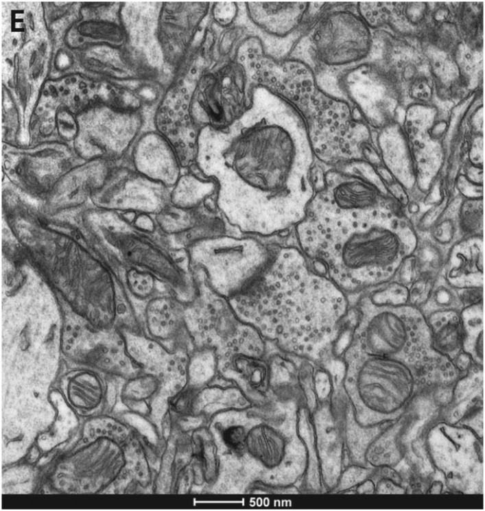
Analysis of brain ultrastructure using electron microscopy typically relies on chemical fixation. However, this is known to cause significant tissue distortion including a reduction in the extracellular space. Cryo fixation is thought to give a truer representation of biological structures, and here we use rapid, high-pressure freezing on adult mouse neocortex to quantify the extent to which these two fixation methods differ in terms of their preservation of the different cellular compartments, and the arrangement of membranes at the synapse and around blood vessels. As well as preserving a physiological extracellular space, cryo fixation reveals larger numbers of docked synaptic vesicles, a smaller glial volume, and a less intimate glial coverage of synapses and blood vessels compared to chemical fixation. The ultrastructure of mouse neocortex therefore differs significantly comparing cryo and chemical fixation conditions.
Overview
The claims and statistics of this paper haven’t been checked yet.
Run the check — free, no account needed. It reads the open-access text and checks the sources it cites.
Integrity signals
Registry
Cites
Statistics
Figures
Verification
Claims the paper makes
The claims and statistics of this paper haven’t been checked yet.
Run the check — free, no account needed. It reads the open-access text and checks the sources it cites.
Claims other papers make about this one
Each is checked against this paper’s own text: does it say what the citing paper says it does?
In the citing paper
“however, recent data with high-pressure cryopreservation show much less astrocytic coverage of both capillaries and synapses than seen with aldehyde fixation, which shrinks the extracellular space (Korogod et al., 2015).”
From Fueling thought: Management of glycolysis and oxidative phosphorylation in neuronal metabolism (2018) · cites this paper as Korogod et al., 2015
Self-check
Percentages, sums and reported differences re-computed from the paper’s own numbers.
The self-check needs the paper’s full text.
Citations
Cites
Calamita, E., Schmid, M., Kunz, M., Ndebele-Murisa, M. R., Magadza, C. H. D., Nyambe, I., & Wehrli, B. (2019) · Figshare · doi:10.26181/6153922fe919c · Cited by 4,239
Glial and neuronal control of brain blood flow
Attwell, D., Buchan, A. M., Charpak, S., Lauritzen, M., MacVicar, B. A., & Newman, E. A. (2010) · Nature · doi:10.1038/nature09613 · Cited by 2,511
Long-term in vivo imaging of experience-dependent synaptic plasticity in adult cortex
Trachtenberg, J. T., Chen, B. E., Knott, G. W., Feng, G., Sanes, J. R., Welker, E., & Svoboda, K. (2002) · Nature · doi:10.1038/nature01273 · Cited by 1,927
Self-citationAxo-somatic and axo-dendritic synapses of the cerebral cortex: an electron microscope study.
Eg, G. (1959) · PubMed · Cited by 1,768
Diffusion in Brain Extracellular Space
Syková, E., & Nicholson, C. (2008) · Physiological Reviews · doi:10.1152/physrev.00027.2007 · Cited by 1,481
Harris, K., & Stevens, J. (1989) · The Journal of Neuroscience · doi:10.1523/jneurosci.09-08-02982.1989 · Cited by 1,073
TrakEM2 Software for Neural Circuit Reconstruction
Cardona, A., Saalfeld, S., Schindelin, J., Arganda‐Carreras, I., Preibisch, S., Longair, M., Tomančák, P., Hartenstein, V., & Douglas, R. J. (2012) · PLoS ONE · doi:10.1371/journal.pone.0038011 · Cited by 1,009
Connectomic reconstruction of the inner plexiform layer in the mouse retina
Helmstaedter, M., Briggman, K. L., Turaga, S. C., Jain, V., Seung, H. S., & Denk, W. (2013) · Nature · doi:10.1038/nature12346 · Cited by 997
Colonnier, M. (1968) · Brain Research · doi:10.1016/0006-8993(68)90234-5 · Cited by 946
Characteristics of Excitatory and Inhibitory Synapses in the Central Nervous System of the Cat
UCHIZONO, K. (1965) · Nature · doi:10.1038/207642a0 · Cited by 917
Cited by
Sorted by citations
Items 301–400 of 408
Yang, C., Xie, J., Huang, X., Tan, H., Li, Q., Tang, Z., Ma, X., Lu, J., He, Q., Fu, W., et al. (2025) · Expert Systems with Applications · doi:10.1016/j.eswa.2024.126370 · Cited by 4
Noradrenaline Drives Structural Changes in Astrocytes and Brain Extracellular Space
Sherpa, A. D., Aoki, C. J., & Hrabětová, S. (2017) · Elsevier eBooks · doi:10.1016/b978-0-12-805088-0.00012-8 · Cited by 4
Multiscale and Multimodal Imaging for Connectomics
Gogoi, A., Keiser, G. E., Kao, F., & Chiang, A. (2018) · Progress in optical science and photonics · doi:10.1007/978-981-10-9020-2_1 · Cited by 4
Numerical simulation study of nanoparticle diffusion in gray matter
Chen, P., Dong, B., & Yao, W. (2024) · Computational and Structural Biotechnology Journal · doi:10.1016/j.csbj.2024.06.002 · Cited by 4
McCoy, J., & Nahmani, M. (2021) · Journal of Visualized Experiments · doi:10.3791/63030 · Cited by 4
Advances in Neuroscience Using Transmission Electron Microscopy: A Historical Perspective
Zhang, J., Reyes, B. A. S., Ross, J. A., Trovillion, V., & Bockstaele, E. J. V. (2016) · Neuromethods · doi:10.1007/7657_2016_101 · Cited by 4
Correlating Data from Imaging Modalities
Paul‐Gilloteaux, P., & Schorb, M. (2019) · doi:10.1002/9781119086420.ch11 · Cited by 4
Dyhr, M. C. A., Sadeghi, M., Moynova, R., Knappe, C., Kepsutlu, B., Werner, S., Schneider, G., McNally, J. G., Noé, F., & Ewers, H. (2022) · bioRxiv (Cold Spring Harbor Laboratory) · doi:10.1101/2022.05.16.492055 · Cited by 4
Kjer, H. M., Andersson, M., He, Y., Pacureanu, A., Daducci, A., Pizzolato, M., Salditt, T., Robisch, A.-L., Eckermann, M., Töpperwien, M., et al. (2024) · eLife · doi:10.7554/elife.94917.1 · Cited by 4
Evaluating ultrastructural preservation quality in banked brain tissuePreprint
Garrood, M., Keberle, A., Sowa, A., Janssen, W. G. M., Thorn, E. L., Sanctis, C. D., Farrell, K. W., Crary, J. F., & McKenzie, A. T. (2025) · bioRxiv (Cold Spring Harbor Laboratory) · doi:10.1101/2025.05.09.652503 · Cited by 6
Nguyen‐Duc, J., Brammerloh, M., Cherchali, M., de Riedmatten, I., Pérot, J., Rafael‐Patiño, J., & Jelescu, I. (2026) · Medical Image Analysis · doi:10.1016/j.media.2026.103946 · Cited by 3
Neuronal synaptic architecture revealed by cryo-correlative light and electron microscopy
Wang, P., Tian, B., Xu, X., Luan, H., Zhang, Y., Sun, W., Hu, L., Li, Y., Yao, Y., Li, W., et al. (2024) · Biophysics Reports · doi:10.52601/bpr.2024.240035 · Cited by 3
Cryo‐SEM in haematological research
Davidovich, I., Levin, C., & Talmon, Y. (2025) · Journal of Microscopy · doi:10.1111/jmi.13424 · Cited by 3
What are memories made of? A survey of neuroscientists on the structural basis of long-term memory
Zeleznikow-Johnston, A., Kendziorra, E., & McKenzie, A. T. (2025) · PLoS ONE · doi:10.1371/journal.pone.0326920 · Cited by 4
Collagens as New Players in Nervous System Diseases
Heikkinen, A., Fox, M. A., & Pihlajaniemi, T. A. (2021) · Biology of extracellular matrix · doi:10.1007/978-3-030-67592-9_8 · Cited by 3
Rothman, J. S., Borges-Merjane, C., Holderith, N. B., Jónás, P., & Silver, R. A. (2023) · PLoS ONE · doi:10.1371/journal.pone.0277148 · Cited by 4
High-Resolution Molecular Imaging and Its Applications in Brain and Synapses
Phan, N. T. N., & Rizzoli, S. O. (2020) · Neuromethods · doi:10.1007/978-1-0716-0691-9_3 · Cited by 3
Dorgans, K., Demais, V., Bailly, Y. J. R., Poulain, B., Isope, P., & Doussau, F. (2018) · bioRxiv (Cold Spring Harbor Laboratory) · doi:10.1101/338152 · Cited by 3
Creekmore, B. C., Kixmoeller, K., Black, B. E., Lee, E. B., & Chang, Y. (2023) · bioRxiv (Cold Spring Harbor Laboratory) · doi:10.1101/2023.09.13.557623 · Cited by 3
Invariant synaptic density across speciesPreprint
Castro, A. F., & Cardona, A. (2024) · bioRxiv (Cold Spring Harbor Laboratory) · doi:10.1101/2024.07.18.604056 · Cited by 3
Memory engram synapse 3D macromolecular architecture visualized by cryoCLEM-guided cryoET
Lovatt, C., O’Sullivan, T., Luis, C. O. S., Ryan, T. J., & Frank, R. A. W. (2025) · Structure · doi:10.1016/j.str.2025.10.014 · Cited by 3
Recent Structural Insights into the Molecular Architecture of Synapses
Brünger, A. T., Held, R. G., Khan, Y. A., Leitz, J., Liang, J., Wang, C., & White, K. I. (2026) · Advances in neurobiology · doi:10.1007/978-3-032-12594-1_2 · Cited by 2
Dynamics of waste proteins in brain tissue: Numerical insights into Alzheimer's risk factors
Watkins, L., Mukherjee, S., & Tithof, J. (2024) · Physical review. E · doi:10.1103/physreve.110.034401 · Cited by 2
Modeling of Blood Flow Dynamics in Rat Somatosensory Cortex
Battini, S., Cantarutti, N., Kotsalos, C., Roussel, Y., Cattabiani, A., Arnaudon, A., Favreau, C., Antonel, S., Markram, H., & Keller, D. X. (2024) · Biomedicines · doi:10.3390/biomedicines13010072 · Cited by 2
Uematsu, M., Mikami, K., Nakamura, A., Takahashi, R., Yokota, T., Hirokawa, K., & Uchihara, T. (2022) · Scientific Reports · doi:10.1038/s41598-022-09849-8 · Cited by 2
Cryo-FIB workflow for imaging brain tissue via in situ cryo-electron microscopyPreprint
Ning, J., Glausier, J. R., Hsieh, C., Schmelzer, T., Buck, S. A., Franks, J. M., Hampton, C. M., Lewis, D. A., Marko, M., & Freyberg, Z. (2023) · bioRxiv (Cold Spring Harbor Laboratory) · doi:10.1101/2023.02.11.528064 · Cited by 2
The impact of chemical fixation on the microanatomy of mouse brain tissuePreprint
Idziak, A., Inavalli, V. V. G. K., Bancelin, S., Arizono, M., & Nägerl, U. V. (2023) · bioRxiv (Cold Spring Harbor Laboratory) · doi:10.1101/2023.02.21.528828 · Cited by 2
Lycas, M. D., Morado, D. R., Gether, U., Briggs, J. A. G., & Erlendsson, S. (2024) · bioRxiv (Cold Spring Harbor Laboratory) · doi:10.1101/2024.04.15.589543 · Cited by 2
Excitability and synaptic transmission after vitrification of mouse corticohippocampal slicesPreprint
German, A., & Akdaş, E. Y. (2024) · bioRxiv (Cold Spring Harbor Laboratory) · doi:10.1101/2024.06.03.597218 · Cited by 3
Rollenhagen, A., Sadeghi, A., Walkenfort, B., Hilgetag, C. C., Sätzler, K., & Lübke, J. H. R. (2024) · eLife · doi:10.7554/elife.99473 · Cited by 3
Connectomic analysis of astrocyte-synapse interactions in the cerebral cortexPreprint
Yener, Y., Motta, A., & Helmstaedter, M. N. (2025) · bioRxiv (Cold Spring Harbor Laboratory) · doi:10.1101/2025.02.20.639274 · Cited by 2
Cryogenic light microscopy of vitrified samples with Ångstrom precisionPreprint
Mazal, H., Wieser, F.-F., Bollschweiler, D., & Sandoghdar, V. (2025) · bioRxiv (Cold Spring Harbor Laboratory) · doi:10.1101/2025.05.27.656160 · Cited by 2
Amick, J., Bhandarkar, S., Wilcox, D. R., Boloker, S., Lu, Y. D., & Gu, C. (2026) · bioRxiv (Cold Spring Harbor Laboratory) · doi:10.64898/2026.05.10.724157 · Cited by 2
Mrestani, A., Lichter, K., Sirén, A., Heckmann, M., Paul, M. M., & Pauli, M. (2023) · International Journal of Molecular Sciences · doi:10.3390/ijms24032128 · Cited by 1
Glymphatic system and aquaporin‑4 in epilepsy
Nowicka, D. (2023) · Acta Neurobiologiae Experimentalis · doi:10.55782/ane-2023-2498 · Cited by 1
A STED Microscope for Multimodal Investigation of Dendritic Spine Structure and Function
Quici, F., Dembitskaya, Y., Bancelin, S., Tønnesen, J., & Nägerl, U. V. (2024) · Neuromethods · doi:10.1007/978-1-0716-4011-1_13 · Cited by 1
Visualizing Postsynaptic Density in Excitatory Synapses with Electron Tomography
Sun, R., & Zhou, Q. (2026) · Advances in neurobiology · doi:10.1007/978-3-032-12594-1_10 · Cited by 2
Gimenez, P., Shakya, R. T., Santamarı́a, F., & Tønnesen, J. (2026) · Fluids and Barriers of the CNS · doi:10.1186/s12987-026-00797-3 · Cited by 1
Weier, A., Gao, F., Morgan, E. T., Liu, P., Melville, Z., Perez, L., Morgan, G., Shi, Q., Vigil, F. A., Eng, E. T., et al. (2026) · Cell Reports Methods · doi:10.1016/j.crmeth.2026.101599 · Cited by 1
Investigate Synaptic Vesicles Mobility in Neuronal Culture Axons by FRAP Imaging
Zhang, X. M., Cordelières, F. P., & Herzog, É. (2021) · BIO-PROTOCOL · doi:10.21769/bioprotoc.3962 · Cited by 1
Brain Interstitial Structure Revealed Through Diffusive Spread of Molecules
Nicholson, C. (2023) · Springer eBooks · doi:10.1007/978-3-031-05946-9_6 · Cited by 1
Rehydration of Freeze Substituted Brain Tissue for Pre-embedding Immunoelectron Microscopy
Pérez-Garza, J., Parrish-Mulliken, E., Deane, Z., & Ostroff, L. E. (2023) · Microscopy and Microanalysis · doi:10.1093/micmic/ozad077 · Cited by 1
3D Quantitative Analysis of Synaptic Ultrastructure in Identified Long-Range Projection Axons
Martin-Correa, P. J., Rodriguez-Moreno, J., Rollenhagen, A., Lübke, J. H. R., & Clascá, F. (2024) · Neuromethods · doi:10.1007/978-1-0716-4019-7_11 · Cited by 1
MBV-Pipe: A One-Stop Toolbox for Assessing Mouse Brain Morphological Changes for Cross-Scale Studies
Jiang, W., Liu, X., Song, M., Yang, Z., Sun, L., & Jiang, T. (2024) · Neuroinformatics · doi:10.1007/s12021-024-09687-1 · Cited by 1
Beyond neurons: computer vision methods for analysis of morphologically complex astrocytes
Syed, T. A., Youssef, M., Schober, A. L., Kubota, Y., Murai, K. K., & Salmon, C. K. (2024) · Frontiers in Computer Science · doi:10.3389/fcomp.2024.1156204 · Cited by 2
General Introduction to Barrier Mechanisms in the Central Nervous System
Saunders, N. R., Dzięgielewska, K. M., Møllgård, K., & Habgood, M. D. (2017) · Springer eBooks · doi:10.1007/978-3-319-45514-3_1 · Cited by 1
Using ephaptic coupling to estimate the synaptic cleft resistivity of the calyx of Held synapse
Sierksma, M. C., & Borst, J. G. G. (2021) · PLoS Computational Biology · doi:10.1371/journal.pcbi.1009527 · Cited by 1
Tracking Photoluminescent Carbon Nanomaterials in Biological Systems
Haziza, S., Cognet, L., & Treussart, F. (2018) · doi:10.1002/9781119373476.ch6 · Cited by 1
Correlating Synaptic Ultrastructure and Function at the NanoscalePreprint
Maus, L., Altas, B., Rhee, J., Brose, N., Imig, C., & Cooper, B. H. (2019) · bioRxiv (Cold Spring Harbor Laboratory) · doi:10.1101/588848 · Cited by 1
Novel Strategies for Glutamate Clearance in the Glia-Deprived Synaptic Hub of C. elegansPreprint
Chan, J., Lee, K. K., Wong, J. C. Y., Morocho, P., & Mano, I. (2019) · bioRxiv (Cold Spring Harbor Laboratory) · doi:10.1101/645812 · Cited by 1
Estimates for the astrocyte endfoot sheath permeability of the extra-cellular pathwayPreprint
Koch, T., Vinje, V., & Mardal, K. (2022) · bioRxiv (Cold Spring Harbor Laboratory) · doi:10.1101/2022.11.16.516727 · Cited by 1
Receptor binding and tissue architecture explain the morphogen local-to-global mobility transitionPreprint
Zhu, S., Loo, Y. T., Veerapathiran, S., Loo, T. Y. J., Trần, B. N., Teh, C., Zhong, J., Matsudaira, P. T., Saunders, T. E., & Wohland, T. (2024) · bioRxiv (Cold Spring Harbor Laboratory) · doi:10.1101/2024.04.28.591267 · Cited by 1
Modeling of blood flow dynamics in the rat somatosensory cortexPreprint
Battini, S., Cantarutti, N., Kotsalos, C., Roussel, Y., Cattabiani, A., Arnaudon, A., Favreau, C., Antonel, S., Markram, H., & Keller, D. X. (2024) · bioRxiv (Cold Spring Harbor Laboratory) · doi:10.1101/2024.11.14.623572 · Cited by 1
Kjer, H. M., Andersson, M., He, Y., Pacureanu, A., Daducci, A., Pizzolato, M., Salditt, T., Robisch, A.-L., Eckermann, M., Töpperwien, M., et al. (2024) · eLife · doi:10.7554/elife.94917.2 · Cited by 1
Verasztó, C., Roussel, Y., Kanari, L., Markram, H., & Keller, D. X. (2024) · bioRxiv (Cold Spring Harbor Laboratory) · doi:10.1101/2024.12.30.630728 · Cited by 1
Ultrafast endocytosis in mouse cortical inhibitory synapsesPreprint
Eddings, C. R., & Watanabe, S. (2025) · bioRxiv (Cold Spring Harbor Laboratory) · doi:10.1101/2025.06.06.658279 · Cited by 1
Simulating calcium signaling in fine astrocytic processes
Denizot, A. (2019) · theses.fr (ABES) · doi:10.70675/970a38b3z8898z4ac0z896ez5ae9336af7f7 · Cited by 1
Iyer, N., & Rivlin, P. K. (2019) · Microscopy and Microanalysis · doi:10.1017/s143192761900597x
Morphologically constrained modeling of spinous inhibition in the somato-sensory cortexPreprint
Gemin, O., Serna, P., Assendorp, N., Fossati, M., Rostaing, P., Triller, A., & Charrier, C. (2020) · bioRxiv (Cold Spring Harbor Laboratory) · doi:10.1101/2020.08.06.230722
Optical analysis of the action range of glutamate in the neuropilPreprint
Matthews, E. A., Sun, W., McMahon, S. M., Doengi, M., Halka, L., Anders, S., Müller, J. A., Steinlein, P., Vana, N., van Dyk, G., et al. (2021) · bioRxiv (Cold Spring Harbor Laboratory) · doi:10.1101/2021.02.05.429974
Investigation of myelin maintenance and turnover by inducible MBP knock-out in adult mice
Meschkat, M. (2019) · eDiss (Georg-August-Universität Göttingen) · doi:10.53846/goediss-7679
Diffusion-weighted magnetic resonance imaging at ultra-high field
Fritz, F. J. (2020) · Research Publications (Maastricht University) · doi:10.26481/dis.20210111flf
Shih, A. Y., Coelho‐Santos, V., & Tieu, T. (2020) · Faculty Opinions – Post-Publication Peer Review of the Biomedical Literature · doi:10.3410/f.738328793.793578304
Sala, C. (2019) · Faculty Opinions – Post-Publication Peer Review of the Biomedical Literature · doi:10.3410/f.735371200.793560713
Nano-Based Probes for the Brain Extracellular Environment
Filteau, J. R., Butler, B., Schimek, N., & Nance, E. A. (2022) · Springer eBooks · doi:10.1007/978-3-031-11409-0_2
Barrière, D. A., Uszynski, I., Rajani, R. M., Gueniot, F., Domenga, V., Boumezbeur, F., Poupon, C., & Joutel, A. (2023) · bioRxiv (Cold Spring Harbor Laboratory) · doi:10.1101/2023.02.27.530286
Astrocytic modulation of the synaptic and the extrasynaptic space
Badia‐Soteras, A. (2023) · doi:10.5463/thesis.127
pan-ECM: live brain extracellular matrix imaging with protein-reactive dyePreprint
Ge, X., Xu, X., Cai, Q., Xiong, H., Xie, C., Hong, Y., Gao, X., Yao, Y., Bachoo, R. M., & Qin, Z. (2023) · bioRxiv (Cold Spring Harbor Laboratory) · doi:10.1101/2023.03.29.534827
Revealing ultrastructural changes that determine the development of a CNS synapse
Hintze, A. (2023) · eDiss (Georg-August-Universität Göttingen) · doi:10.53846/goediss-9828
Liu, J., Kumar, M., Guo, J., Huang, L., Zhang, Z., Wang, Y., Luo, R., chu, S., Chen, S., Liu, R., et al. (2023) · Research Square · doi:10.21203/rs.3.rs-3028525/v1
Rollenhagen, A., Sadeghi, A., Walkenfort, B., Hilgetag, C. C., Sätzler, K., & Lübke, J. H. R. (2025) · eLife · doi:10.1101/2024.06.14.599047
Quantitative Analysis of the Synaptic Organization of the Human Temporal Lobe Neocortex
Rollenhagen, A., Sätzler, K., & Lübke, J. H. R. (2024) · Neuromethods · doi:10.1007/978-1-0716-4019-7_12
Rollenhagen, A., Dastjerdi, A. S., Walkenfort, B., Hilgetag, C. C., Sätzler, K., & Lübke, J. H. R. (2024) · eLife · doi:10.7554/elife.99473.1
Rollenhagen, A., Sadeghi, A., Walkenfort, B., Hilgetag, C. C., Sätzler, K., & Lübke, J. H. R. (2025) · eLife · doi:10.7554/elife.99473.2
Super-resolution Analysis of Astrocyte Morphology Using Expansion Microscopy
Seifert, G., Sommer, E., Passlick, S., & Henneberger, C. (2025) · Methods in molecular biology · doi:10.1007/978-1-0716-4366-2_12
Rollenhagen, A., Sadeghi, A., Walkenfort, B., Hilgetag, C. C., Sätzler, K., & Lübke, J. H. R. (2025) · eLife · doi:10.7554/elife.99473.3
Rollenhagen, A., Sadeghi, A., Walkenfort, B., Hilgetag, C. C., Sätzler, K., & Lübke, J. H. R. (2025) · eLife · doi:10.7554/elife.99473.4
Effect of Dead-Space Microdomain Entrance Size and Volume on Brain Extracellular Space Diffusion
Xiao, E., Hrabětová, S., & Hrabě, J. (2025) · Diffusion fundamentals. · doi:10.62721/diffusion-fundamentals.39.1266
Biophysics of intracellular and extracellular diffusion
Alvarez-Leefmans, F. J. (2025) · Elsevier eBooks · doi:10.1016/b978-0-12-811114-7.00002-5
Martin‐Solana, E., Casado-Zueras, L., Torres, T. E., Goya, G. F., Fernandez-Fernandez, M.-R., & Fernandez, J.-J. (2024) · Figshare · doi:10.6084/m9.figshare.26729824
Kedarasetti, R. T., Drew, P. J., & Costanzo, F. (2022) · Figshare · doi:10.6084/m9.figshare.19771061
Verasztó, C., Roussel, Y., Kanari, L., Piluso, S., Markram, H., & Keller, D. X. (2026) · PLoS Computational Biology · doi:10.1371/journal.pcbi.1014106 · Cited by 1
Direct Reconstruction of DC Cortical Conductivity from Large-Scale Electron Microscopy DataPreprint
Noetscher, G. M., Miles, A., Danskin, B. P., Tang, D., Ingersoll, M., Ponasso, G. N., Paxton, C., Ludwig, R., Burnham, E. H., Deng, Z., et al. (2026) · bioRxiv (Cold Spring Harbor Laboratory) · doi:10.64898/2026.03.23.713806 · Cited by 1
Mansouri-Guilani, N. (2017) · theses.fr (ABES) · doi:10.70675/99ced3b5zeac6z4eb2z8e6cz01bcad7cf2d9
Dorgans, K. (2017) · theses.fr (ABES) · doi:10.70675/031e34e5zd5b7z4debzab0dzcd77f69b8c71
Idziak, A. (2022) · theses.fr (ABES) · doi:10.70675/91c65c6ez289bz41b2za50azc10a875e4675
Ultrastructural and molecular analysis of cortico-striatal dopamine hub synapses
Lapios, P. (2024) · theses.fr (ABES) · doi:10.70675/527646a6zd9b5z4935z80ebz0950bba9d08e
Molecular impact of anti-NMDA receptor autoantibodies
Jamet, Z. (2023) · theses.fr (ABES) · doi:10.70675/87243db0za802z49e1zbd1ez607bb3d60120
Role of extracellular space in neurodegenerative diseases
Estaún‐Panzano, J. (2024) · theses.fr (ABES) · doi:10.70675/98d1d96eza025z406ezba93z33e0820c3cf3
Rôle des astrocytes dans la période critique de la plasticité visuelle
Breton, R. (2023) · theses.fr (ABES) · doi:10.70675/9140a00az6c88z4fb6zaabfz9eb16e7593ca
Wulffelé, J. (2023) · theses.fr (ABES) · doi:10.70675/e14a5d84zba60z4abfza023z81a1de13fb51
Lycas, M. D., Morado, D. R., Gether, U., Briggs, J. A. G., & Erlendsson, S. (2026) · The Journal of Cell Biology · doi:10.1083/jcb.202503059 · Cited by 3
Lv, Z.-Y., Lu, Z.-H., Liu, S., Zhou, P., Lau, P.-M., Bi, G., & Tao, C.-L. (2026) · Neuroscience Bulletin · doi:10.1007/s12264-026-01646-x
Kopach, O., Reynolds, J. R., Jensen, T. P., Zheng, K., Savtchenko, L. P., & Rusakov, D. A. (2026) · bioRxiv (Cold Spring Harbor Laboratory) · doi:10.2139/ssrn.7135125
Metadata
- Authors
- Korogod, N., Petersen, C. C., & Knott, G. W.
- Affiliations
- École Polytechnique Fédérale de Lausanne (CH)
- Corresponding author
- Petersen, Carl CH
- Knott, Graham W
- Venue
- eLife
- Publisher
- eLife Sciences Publications, Ltd
- Published
- 2015-08-11
- Volume
- 4
- ISSN
- 2050-084X (ISSN-L)
- PMC
- PMC4530226
- OpenAlex
- W1926373965
- Cited by
- 408
- References
- 63
- Citations by year
- 2026: 22 · 2025: 38 · 2024: 55 · 2023: 54 · 2022: 38 · 2021: 48 · 2020: 41 · 2019: 32 · 2018: 28 · 2017: 27 · 2016: 20 · 2015: 5
- FWCI
- 12.38
- Type
- journal article
- Language
- en
- Open access
- gold
- doi.org
- Licence
- Locations
- eLife (publishedVersion, cc-by)
- PubMed Central (publishedVersion, cc-by)
- PubMed (publishedVersion)
- DOAJ (DOAJ: Directory of Open Access Journals) (submittedVersion, cc-by-sa)
- Infoscience (Ecole Polytechnique Fédérale de Lausanne) (submittedVersion)
- Infoscience (Ecole Polytechnique Fédérale de Lausanne) (submittedVersion, cc-by-nc-nd)
- Funders
- Schweizerische Nationalfonds zur Förderung der Wissenschaftlichen Forschung
- Human Frontier Science Program (HFSP)
- European Research Council (ERC)
- Topics
- Life Sciences › Neuroscience › Cellular and Molecular Neuroscience › Neuroscience and Neuropharmacology Research
- Life Sciences › Biochemistry, Genetics and Molecular Biology › Structural Biology › Advanced Electron Microscopy Techniques and Applications
- Health Sciences › Medicine › Anatomy › Classical Anatomy and Histology
- Keywords
- neocortex, cryofixation, chemical fixation, high-pressure freezing, extracellular space, synaptic vesicles, synapses, blood vessels, tissue deformation
- MeSH terms
- Aldehydes, Animals, Fixatives, Freezing, Microscopy, Electron, Perfusion, Tissue Fixation, Neocortex, Mice
About these checks
Registry, retraction, Cites and statistics results are computed, not judged, with one exception: a printed reference that no index lists is read once by a model before it counts as a citation error. Claims are extracted by a model and kept only when their quote appears verbatim in the text; each cited source is then checked, with a model’s help, for whether it says what the claim relies on. Those verdicts can be wrong — read the claim’s quote and the cited source before relying on one.
Statistics and arithmetic are recomputed, not judged.
Confidence bands are shown only for fields calibrated on our annotated test set. None is calibrated yet, so no band is shown.
What counts as a claim ›How we measure confidence ›How we check this ›
Are you an author, or do you see a problem with this page? Request a review
