

The DOI matches its Crossref record; 63 of the 63 references it cites were found in a registry or an index.
Ultrastructural analysis of adult mouse neocortex comparing aldehyde perfusion with cryo fixation
Open accessAbstract
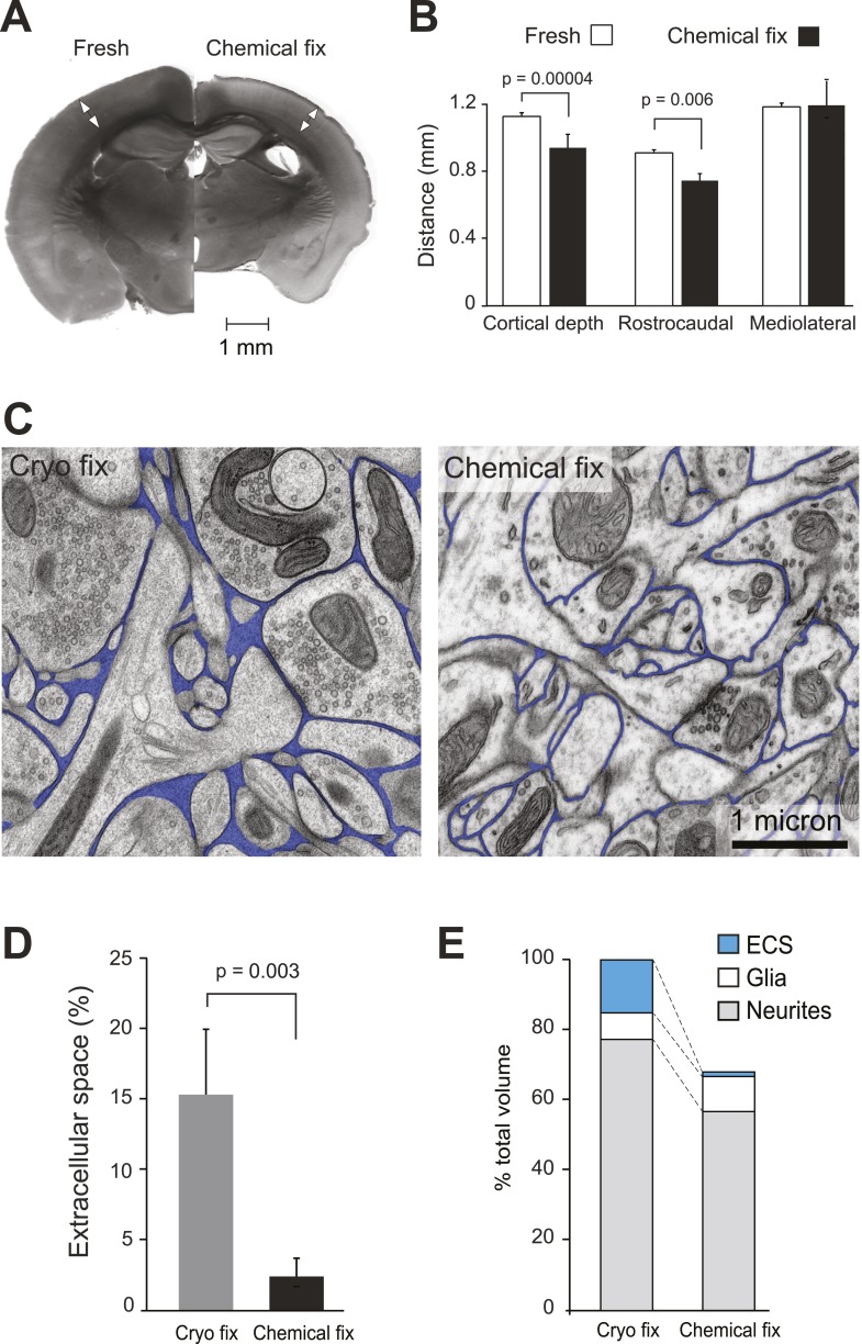
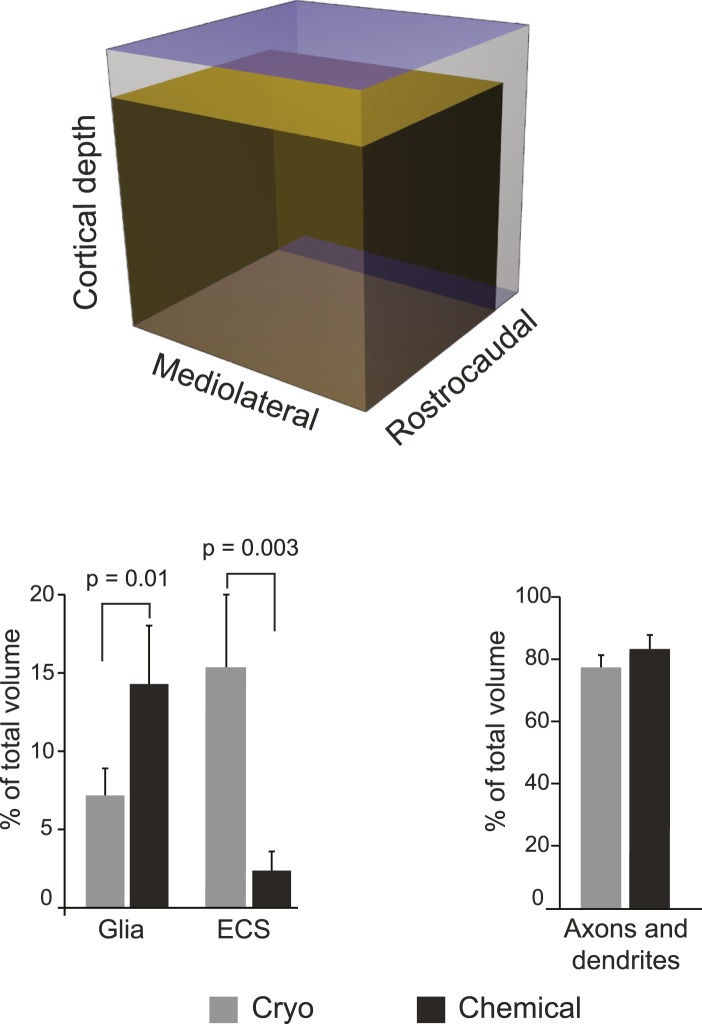
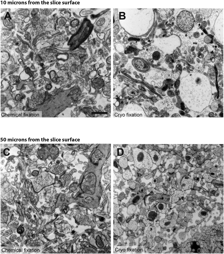
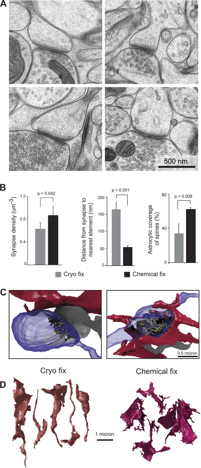
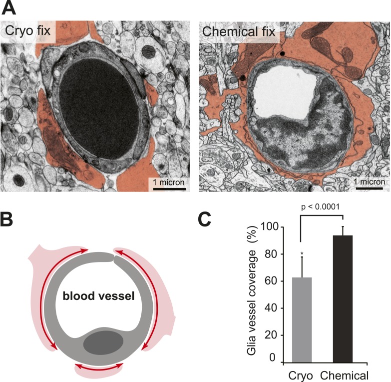
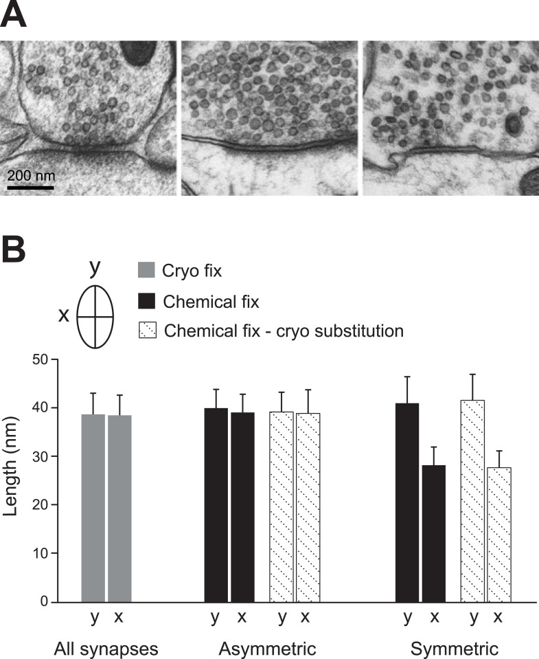
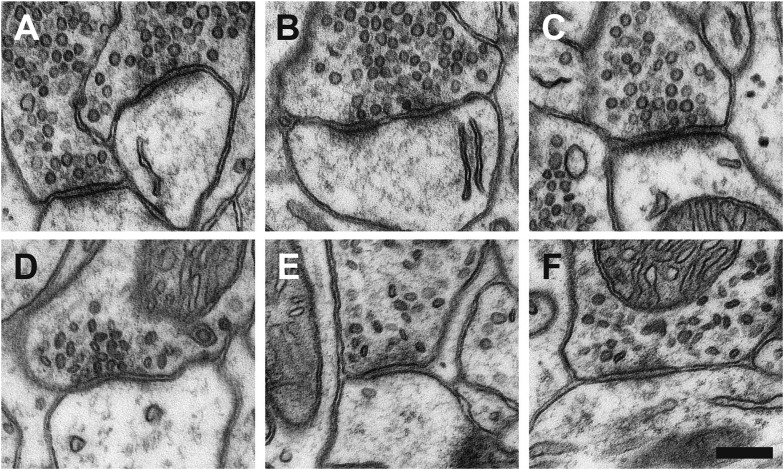
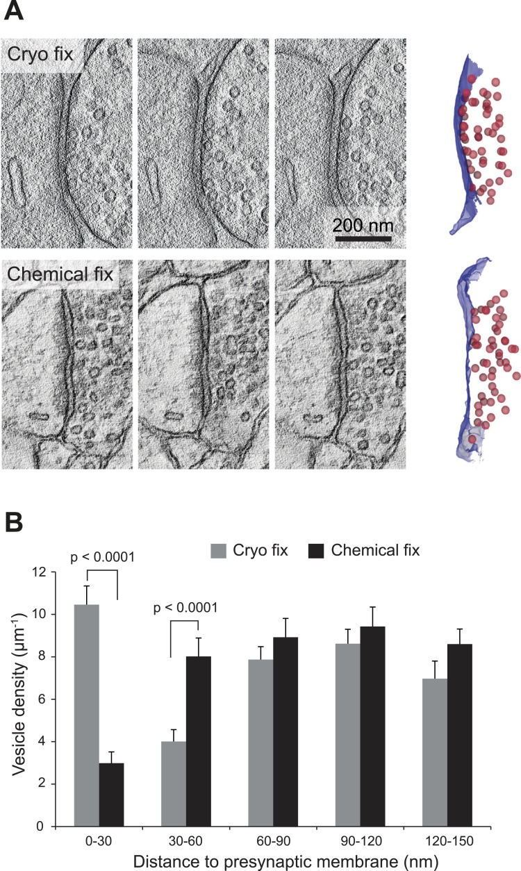
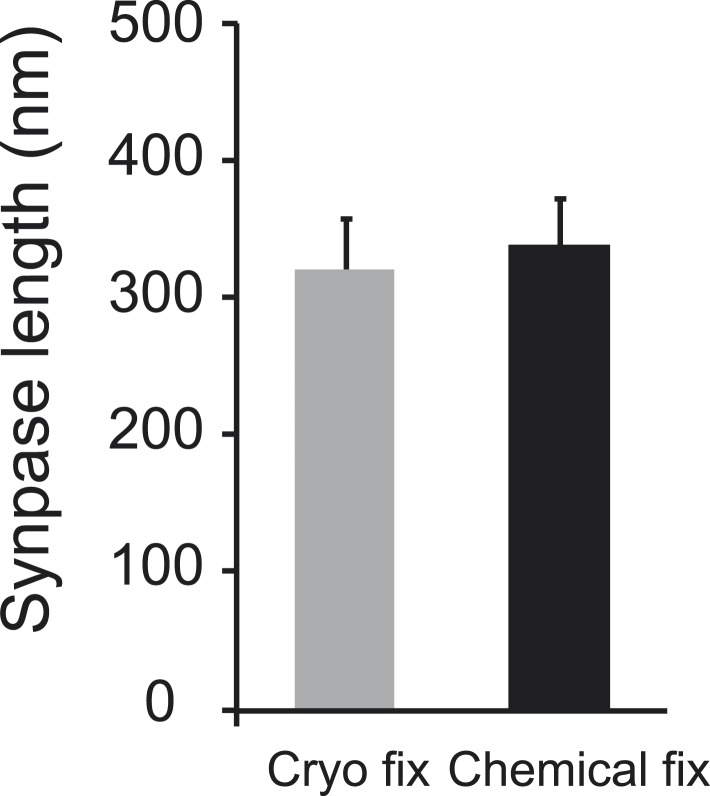
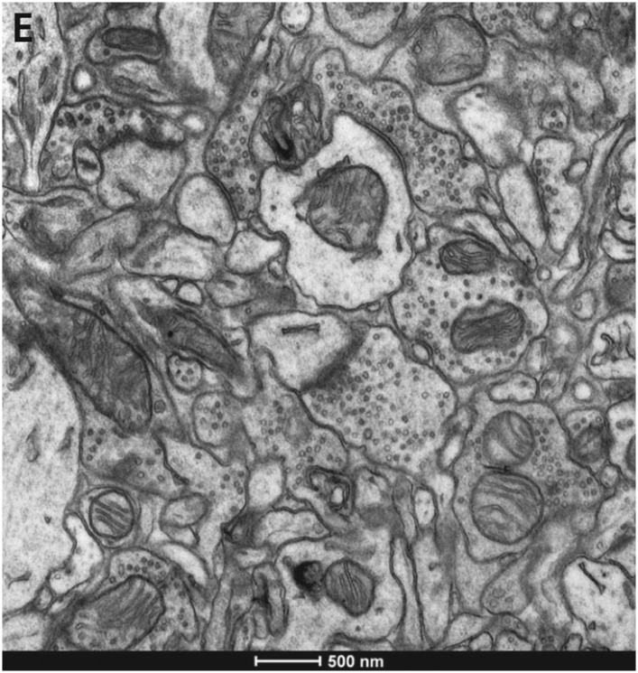
Analysis of brain ultrastructure using electron microscopy typically relies on chemical fixation. However, this is known to cause significant tissue distortion including a reduction in the extracellular space. Cryo fixation is thought to give a truer representation of biological structures, and here we use rapid, high-pressure freezing on adult mouse neocortex to quantify the extent to which these two fixation methods differ in terms of their preservation of the different cellular compartments, and the arrangement of membranes at the synapse and around blood vessels. As well as preserving a physiological extracellular space, cryo fixation reveals larger numbers of docked synaptic vesicles, a smaller glial volume, and a less intimate glial coverage of synapses and blood vessels compared to chemical fixation. The ultrastructure of mouse neocortex therefore differs significantly comparing cryo and chemical fixation conditions.
Overview
The claims and statistics of this paper haven’t been checked yet.
Run the check — free, no account needed. It reads the open-access text and checks the sources it cites.
Integrity signals
Registry
Cites
Statistics
Figures
Verification
Claims the paper makes
The claims and statistics of this paper haven’t been checked yet.
Run the check — free, no account needed. It reads the open-access text and checks the sources it cites.
Claims other papers make about this one
Each is checked against this paper’s own text: does it say what the citing paper says it does?
In the citing paper
“however, recent data with high-pressure cryopreservation show much less astrocytic coverage of both capillaries and synapses than seen with aldehyde fixation, which shrinks the extracellular space (Korogod et al., 2015).”
From Fueling thought: Management of glycolysis and oxidative phosphorylation in neuronal metabolism (2018) · cites this paper as Korogod et al., 2015
Self-check
Percentages, sums and reported differences re-computed from the paper’s own numbers.
The self-check needs the paper’s full text.
Citations
Cites
Calamita, E., Schmid, M., Kunz, M., Ndebele-Murisa, M. R., Magadza, C. H. D., Nyambe, I., & Wehrli, B. (2019) · Figshare · doi:10.26181/6153922fe919c · Cited by 4,239
Glial and neuronal control of brain blood flow
Attwell, D., Buchan, A. M., Charpak, S., Lauritzen, M., MacVicar, B. A., & Newman, E. A. (2010) · Nature · doi:10.1038/nature09613 · Cited by 2,511
Long-term in vivo imaging of experience-dependent synaptic plasticity in adult cortex
Trachtenberg, J. T., Chen, B. E., Knott, G. W., Feng, G., Sanes, J. R., Welker, E., & Svoboda, K. (2002) · Nature · doi:10.1038/nature01273 · Cited by 1,927
Self-citationAxo-somatic and axo-dendritic synapses of the cerebral cortex: an electron microscope study.
Eg, G. (1959) · PubMed · Cited by 1,768
Diffusion in Brain Extracellular Space
Syková, E., & Nicholson, C. (2008) · Physiological Reviews · doi:10.1152/physrev.00027.2007 · Cited by 1,481
Harris, K., & Stevens, J. (1989) · The Journal of Neuroscience · doi:10.1523/jneurosci.09-08-02982.1989 · Cited by 1,073
TrakEM2 Software for Neural Circuit Reconstruction
Cardona, A., Saalfeld, S., Schindelin, J., Arganda‐Carreras, I., Preibisch, S., Longair, M., Tomančák, P., Hartenstein, V., & Douglas, R. J. (2012) · PLoS ONE · doi:10.1371/journal.pone.0038011 · Cited by 1,009
Connectomic reconstruction of the inner plexiform layer in the mouse retina
Helmstaedter, M., Briggman, K. L., Turaga, S. C., Jain, V., Seung, H. S., & Denk, W. (2013) · Nature · doi:10.1038/nature12346 · Cited by 997
Colonnier, M. (1968) · Brain Research · doi:10.1016/0006-8993(68)90234-5 · Cited by 946
Characteristics of Excitatory and Inhibitory Synapses in the Central Nervous System of the Cat
UCHIZONO, K. (1965) · Nature · doi:10.1038/207642a0 · Cited by 917
Cited by
Sorted by citations
Items 101–200 of 408
All-optical visualization of specific molecules in the ultrastructural context of brain tissue
M’Saad, O., Cairns, A., Gulcicek, J., Kasula, R., Liao, J., Kondratiuk, I., Bewersdorf, E. H., Kidd, P., Falahati, H., Gentile, J. E., et al. (2025) · Nature Biotechnology · doi:10.1038/s41587-025-02905-4 · Cited by 52
Three-dimensional synaptic Organization of Layer III of the human temporal neocortex
Cano-Astorga, N., DeFelipe, J., & Alonso-Nanclares, L. (2021) · Cerebral Cortex · doi:10.1093/cercor/bhab120 · Cited by 49
Dense 4D nanoscale reconstruction of living brain tissue
Velicky, P., Miguel, E., Michalska, J. M., Lyudchik, J., Wei, D., Lin, Z., Watson, J. F., Troidl, J., Beyer, J., Ben‐Simon, Y., et al. (2023) · Nature Methods · doi:10.1038/s41592-023-01936-6 · Cited by 43
Near‐critical GLUT1 and Neurodegeneration
Barros, L. F., Martín, A. S., Ruminot, I., Sandoval, P. Y., Fernández‐Moncada, I., Baeza‐Lehnert, F., Arce‐Molina, R., Contreras‐Baeza, Y., Cortés‐Molina, F., Galaz, A., et al. (2017) · Journal of Neuroscience Research · doi:10.1002/jnr.23998 · Cited by 47
Structure, biophysics, and circuit function of a “giant” cortical presynaptic terminal
Vandael, D., & Jonas, P. (2024) · Science · doi:10.1126/science.adg6757 · Cited by 45
Astrocytic glutamate transport regulates a Drosophila CNS synapse that lacks astrocyte ensheathment
MacNamee, S. E., Liu, K. E., Gerhard, S., Tran, C. T., Fetter, R. D., Cardona, A., Tolbert, L. P., & Oland, L. A. (2016) · The Journal of Comparative Neurology · doi:10.1002/cne.24016 · Cited by 45
Metabolic Recruitment in Brain Tissue
Barros, L. F., Ruminot, I., Sotelo-Hitschfeld, T., Lerchundi, R., & Fernández‐Moncada, I. (2022) · Annual Review of Physiology · doi:10.1146/annurev-physiol-021422-091035 · Cited by 43
Creekmore, B. C., Kixmoeller, K., Black, B. E., Lee, E. B., & Chang, Y. (2024) · Nature Communications · doi:10.1038/s41467-024-47066-1 · Cited by 48
Axon tension regulates fasciculation/defasciculation through the control of axon shaft zippering
Šmít, D., Fouquet, C., Pincet, F., Zapotocky, M., & Trembleau, A. (2017) · eLife · doi:10.7554/elife.19907 · Cited by 44
Astrocytes and neurons communicate via a monocarboxylic acid shuttle
Roosterman, D., & Cottrell, G. (2020) · AIMS Neuroscience · doi:10.3934/neuroscience.2020007 · Cited by 34
Ultrastructural comparison of dendritic spine morphology preserved with cryo and chemical fixation
Tamada, H., Blanc, J., Korogod, N., Petersen, C. C. H., & Knott, G. W. (2020) · eLife · doi:10.7554/elife.56384 · Cited by 44
Guma, E., Bordeleau, M., Ibáñez, F. G., Picard, K., Snook, E., Desrosiers-Grégoire, G., Spring, S., Lerch, J. P., Nieman, B. J., Devenyi, G. A., et al. (2022) · Proceedings of the National Academy of Sciences · doi:10.1073/pnas.2114545119 · Cited by 42
Public Volume Electron Microscopy Data: An Essential Resource to Study the Brain Microvasculature
Bonney, S. K., Coelho‐Santos, V., Huang, S., Takeno, M. M., Kornfeld, J. M. R., Keller, A., & Shih, A. Y. (2022) · Frontiers in Cell and Developmental Biology · doi:10.3389/fcell.2022.849469 · Cited by 43
The structural and functional evidence for vesicular release from astrocytes in situ
Bohmbach, K., Schwarz, M. K., Schoch, S., & Henneberger, C. (2017) · Brain Research Bulletin · doi:10.1016/j.brainresbull.2017.01.015 · Cited by 47
Getting sharper: the brain under the spotlight of super-resolution microscopy
Arizono, M., Idziak, A., Quici, F., & Nägerl, U. V. (2022) · Trends in Cell Biology · doi:10.1016/j.tcb.2022.06.011 · Cited by 43
Rollenhagen, A., Ohana, O., Sätzler, K., Hilgetag, C. C., Kuhl, D., & Lübke, J. H. R. (2018) · Frontiers in Synaptic Neuroscience · doi:10.3389/fnsyn.2018.00024 · Cited by 45
Glymphatic System Dysfunction in Central Nervous System Diseases and Mood Disorders
Zhang, D., Li, X., & Li, B. (2022) · Frontiers in Aging Neuroscience · doi:10.3389/fnagi.2022.873697 · Cited by 40
Sensitivity analysis on a network model of glymphatic flow
Stevens, K. A., Tithof, J., Cook, D. D., Thomas, J. H., & Kelley, D. H. (2022) · Journal of The Royal Society Interface · doi:10.1098/rsif.2022.0257 · Cited by 39
Pleomorphic linkers as ubiquitous structural organizers of vesicles in axons
Schrod, N., Vanhecke, D., Laugks, U., Stein, V., Fukuda, Y., Schaffer, M., Baumeister, W. P., & Lučić, V. (2018) · PLoS ONE · doi:10.1371/journal.pone.0197886 · Cited by 38
Using high-throughput barcode sequencing to efficiently map connectomes
Peikon, I. D., Kebschull, J. M., Vagin, V. V., Ravens, D. I., Sun, Y.-C., Brouzes, E. R., Junior, I. R. C., Bressan, D., & Zador, A. M. (2017) · Nucleic Acids Research · doi:10.1093/nar/gkx292 · Cited by 39
Minge, D., Domingos, C., Unichenko, P., Behringer, C., Pauletti, A., Anders, S., Herde, M. K., Delekate, A., Gulakova, P. E., Schoch, S., et al. (2021) · Frontiers in Cellular Neuroscience · doi:10.3389/fncel.2021.669280 · Cited by 36
Bork, P. A. R., Ladrón-de-Guevara, A., Christensen, A. H., Jensen, K. H., Nedergaard, M., & Bohr, T. (2023) · Journal of The Royal Society Interface · doi:10.1098/rsif.2023.0050 · Cited by 37
Dorgans, K., Demais, V., Bailly, Y. J. R., Poulain, B., Isope, P., & Doussau, F. (2019) · eLife · doi:10.7554/elife.41586 · Cited by 44
Ultrastructural and dynamic studies of the endosomal compartment in Down syndrome
Botté, A., Lainé, J., Xicota, L., Heiligenstein, X., Fontaine, G., Kasri, A., Rivals, I., Goh, P. A., Faklaris, O., Cossec, J., et al. (2020) · Acta Neuropathologica Communications · doi:10.1186/s40478-020-00956-z · Cited by 35
Layer-specific anatomical and physiological features of the retina’s neurovascular unit
Grimes, W. N., Berson, D. M., Sabnis, A., Hoon, M., Sinha, R., Tian, H., & Diamond, J. S. (2024) · Current Biology · doi:10.1016/j.cub.2024.11.023 · Cited by 46
Pyramidal neuron growth and increased hippocampal volume during labor and birth in autism
Cloarec, R., Riffault, B., Dufour, A., Rabiei, H., Gouty‐Colomer, L., Dumon, C., Guimond, D., Bonifazi, P., Eftekhari, S., Lozovaya, N., et al. (2019) · Science Advances · doi:10.1126/sciadv.aav0394 · Cited by 32
Pardridge, W. M., & Chou, T. (2021) · Pharmaceuticals · doi:10.3390/ph14060535 · Cited by 35
Wildenberg, G. A., Sorokina, A. M., Koranda, J. L., Monical, A., Heer, C., Sheffield, M. E., Zhuang, X., McGehee, D., & Kasthuri, N. (2021) · eLife · doi:10.7554/elife.71981 · Cited by 39
Neural Metabolic Networks: Key Elements of Healthy Brain Function
Madrer, N., Perera, N. D., Uccelli, N. A., Abbondanza, A., Andersen, J. V., Carsana, E. V., Demmings, M. D., Fernandez, R. F., de Fragas, M. G., Gbadamosi, I., et al. (2025) · Journal of Neurochemistry · doi:10.1111/jnc.70084 · Cited by 37
Su, J., Cole, J., & Fox, M. A. (2017) · ASN NEURO · doi:10.1177/1759091416689020 · Cited by 36
Paviolo, C., & Cognet, L. (2021) · Neurobiology of Disease · doi:10.1016/j.nbd.2021.105328 · Cited by 35
Membrane mechanics dictate axonal pearls-on-a-string morphology and function
Griswold, J. M., Bonilla-Quintana, M., Pepper, R., Lee, C. T., Raychaudhuri, S., Ma, S., Gan, Q., Syed, S., Zhu, C., Bell, M. K., et al. (2024) · Nature Neuroscience · doi:10.1038/s41593-024-01813-1 · Cited by 43
Molecular architecture of the presynaptic terminal
Zuber, B., & Lučić, V. (2019) · Current Opinion in Structural Biology · doi:10.1016/j.sbi.2019.01.008 · Cited by 33
Mlc1-Expressing Perivascular Astrocytes Promote Blood–Brain Barrier Integrity
Morales, J. E., De, A., Miller, A. A., Chen, Z., & McCarty, J. H. (2021) · Journal of Neuroscience · doi:10.1523/jneurosci.1918-21.2021 · Cited by 33
McKenna, M., Shackelford, D. B., Pontes, C., Ball, B. K., Gao, T., & Nance, E. A. (2021) · ACS Nano · doi:10.1021/acsnano.1c00394 · Cited by 32
Ng, J., Browning, A., Lechner, L., Terada, M., Howard, G., & Jefferis, G. S. X. E. (2016) · Scientific Reports · doi:10.1038/srep38863 · Cited by 32
The Astrocyte: Metabolic Hub of the BrainPreprint
Barros, L. F., Schirmeier, S., & Weber, B. (2024) · Cold Spring Harbor Perspectives in Biology · doi:10.1101/cshperspect.a041355 · Cited by 34
Dufour, A., Rollenhagen, A., Sätzler, K., & Lübke, J. H. R. (2015) · Cerebral Cortex · doi:10.1093/cercor/bhv270 · Cited by 39
Engineered cell culture microenvironments for mechanobiology studies of brain neural cells
Ransanz, L. C., van Altena, P. F. J., Heine, V. M., & Accardo, A. (2022) · Frontiers in Bioengineering and Biotechnology · doi:10.3389/fbioe.2022.1096054 · Cited by 31
Song, K., Feng, Z., & Helmstaedter, M. N. (2023) · Nature Methods · doi:10.1038/s41592-023-01866-3 · Cited by 31
Circadian Mechanisms in Brain Fluid Biology
Vizcarra, V. S., Fame, R. M., & Hablitz, L. M. (2024) · Circulation Research · doi:10.1161/circresaha.123.323516 · Cited by 31
The effect of progressive hearing loss on the morphology of endbulbs of Held and bushy cells
Connelly, C. J., Ryugo, D. K., & Muniak, M. A. (2016) · Hearing Research · doi:10.1016/j.heares.2016.07.004 · Cited by 30
Innocenti, G. M., Caminiti, R., Rouiller, E. M., Knott, G. W., Dyrby, T. B., Descoteaux, M., & Thiran, J. (2017) · Cerebral Cortex · doi:10.1093/cercor/bhx363 · Cited by 30
Palhol, J. S. C., Balia, M., Terán, F. S.-R., Labarchède, M., Gontier, É., & Battefeld, A. (2023) · Fluids and Barriers of the CNS · doi:10.1186/s12987-023-00425-4 · Cited by 30
Sele, M., Wernitznig, S., Lipovšek, S., Radulović, S., Haybaeck, J., Birkl-Toeglhofer, A. M., Wodlej, C., Kleinegger, F., Sygulla, S., Leoni, M., et al. (2019) · Acta Neuropathologica Communications · doi:10.1186/s40478-019-0794-3 · Cited by 29
En bloc preparation of Drosophila brains enables high-throughput FIB-SEM connectomics
Lu, Z., Xu, C. S., Hayworth, K. J., Pang, S., Shinomiya, K., Plaza, S. M., Scheffer, L. K., Rubin, G. M., Hess, H. F., Rivlin, P. K., et al. (2022) · Frontiers in Neural Circuits · doi:10.3389/fncir.2022.917251 · Cited by 29
Permeabilization-free en bloc immunohistochemistry for correlative microscopy
Fulton, K. A., & Briggman, K. L. (2021) · eLife · doi:10.7554/elife.63392 · Cited by 27
Inter-neuronal signaling mediated by small extracellular vesicles: wireless communication?
Torres, D. N., & Lee, S. H. (2023) · Frontiers in Molecular Neuroscience · doi:10.3389/fnmol.2023.1187300 · Cited by 30
Glynn, C. A., Smith, J. L. R., Case, M. J., Csöndör, R., Katsini, A., Sanita, M. E., Glen, T. S., Pennington, A., & Grange, M. (2025) · Cell Reports Methods · doi:10.1016/j.crmeth.2025.101080 · Cited by 27
The two-cell model of glucose metabolism: a hypothesis of schizophrenia
Roosterman, D., & Cottrell, G. S. (2021) · Molecular Psychiatry · doi:10.1038/s41380-020-00980-4 · Cited by 27
Cytonemes with complex geometries and composition extend into invaginations of target cells
Wood, B. M., Baena, V., Huang, H., Jorgens, D. M., Terasaki, M., & Kornberg, T. B. (2021) · The Journal of Cell Biology · doi:10.1083/jcb.202101116 · Cited by 27
Arizono, M., & Nägerl, U. V. (2021) · Glia · doi:10.1002/glia.24103 · Cited by 28
ECS Dynamism and Its Influence on Neuronal Excitability and Seizures
Colbourn, R., Naik, A. A., & Hrabětová, S. (2019) · Neurochemical Research · doi:10.1007/s11064-019-02773-w · Cited by 27
Schmuhl-Giesen, S. F., Rollenhagen, A., Walkenfort, B., Yakoubi, R., Sätzler, K., Miller, D., von Lehe, M., Hasenberg, M., & Lübke, J. H. R. (2021) · Cerebral Cortex · doi:10.1093/cercor/bhab315 · Cited by 29
Shadow imaging for panoptical visualization of brain tissue in vivo
Dembitskaya, Y., Boyce, A. K. J., Idziak, A., Langeroudi, A. P., Arizono, M., Girard, J., Bourdellès, G. L., Ducros, M. G., Sato-Fitoussi, M., Otxoa-de-Amezaga, A., et al. (2023) · Nature Communications · doi:10.1038/s41467-023-42055-2 · Cited by 24
Piantino, M., Figarol, A., & Matsusaki, M. (2021) · Frontiers in Toxicology · doi:10.3389/ftox.2021.656254 · Cited by 25
Wildenberg, G. A., Sorokina, A., Koranda, J. L., Monical, A., Heer, C. M., Sheffield, M. E. J., Zhuang, X., McGehee, D. S., & Kasthuri, B. (2021) · University of Chicago · doi:10.6082/232sj-vk702 · Cited by 27
Terstege, D. J., & Epp, J. R. (2022) · Biology · doi:10.3390/biology12010034 · Cited by 23
CtBP1-Mediated Membrane Fission Contributes to Effective Recycling of Synaptic Vesicles
Ivanova, D., Imig, C., Camacho, M., Reinhold, A. S., Guhathakurta, D., Montenegro‐Venegas, C., Cousin, M. A., Gundelfinger, E. D., Rosenmund, C., Cooper, B. H., et al. (2020) · Cell Reports · doi:10.1016/j.celrep.2020.01.079 · Cited by 23
Paracrine Role for Somatostatin Interneurons in the Assembly of Perisomatic Inhibitory Synapses
Su, J., Basso, D., Iyer, S., Su, K., Wei, J., & Fox, M. A. (2020) · Journal of Neuroscience · doi:10.1523/jneurosci.0613-20.2020 · Cited by 26
Monai, H., Koketsu, S., Shinohara, Y., Ueki, T., Kusk, P., Hauglund, N. L., Samson, A. J., Nedergaard, M., & Hirase, H. (2021) · Scientific Reports · doi:10.1038/s41598-021-87609-w · Cited by 24
Micro- and nano-electrodes for neurotransmitter monitoring
Marinesco, S. (2021) · Current Opinion in Electrochemistry · doi:10.1016/j.coelec.2021.100746 · Cited by 23
Synaptic environment and extrasynaptic glutamate signals: The quest continues
Rusakov, D. A., & Stewart, M. G. (2021) · Neuropharmacology · doi:10.1016/j.neuropharm.2021.108688 · Cited by 23
In situ X-ray-assisted electron microscopy staining for large biological samples
Ströh, S., Hammerschmith, E. W., Tank, D. W., Seung, H. S., & Wanner, A. A. (2022) · eLife · doi:10.7554/elife.72147 · Cited by 23
Developmental transformation of Ca2+ channel-vesicle nanotopography at a central GABAergic synapse
Chen, J., Kaufmann, W. A., Chen, C., Arai, I., Kim, O., Shigemoto, R., & Jónás, P. (2024) · Neuron · doi:10.1016/j.neuron.2023.12.002 · Cited by 24
Lemelle, L., Simionovici, A., Colin, P., Knott, G. W., Bohic, S., Cloetens, P., & Schneider, B. L. (2020) · Communications Biology · doi:10.1038/s42003-020-1084-0 · Cited by 23
McKenzie, A. T. (2019) · OSF Preprints (OSF Preprints) · doi:10.31219/osf.io/8zd4e · Cited by 25
Microglia contact cerebral vasculature through gaps between astrocyte endfeet
Morris, G. P., Foster, C. G., Sutherland, B. A., & Grubb, S. (2024) · Journal of Cerebral Blood Flow & Metabolism · doi:10.1177/0271678x241280775 · Cited by 23
Hutchinson, E. B., Schwerin, S. C., Radomski, K. L., Sadeghi, N., Komlosh, M. E., Irfanoglu, M. O., Juliano, S. L., & Pierpaoli, C. M. (2018) · Frontiers in Neuroscience · doi:10.3389/fnins.2018.00573 · Cited by 22
Gemin, O., Serna, P., Zamith, J., Assendorp, N., Fossati, M., Rostaing, P., Triller, A., & Charrier, C. (2021) · PLoS Biology · doi:10.1371/journal.pbio.3001375 · Cited by 25
Dendritic Spines in Learning and Memory: From First Discoveries to Current Insights
Heck, N., & Santos, M. D. (2023) · Advances in neurobiology · doi:10.1007/978-3-031-36159-3_7 · Cited by 24
The role of astrocytes in the glymphatic network: a narrative review
Das, N., Dhamija, R., & Sarkar, S. (2023) · Metabolic Brain Disease · doi:10.1007/s11011-023-01327-y · Cited by 23
Correlative light and electron microscopy for human brain and other biological models
Shafiei, N., Stähli, D. A., Burger, D., Fabrizio, M. D., van den Heuvel, L., Daraspe, J., Böing, C., Shahmoradian, S. H., van de Berg, W. D. J., Genoud, C., et al. (2025) · Nature Protocols · doi:10.1038/s41596-025-01153-9 · Cited by 21
Estimates of the permeability of extra-cellular pathways through the astrocyte endfoot sheath
Koch, T., Vinje, V., & Mardal, K. (2023) · Fluids and Barriers of the CNS · doi:10.1186/s12987-023-00421-8 · Cited by 24
Nanoscale and functional heterogeneity of the hippocampal extracellular space
Grassi, D., Idziak, A., Lee, A., Calaresu, I., Sibarita, J.-B., Cognet, L., Nägerl, U. V., & Groc, L. (2023) · Cell Reports · doi:10.1016/j.celrep.2023.112478 · Cited by 25
Dyhr, M. C. A., Sadeghi, M., Moynova, R., Knappe, C., Kepsutlu, B., Werner, S., Schneider, G., McNally, J. G., Noé, F., & Ewers, H. (2023) · Proceedings of the National Academy of Sciences · doi:10.1073/pnas.2209938120 · Cited by 21
Big data in nanoscale connectomics, and the greed for training labels
Motta, A., Sievers, M., Staffler, B., & Helmstaedter, M. N. (2019) · Current Opinion in Neurobiology · doi:10.1016/j.conb.2019.03.012 · Cited by 21
Abalo, X. M., Lagman, D., Heras, G., del Pozo, A., Eggert, J., & Larhammar, D. (2019) · Vision Research · doi:10.1016/j.visres.2019.11.001 · Cited by 21
Han, M., Seo, H., Choi, H., Lee, E., & Park, J. (2021) · Frontiers in Neuroscience · doi:10.3389/fnins.2021.685977 · Cited by 22
Three-Dimensional Structure of Dendritic Spines Revealed by Volume Electron Microscopy Techniques
Parajuli, L. K., & Koike, M. (2021) · Frontiers in Neuroanatomy · doi:10.3389/fnana.2021.627368 · Cited by 23
Brain structure and structural basis of neurodegenerative diseases
Yang, J., Sui, S., & Liu, Z. (2022) · Biophysics Reports · doi:10.52601/bpr.2022.220013 · Cited by 20
CACTUS: a computational framework for generating realistic white matter microstructure substrates
Haro, J. L. V., Gardier, R. M. M., Canales-Rodríguez, E. J., Fischi-Gómez, E., Girard, G., Thiran, J., & Rafael‐Patiño, J. (2023) · Frontiers in Neuroinformatics · doi:10.3389/fninf.2023.1208073 · Cited by 20
Expression and cellular function of vSNARE proteins in brain astrocytes
Ropert, N., Jalil, A., & Li, D. (2015) · Neuroscience · doi:10.1016/j.neuroscience.2015.10.036 · Cited by 21
Danbolt, N. C., Zhou, Y., Furness, D. N., & Holmseth, S. (2016) · Glia · doi:10.1002/glia.23027 · Cited by 19
Brain tumor vessels—a barrier for drug delivery
Hempel, C., Johnsen, K. B., Kostrikov, S., Hamerlik, P., & Andresen, T. L. (2020) · Cancer and Metastasis Reviews · doi:10.1007/s10555-020-09877-8 · Cited by 18
Coronado‐Leija, R., Abdollahzadeh, A., Lee, H., Coelho, S., Ades‐Aron, B., Liao, Y., Salo, R. A., Tohka, J., Sierra, A., Novikov, D. S., et al. (2024) · Imaging Neuroscience · doi:10.1162/imag_a_00212 · Cited by 19
Fernández-Fernández, M. R., Ruiz-Garcia, D., Martin‐Solana, E., Chichón, F. J., Carrascosa, J. L., & Fernández, J. (2016) · Journal of Cell Science · doi:10.1242/jcs.188060 · Cited by 19
Astrocyte-Synapse Structural Plasticity in Neurodegenerative and Neuropsychiatric Diseases
Badia‐Soteras, A., Mak, A., Blok, T. M., Boers-Escuder, C., van den Oever, M. C., Min, R., Smit, A. B., & Verheijen, M. H. G. (2025) · Biological Psychiatry · doi:10.1016/j.biopsych.2025.04.011 · Cited by 21
Brymer, K. J., Hurley, E. P., Barron, J. C., Mukherjee, B., Barnes, J. R., Nafar, F., & Parsons, M. P. (2023) · Acta Neuropathologica Communications · doi:10.1186/s40478-023-01524-x · Cited by 18
Verkhratsky, A., & Semyanov, A. V. (2023) · Sub-cellular biochemistry/Subcellular biochemistry · doi:10.1007/978-3-031-26576-1_11 · Cited by 20
Koppensteiner, P., Bhandari, P., Önal, C., Borges-Merjane, C., Monnier, E. L., Roy, U., Nakamura, Y., Sadakata, T., Sanbo, M., Hirabayashi, M., et al. (2024) · Proceedings of the National Academy of Sciences · doi:10.1073/pnas.2301449121 · Cited by 25
Zhan, H., Bruckner, J. J., Zhang, Z., & O’Connor-Giles, K. M. (2016) · Journal of Neurogenetics · doi:10.1080/01677063.2016.1253693 · Cited by 20
Spatial Structure of Synchronized Inhibition in the Olfactory Bulb
Arnson, H. A., & Strowbridge, B. W. (2017) · Journal of Neuroscience · doi:10.1523/jneurosci.1004-17.2017 · Cited by 17
Spatial Organization and Dynamics of the Extracellular Space in the Mouse Retina
Kuo, S. P., Chiang, P.-P., Nippert, A. R., & Newman, E. A. (2020) · Journal of Neuroscience · doi:10.1523/jneurosci.1717-20.2020 · Cited by 20
Mazal, H., Wieser, F.-F., Bollschweiler, D., Schambony, A., & Sandoghdar, V. (2025) · Science Advances · doi:10.1126/sciadv.adw4402 · Cited by 17
Molecular profiling of brain endothelial cell to astrocyte endfoot communication in mouse and human
Hill, S. A., Bravo‐Ferrer, I., Čiulkinytė, A., Ramos, N. P., Rossetti, I., Colvin, C., Beltran-Lobo, P., Parra-Pérez, C., Emelianova, K., Dando, O., et al. (2025) · Nature Communications · doi:10.1038/s41467-025-65487-4 · Cited by 21
Lu, X., Han, X., Meirovitch, Y., Sjöstedt, E., Schalek, R. L., & Lichtman, J. W. (2023) · Cell Reports Methods · doi:10.1016/j.crmeth.2023.100520 · Cited by 16
Synaptic-resolution connectomics: towards large brains and connectomic screening
Helmstaedter, M. N. (2025) · Nature reviews. Neuroscience · doi:10.1038/s41583-025-00998-z · Cited by 25
Costanzo, F., & Miller, S. (2017) · Computer Methods in Applied Mechanics and Engineering · doi:10.1016/j.cma.2017.05.006 · Cited by 15
Hwang, Y.-S., Maclachlan, C., Blanc, J., Dubois, A., Petersen, C. C. H., Knott, G. W., & Lee, S. (2020) · Cerebral Cortex · doi:10.1093/cercor/bhaa378 · Cited by 18
Metadata
- Authors
- Korogod, N., Petersen, C. C., & Knott, G. W.
- Affiliations
- École Polytechnique Fédérale de Lausanne (CH)
- Corresponding author
- Petersen, Carl CH
- Knott, Graham W
- Venue
- eLife
- Publisher
- eLife Sciences Publications, Ltd
- Published
- 2015-08-11
- Volume
- 4
- ISSN
- 2050-084X (ISSN-L)
- PMC
- PMC4530226
- OpenAlex
- W1926373965
- Cited by
- 408
- References
- 63
- Citations by year
- 2026: 22 · 2025: 38 · 2024: 55 · 2023: 54 · 2022: 38 · 2021: 48 · 2020: 41 · 2019: 32 · 2018: 28 · 2017: 27 · 2016: 20 · 2015: 5
- FWCI
- 12.38
- Type
- journal article
- Language
- en
- Open access
- gold
- doi.org
- Licence
- Locations
- eLife (publishedVersion, cc-by)
- PubMed Central (publishedVersion, cc-by)
- PubMed (publishedVersion)
- DOAJ (DOAJ: Directory of Open Access Journals) (submittedVersion, cc-by-sa)
- Infoscience (Ecole Polytechnique Fédérale de Lausanne) (submittedVersion)
- Infoscience (Ecole Polytechnique Fédérale de Lausanne) (submittedVersion, cc-by-nc-nd)
- Funders
- Schweizerische Nationalfonds zur Förderung der Wissenschaftlichen Forschung
- Human Frontier Science Program (HFSP)
- European Research Council (ERC)
- Topics
- Life Sciences › Neuroscience › Cellular and Molecular Neuroscience › Neuroscience and Neuropharmacology Research
- Life Sciences › Biochemistry, Genetics and Molecular Biology › Structural Biology › Advanced Electron Microscopy Techniques and Applications
- Health Sciences › Medicine › Anatomy › Classical Anatomy and Histology
- Keywords
- neocortex, cryofixation, chemical fixation, high-pressure freezing, extracellular space, synaptic vesicles, synapses, blood vessels, tissue deformation
- MeSH terms
- Aldehydes, Animals, Fixatives, Freezing, Microscopy, Electron, Perfusion, Tissue Fixation, Neocortex, Mice
About these checks
Registry, retraction, Cites and statistics results are computed, not judged, with one exception: a printed reference that no index lists is read once by a model before it counts as a citation error. Claims are extracted by a model and kept only when their quote appears verbatim in the text; each cited source is then checked, with a model’s help, for whether it says what the claim relies on. Those verdicts can be wrong — read the claim’s quote and the cited source before relying on one.
Statistics and arithmetic are recomputed, not judged.
Confidence bands are shown only for fields calibrated on our annotated test set. None is calibrated yet, so no band is shown.
What counts as a claim ›How we measure confidence ›How we check this ›
Are you an author, or do you see a problem with this page? Request a review
