

The DOI matches its Crossref record; 63 of the 63 references it cites were found in a registry or an index.
Ultrastructural analysis of adult mouse neocortex comparing aldehyde perfusion with cryo fixation
Open accessAbstract
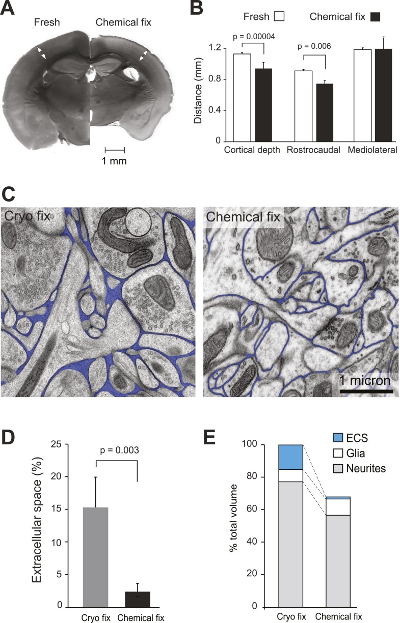
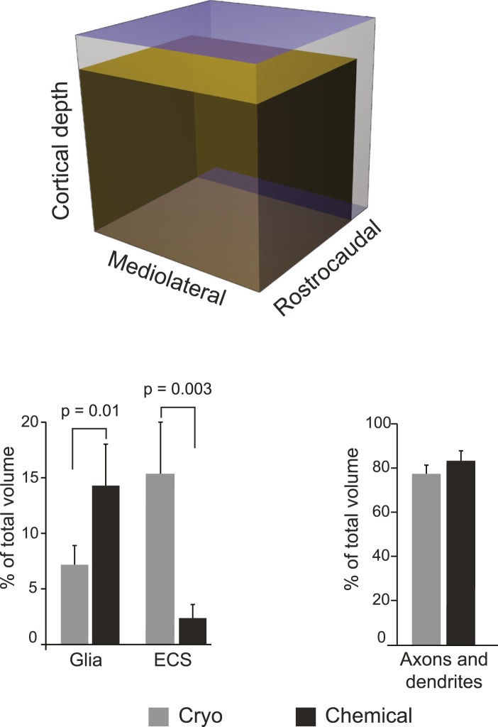
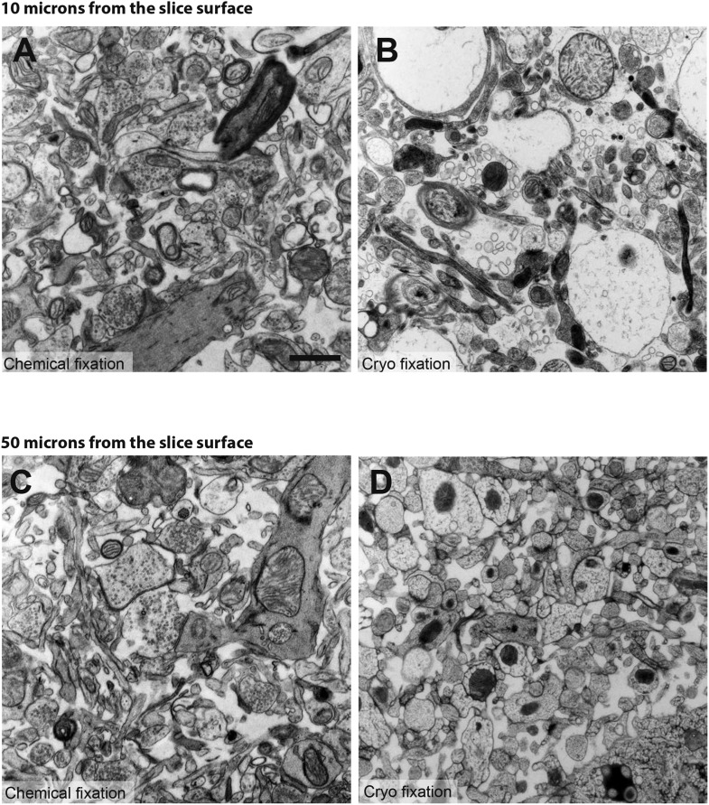
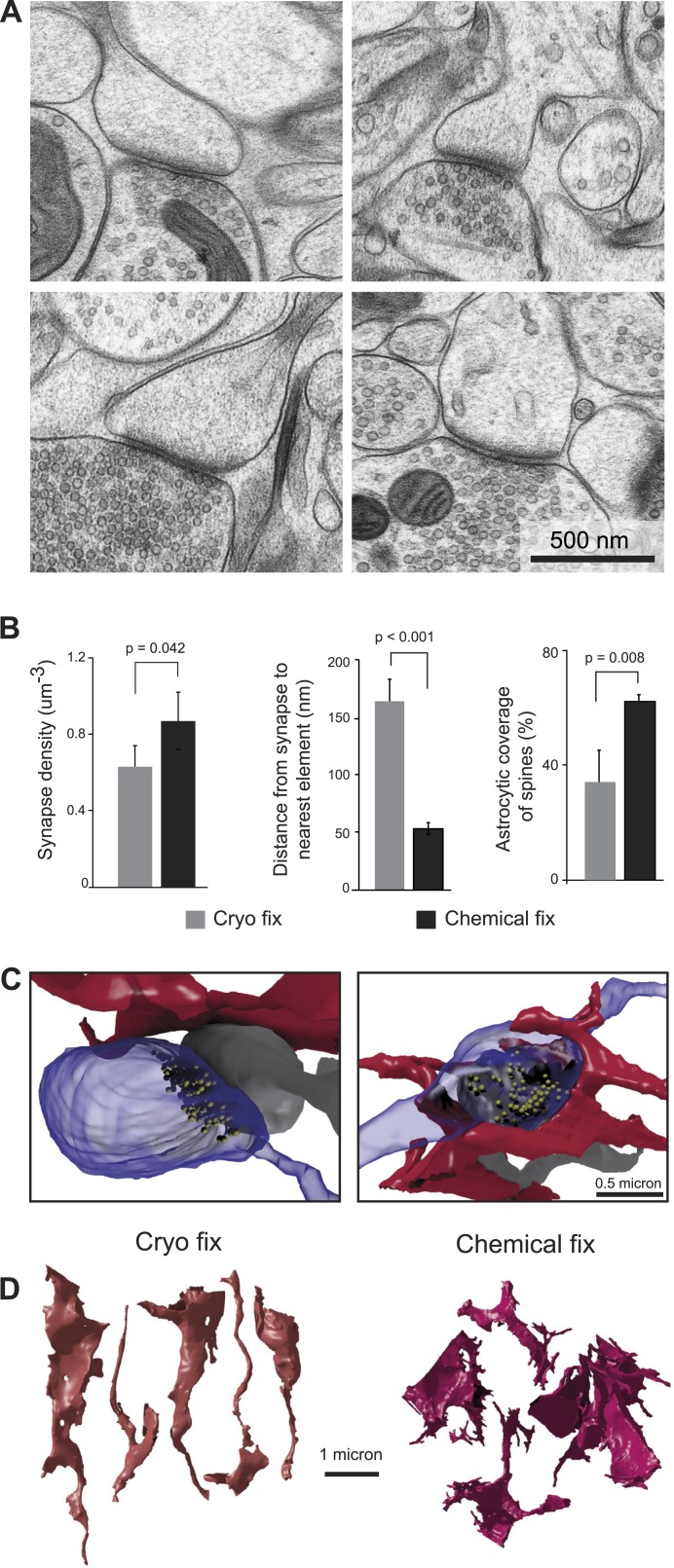
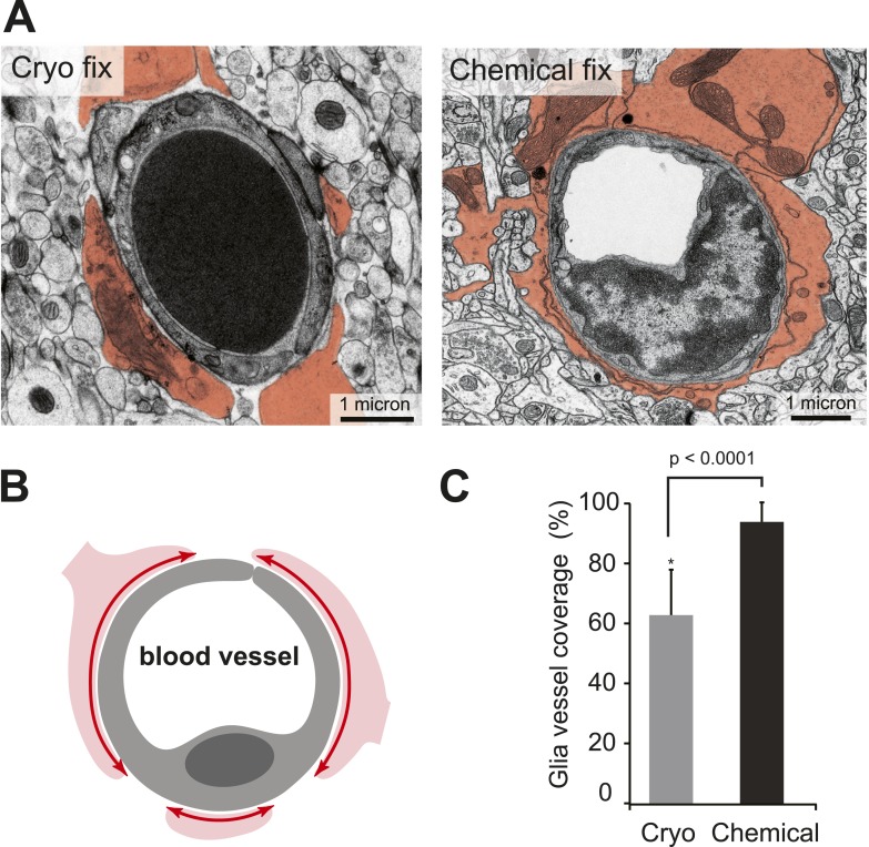
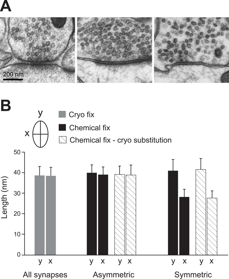
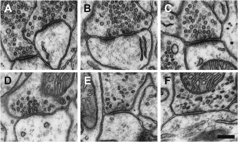
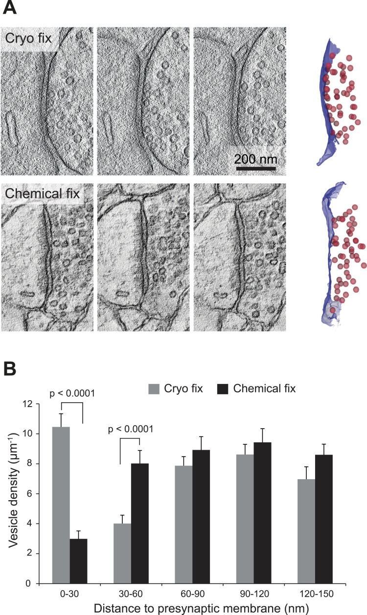
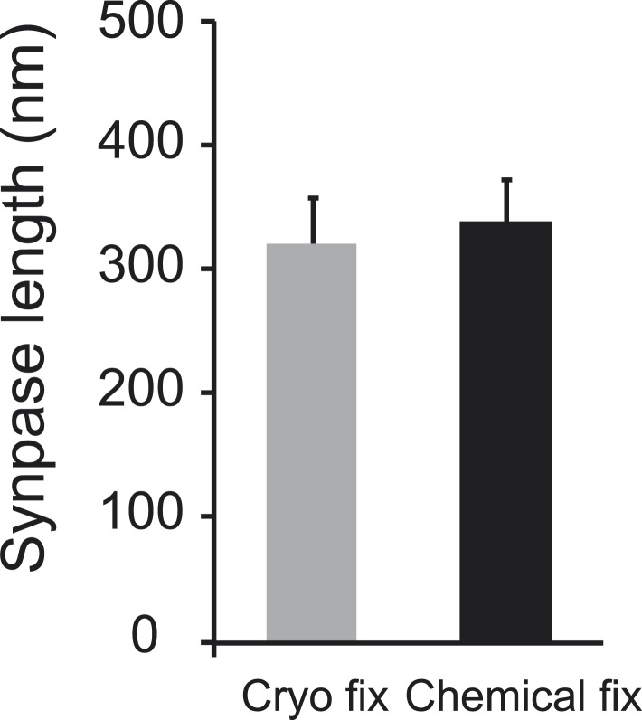
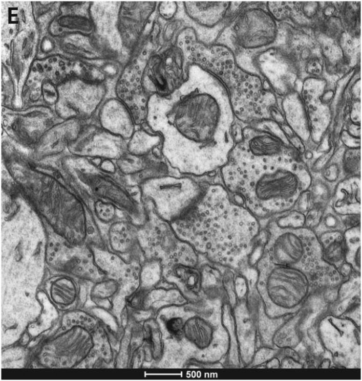
Analysis of brain ultrastructure using electron microscopy typically relies on chemical fixation. However, this is known to cause significant tissue distortion including a reduction in the extracellular space. Cryo fixation is thought to give a truer representation of biological structures, and here we use rapid, high-pressure freezing on adult mouse neocortex to quantify the extent to which these two fixation methods differ in terms of their preservation of the different cellular compartments, and the arrangement of membranes at the synapse and around blood vessels. As well as preserving a physiological extracellular space, cryo fixation reveals larger numbers of docked synaptic vesicles, a smaller glial volume, and a less intimate glial coverage of synapses and blood vessels compared to chemical fixation. The ultrastructure of mouse neocortex therefore differs significantly comparing cryo and chemical fixation conditions.
Overview
The claims and statistics of this paper haven’t been checked yet.
Run the check — free, no account needed. It reads the open-access text and checks the sources it cites.
Integrity signals
Registry
Cites
Statistics
Figures
Verification
Claims the paper makes
The claims and statistics of this paper haven’t been checked yet.
Run the check — free, no account needed. It reads the open-access text and checks the sources it cites.
Claims other papers make about this one
Each is checked against this paper’s own text: does it say what the citing paper says it does?
In the citing paper
“however, recent data with high-pressure cryopreservation show much less astrocytic coverage of both capillaries and synapses than seen with aldehyde fixation, which shrinks the extracellular space (Korogod et al., 2015).”
From Fueling thought: Management of glycolysis and oxidative phosphorylation in neuronal metabolism (2018) · cites this paper as Korogod et al., 2015
Self-check
Percentages, sums and reported differences re-computed from the paper’s own numbers.
The self-check needs the paper’s full text.
Citations
Cites
Calamita, E., Schmid, M., Kunz, M., Ndebele-Murisa, M. R., Magadza, C. H. D., Nyambe, I., & Wehrli, B. (2019) · Figshare · doi:10.26181/6153922fe919c · Cited by 4,239
Glial and neuronal control of brain blood flow
Attwell, D., Buchan, A. M., Charpak, S., Lauritzen, M., MacVicar, B. A., & Newman, E. A. (2010) · Nature · doi:10.1038/nature09613 · Cited by 2,511
Long-term in vivo imaging of experience-dependent synaptic plasticity in adult cortex
Trachtenberg, J. T., Chen, B. E., Knott, G. W., Feng, G., Sanes, J. R., Welker, E., & Svoboda, K. (2002) · Nature · doi:10.1038/nature01273 · Cited by 1,927
Self-citationAxo-somatic and axo-dendritic synapses of the cerebral cortex: an electron microscope study.
Eg, G. (1959) · PubMed · Cited by 1,768
Diffusion in Brain Extracellular Space
Syková, E., & Nicholson, C. (2008) · Physiological Reviews · doi:10.1152/physrev.00027.2007 · Cited by 1,481
Harris, K., & Stevens, J. (1989) · The Journal of Neuroscience · doi:10.1523/jneurosci.09-08-02982.1989 · Cited by 1,073
TrakEM2 Software for Neural Circuit Reconstruction
Cardona, A., Saalfeld, S., Schindelin, J., Arganda‐Carreras, I., Preibisch, S., Longair, M., Tomančák, P., Hartenstein, V., & Douglas, R. J. (2012) · PLoS ONE · doi:10.1371/journal.pone.0038011 · Cited by 1,009
Connectomic reconstruction of the inner plexiform layer in the mouse retina
Helmstaedter, M., Briggman, K. L., Turaga, S. C., Jain, V., Seung, H. S., & Denk, W. (2013) · Nature · doi:10.1038/nature12346 · Cited by 997
Colonnier, M. (1968) · Brain Research · doi:10.1016/0006-8993(68)90234-5 · Cited by 946
Characteristics of Excitatory and Inhibitory Synapses in the Central Nervous System of the Cat
UCHIZONO, K. (1965) · Nature · doi:10.1038/207642a0 · Cited by 917
Cited by
Sorted by citations
Items 201–300 of 408
Imaging of Virus-Infected Cells with Soft X-ray Tomography
Garriga, D., Chichón, F. J., Calisto, B. M., Ferrero, D. S., Gastaminza, P., Pereiro, E., & Perez-Berna, A. J. (2021) · Viruses · doi:10.3390/v13112109 · Cited by 16
Nondestructive X-ray tomography of brain tissue ultrastructure
Bosch, C., Aidukas, T., Holler, M., Pacureanu, A., Müller, E. A., Peddie, C. J., Zhang, Y., Cook, P., Collinson, L., Bunk, O., et al. (2025) · Nature Methods · doi:10.1038/s41592-025-02891-0 · Cited by 15
Structural Heterogeneity of the GABAergic Tripartite Synapse
Brunskine, C., Passlick, S., & Henneberger, C. (2022) · Cells · doi:10.3390/cells11193150 · Cited by 15
Gan, Y., Thomas, J. H., & Kelley, D. H. (2024) · Journal of The Royal Society Interface · doi:10.1098/rsif.2023.0659 · Cited by 14
Ultrastructural analysis of wild-type and RIM1α knockout active zones in a large cortical synapse
Lichter, K., Paul, M. M., Pauli, M., Schoch, S., Kollmannsberger, P., Stigloher, C., Heckmann, M., & Sirén, A.-L. (2022) · Cell Reports · doi:10.1016/j.celrep.2022.111382 · Cited by 15
Spontaneous Ca2+ Fluctuations Arise in Thin Astrocytic Processes With Real 3D Geometry
Héja, L., Szabó, Z. M., Péter, M., & Kardos, J. (2021) · Frontiers in Cellular Neuroscience · doi:10.3389/fncel.2021.617989 · Cited by 15
Schilling, K. G., Grussu, F., Ianuș, A., Hansen, B., Howard, A. F. D., Barrett, R. L. C., Aggarwal, M., Michielse, S., Nasrallah, F. A., Syeda, W. T., et al. (2025) · Magnetic Resonance in Medicine · doi:10.1002/mrm.30435 · Cited by 29
Han, Z., & Porter, A. E. (2020) · Frontiers in Nanotechnology · doi:10.3389/fnano.2020.606253 · Cited by 14
Lessons on brain edema in HE: from cellular to animal models and clinical studies
Pierzchała, K., Hadjihambi, A., Mosso, J., Jalan, R., Rose, C. F., & Cudalbu, C. (2023) · Metabolic Brain Disease · doi:10.1007/s11011-023-01269-5 · Cited by 16
Insights into protein structure using cryogenic light microscopy
Mazal, H., Wieser, F.-F., & Sandoghdar, V. (2023) · Biochemical Society Transactions · doi:10.1042/bst20221246 · Cited by 14
Sparse force-bearing bridges between neighboring synaptic vesicles
Wesseling, J. F., Phan, S., Bushong, E. A., Siksou, L., Marty, S., Pérez‐Otaño, I., & Ellisman, M. H. (2019) · Brain Structure and Function · doi:10.1007/s00429-019-01966-x · Cited by 16
Hempel, C., Kapishnikov, S., Perez-Berna, A. J., Werner, S., Guttmann, P., Pereiro, E., Qvortrup, K., & Andresen, T. L. (2020) · Microcirculation · doi:10.1111/micc.12643 · Cited by 14
Ghali, M. G. Z., Marchenko, V., Yasargil, M. G., & Ghali, G. Z. (2020) · Neurobiology of Disease · doi:10.1016/j.nbd.2020.105022 · Cited by 15
The secret world in the gaps between brain cells
Nicholson, C. (2022) · Physics Today · doi:10.1063/pt.3.4999 · Cited by 14
Reconstruction of ovine axonal cytoarchitecture enables more accurate models of brain biomechanics
Bernardini, A., Trovatelli, M., Kłosowski, M. M., Pederzani, M., Zani, D. D., Brizzola, S., Porter, A. E., y Baena, F. R., & Dini, D. (2022) · Communications Biology · doi:10.1038/s42003-022-04052-x · Cited by 14
Recent advancement in the challenges to connectomics
Ohno, N., Katoh, M., Saitoh, Y., & Saitoh, S. (2015) · Microscopy · doi:10.1093/jmicro/dfv371 · Cited by 15
Imig, C., & Cooper, B. H. (2016) · Methods in molecular biology · doi:10.1007/978-1-4939-6688-2_15 · Cited by 13
An optimized approach using cryofixation for high-resolution 3D analysis by FIB-SEM
Guo, J., Wang, G., Tang, W., Song, D., Wang, X., Hong, J., & Zhang, Z. (2020) · Journal of Structural Biology · doi:10.1016/j.jsb.2020.107600 · Cited by 14
The Impact of Chemical Fixation on the Microanatomy of Mouse Organotypic Hippocampal Slices
Idziak, A., Inavalli, V. V. G. K., Bancelin, S., Arizono, M., & Nägerl, U. V. (2023) · eNeuro · doi:10.1523/eneuro.0104-23.2023 · Cited by 13
Thomas, C. I., Ryan, M. A., McNabb, M. C., Kamasawa, N., & Scholl, B. (2024) · Glia · doi:10.1002/glia.24582 · Cited by 19
Artificial Intelligence in Diagnostic Medical Image Processing for Advanced Healthcare Applications
Kalita, A. J., Boruah, A., Das, T., Mazumder, N., Jaiswal, S. K., Zhuo, G., Gogoi, A., Kakoty, N. M., & Kao, F. (2024) · Biological and medical physics, biomedical engineering · doi:10.1007/978-981-97-5345-1_1 · Cited by 13
Probabilistic fluorescence-based synapse detection
Simhal, A. K., Aguerrebere, C., Collman, F., Vogelstein, J. T., Micheva, K. D., Weinberg, R. J., Smith, S. J., & Sapiro, G. (2017) · PLoS Computational Biology · doi:10.1371/journal.pcbi.1005493 · Cited by 15
Exploring the interstitial system in the brain: the last mile of drug delivery
Sun, Y. X., & Sun, X. (2021) · Reviews in the Neurosciences · doi:10.1515/revneuro-2020-0057 · Cited by 14
Morphology of Invertebrate Neurons and Synapses
Meinertzhagen, I. A. (2017) · Oxford University Press eBooks · doi:10.1093/oxfordhb/9780190456757.013.9 · Cited by 14
Structural Basis of Astrocytic Ca 2 Signals at Tripartite SynapsesPreprint
Arizono, M., Panatier, A., Inavalli, V. V. G. K., Pfeiffer, T., Angibaud, J., Stobart, J. L., Bellocchio, L., Marsicano, G., Mikoshiba, K., Oliet, S. H. R., et al. (2018) · SSRN Electronic Journal · doi:10.2139/ssrn.3287791 · Cited by 13
Tian, Q., Ngamsombat, C., Lee, H., Berger, D. R., Wu, Y., Fan, Q., Bilgiç, B., Li, Z., Novikov, D. S., Fieremans, E., et al. (2025) · NeuroImage · doi:10.1016/j.neuroimage.2025.121212 · Cited by 12
Identification of PSD-95 in the Postsynaptic Density Using MiniSOG and EM Tomography
Chen, X., Winters, C. A., Crocker, V. T., Lazarou, M., Sousa, A. A., Leapman, R. D., & Reese, T. S. (2018) · Frontiers in Neuroanatomy · doi:10.3389/fnana.2018.00107 · Cited by 12
Schifferer, M., Snaidero, N., Djannatian, M., Kerschensteiner, M., & Misgeld, T. (2021) · Frontiers in Neuroanatomy · doi:10.3389/fnana.2021.732506 · Cited by 9
Pipkin, J. E., Bushong, E. A., Ellisman, M. H., & Kristan, W. B. (2016) · The Journal of Comparative Neurology · doi:10.1002/cne.24120 · Cited by 11
Kjer, H. M., Andersson, M., He, Y., Pacureanu, A., Daducci, A., Pizzolato, M., Salditt, T., Robisch, A.-L., Eckermann, M., Töpperwien, M., et al. (2024) · eLife · doi:10.7554/elife.94917 · Cited by 12
Huang, X., Li, K., Liu, Y., Yang, C., & Han, H. (2023) · Sensing and Imaging · doi:10.1007/s11220-022-00408-z · Cited by 12
Huang, S.-H., Shmoel, N., Jankowski, M. M., Erez, H., Sharon, A., Abu-Salah, W., Nelken, I., Weiss, A. M., & Spira, M. E. (2020) · Frontiers in Neuroscience · doi:10.3389/fnins.2020.00926 · Cited by 10
Sharon, A., Shmoel, N., Erez, H., Jankowski, M. M., Friedmann, Y., & Spira, M. E. (2021) · Frontiers in Neuroscience · doi:10.3389/fnins.2021.764448 · Cited by 10
Current molecular approaches to investigate pre‐synaptic dysfunction
Harper, C. B., & Smillie, K. J. (2021) · Journal of Neurochemistry · doi:10.1111/jnc.15316 · Cited by 11
Delling, J. P., Bauer, H. F., Gerlach-Arbeiter, S., Schön, M., Jacob, C., Wagner, J., Pedro, M. T., Knöll, B., & Boeckers, T. M. (2024) · Molecular Psychiatry · doi:10.1038/s41380-024-02559-9 · Cited by 10
Martin‐Solana, E., Casado-Zueras, L., Torres, T. E., Goya, G. F., Fernández-Fernández, M. R., & Fernández, J. (2024) · Acta Neuropathologica Communications · doi:10.1186/s40478-024-01802-2 · Cited by 10
A brain-wide solute transport model of the glymphatic system
Quirk, K., Stevens, K. A., Tithof, J., & Kelley, D. H. (2024) · Journal of The Royal Society Interface · doi:10.1098/rsif.2024.0369 · Cited by 10
Advection and diffusion in perivascular and extracellular spaces in the brain
Guo, Y., Quirk, K., Kelley, D. H., & Thomas, J. H. (2025) · Journal of The Royal Society Interface · doi:10.1098/rsif.2025.0010 · Cited by 12
Axonal structure-function relationships across experimental modalities
Skoven, C. S., Andersson, M., Torre, M. L. B., Pizzolato, M., Siebner, H. R., & Dyrby, T. B. (2025) · Imaging Neuroscience · doi:10.1162/imag.a.1058 · Cited by 10
Handling Difficult Cryo-ET Samples: A Study with Primary Neurons from Drosophila melanogaster
Kim, J. Y., Yang, J. E., Mitchell, J. W., English, L. A., Yang, S. Z., Tenpas, T., Dent, E. W., Wildonger, J., & Wright, E. (2023) · Microscopy and Microanalysis · doi:10.1093/micmic/ozad125 · Cited by 10
Salo, R. A., Belevich, I. N., Jokitalo, E., Gröhn, O. H. J., & Sierra, A. (2020) · NeuroImage · doi:10.1016/j.neuroimage.2020.117529 · Cited by 13
Ozsvár, A., Komlósi, G., Oláh, G., Baka, J., Molnár, G., & Tamás, G. (2021) · eLife · doi:10.7554/elife.65634 · Cited by 8
Bridging length scales from molecules to the whole organism by cryoCLEM and cryoET
Lovatt, C. A., Leistner, C., & Frank, R. A. W. (2022) · Faraday Discussions · doi:10.1039/d2fd00081d · Cited by 12
Cryo‐Correlative Light and Electron Microscopy
Bharat, T. A. M., & Kukulski, W. (2019) · doi:10.1002/9781119086420.ch8 · Cited by 10
A Scalable Staining Strategy for Whole-Brain ConnectomicsPreprint
Lu, X., Wu, Y., Schalek, R. L., Meirovitch, Y., Berger, D. R., & Lichtman, J. W. (2023) · bioRxiv (Cold Spring Harbor Laboratory) · doi:10.1101/2023.09.26.558265 · Cited by 10
Three-dimensional ultrastructure analysis of organelles in injured motor neuron
Tamada, H. (2023) · Anatomical Science International · doi:10.1007/s12565-023-00720-y · Cited by 10
Regulated exocytosis from astrocytes: a matter of vesicles?
Calì, C. (2024) · Frontiers in Neuroscience · doi:10.3389/fnins.2024.1393165 · Cited by 9
Ultrastructure of astrocytes using volume electron microscopy: A scoping review
Chiappini, V., Castillo, M. F. V., Biancardi, F., Cunto, F. D., Magistretti, P. J., Vercelli, A., Agus, M., & Calì, C. (2025) · The Journal of Physiology · doi:10.1113/jp287455 · Cited by 9
Functional recovery of the adult murine hippocampus after cryopreservation by vitrification
German, A., Akdaş, E. Y., Flügel‐Koch, C., Erterek, E., Frischknecht, R., Fejtová, A., Winkler, J., Alzheimer, C., & Zheng, F. (2026) · Proceedings of the National Academy of Sciences · doi:10.1073/pnas.2516848123 · Cited by 10
Cellular and signalling mechanisms that regulate the blood–brain barrier
Amick, J., Gordon, L., & Gu, C. (2026) · Nature Reviews Molecular Cell Biology · doi:10.1038/s41580-026-01000-z · Cited by 12
Induced Remodelling of Astrocytes In Vitro and In Vivo by Manipulation of Astrocytic RhoA Activity
Domingos, C., Müller, F. E., Passlick, S., Wachten, D., Ponimaskin, E. G., Schwarz, M. K., Schoch, S., Zeug, A., & Henneberger, C. (2023) · Cells · doi:10.3390/cells12020331 · Cited by 10
Understanding Neuropeptide Transmission in the Brain by Optical Uncaging and Release
Xiong, H., Wilson, B. A., Slesinger, P. A., & Qin, Z. (2023) · ACS Chemical Neuroscience · doi:10.1021/acschemneuro.2c00684 · Cited by 10
Sheet and void porous media models for brain interstitial space
Nicholson, C. (2023) · Journal of The Royal Society Interface · doi:10.1098/rsif.2023.0223 · Cited by 9
Receptor binding and tortuosity explain morphogen local-to-global diffusion coefficient transition
Zhu, S., Loo, Y. T., Veerapathiran, S., Loo, T. Y. J., Trần, B. N., Teh, C., Zhong, J., Matsudaira, P. T., Saunders, T. E., & Wohland, T. (2024) · Biophysical Journal · doi:10.1016/j.bpj.2024.07.024 · Cited by 9
Hoang, T. V., Kizilyaprak, C., Spehner, D., Humbel, B. M., & Schultz, P. (2016) · Journal of Structural Biology · doi:10.1016/j.jsb.2016.10.005 · Cited by 10
To, X. V., Benetatos, J., Soni, N., Liu, D., Abraha, H. M., Yan, W., Panagiotopoulou, O., & Nasrallah, F. A. (2020) · Journal of Neurotrauma · doi:10.1089/neu.2019.6944 · Cited by 9
Shaaban, A., Maaß, F., Schwarze, V., Lund, M. L., Beuermann, S., Chan, M., Harenberg, C., Bewick, G. A., Keating, D. J., Benseler, F., et al. (2021) · bioRxiv (Cold Spring Harbor Laboratory) · doi:10.1101/2021.05.28.446100 · Cited by 8
Yong, Y., Cai, Y., Lin, J., Ma, L., Han, H., & Li, F. (2024) · Med-X · doi:10.1007/s44258-024-00021-7 · Cited by 10
Live STED imaging of functional neuroanatomy
Arizono, M., Idziak, A., & Nägerl, U. V. (2025) · Nature Protocols · doi:10.1038/s41596-024-01132-6 · Cited by 8
Lapios, P., Anger, R., Paget-Blanc, V., Marza, E., Lučić, V., Fronzes, R., Herzog, É., & Perrais, D. (2025) · Nature Communications · doi:10.1038/s41467-025-66355-x · Cited by 8
Denizot, A., Castillo, M. F. V., Puchenkov, P., Calì, C., & Schutter, E. D. (2025) · Glia · doi:10.1002/glia.70091 · Cited by 8
Turk, A. Z., & SheikhBahaei, S. (2021) · The Journal of Comparative Neurology · doi:10.1002/cne.25230 · Cited by 7
Plasticity of perisynaptic astroglia during ischemia-induced spreading depolarization
Fomitcheva, I. V., Sword, J., Shi, Y., & Kirov, S. A. (2022) · Cerebral Cortex · doi:10.1093/cercor/bhac434 · Cited by 8
Quantitation of the physicochemical properties of myelin using Nile Red fluorescence spectroscopy
Teo, W., Morgan, M. L., & Stys, P. K. (2024) · Journal of Neurochemistry · doi:10.1111/jnc.16203 · Cited by 7
Epileptic activity triggers rapid ROCK1‐dependent astrocyte morphology changes
Anders, S., Breithausen, B., Unichenko, P., Herde, M. K., Minge, D., Abramian, A., Behringer, C., Deshpande, T., Boehlen, A., Domingos, C., et al. (2023) · Glia · doi:10.1002/glia.24495 · Cited by 8
Fluorescence microscopy shadow imaging for neuroscience
Inavalli, V. V. G. K., Puente‐Muñoz, V., Draffin, J. E., & Tønnesen, J. (2024) · Frontiers in Cellular Neuroscience · doi:10.3389/fncel.2024.1330100 · Cited by 7
Dehghani, H., Holzapfel, G. A., Mittelbronn, M. G. A., & Zilian, A. (2024) · Journal of the mechanical behavior of biomedical materials/Journal of mechanical behavior of biomedical materials · doi:10.1016/j.jmbbm.2024.106486 · Cited by 7
Intracerebral Injections and Ultrastructural Analysis of High‐Pressure Frozen Brain Tissue
Weil, M., Ruhwedel, T., Möbius, W., & Simons, M. (2017) · Current Protocols in Neuroscience · doi:10.1002/cpns.22 · Cited by 7
Xu, X., Ge, X., Xiong, H., & Qin, Z. (2022) · Neurophotonics · doi:10.1117/1.nph.9.3.032210 · Cited by 7
New Insights in the Complexity and Functionality of the Neurovascular Unit
Schaffenrath, J., & Keller, A. (2020) · Handbook of experimental pharmacology · doi:10.1007/164_2020_424 · Cited by 8
Cortical Presynaptic Boutons Progressively Engulf Spinules as They Mature
Campbell, C., Lindhartsen, S., Knyaz, A., Erişir, A., & Nahmani, M. (2020) · eNeuro · doi:10.1523/eneuro.0426-19.2020 · Cited by 7
Whitening of odor representations by the wiring diagram of the olfactory bulbPreprint
Wanner, A., & Friedrich, R. W. (2019) · bioRxiv (Cold Spring Harbor Laboratory) · doi:10.1101/515411 · Cited by 7
The retina’s neurovascular unit: Müller glial sheaths and neuronal contactsPreprint
Grimes, W. N., Berson, D. M., Sabnis, A., Hoon, M., Sinha, R., Tian, H., & Diamond, J. S. (2024) · bioRxiv (Cold Spring Harbor Laboratory) · doi:10.1101/2024.04.30.591885 · Cited by 7
Santini, T., Shim, A., Liou, J., Rahman, N., Varela-Mattatall, G. E., Budde, M. D., Inoue, W., Everling, S., & Baron, C. A. (2024) · Magnetic Resonance in Medicine · doi:10.1002/mrm.30298 · Cited by 6
Nam, A. J., Kuwajima, M., Parker, P. H., Bowden, J. B., Abraham, W. C., & Harris, K. M. (2025) · Journal of Neuroscience · doi:10.1523/jneurosci.0943-25.2025 · Cited by 6
Alcaide, D., Cacheux, J., Bancaud, A., Muramatsu, R., & Matsunaga, Y. T. (2023) · Biomaterials Science · doi:10.1039/d3bm00027c · Cited by 7
Knopper, R. W., Skoven, C. S., Eskildsen, S. F., Østergaard, L., & Hansen, B. (2024) · Frontiers in Cellular Neuroscience · doi:10.3389/fncel.2024.1498133 · Cited by 7
Super-resolution imaging to reveal the nanostructure of tripartite synapses
Aleksejenko, N., & Heller, J. P. (2021) · Neuronal Signaling · doi:10.1042/ns20210003 · Cited by 7
Kjer, H. M., Andersson, M., He, Y., Pacureanu, A., Daducci, A., Pizzolato, M., Salditt, T., Robisch, A.-L., Eckermann, M., Töpperwien, M., et al. (2025) · eLife · doi:10.7554/elife.94917.3 · Cited by 6
Eid, L., & Parent, M. (2017) · Journal of Visualized Experiments · doi:10.3791/55397 · Cited by 6
FIB‐SEM imaging properties of Drosophila melanogaster tissues embedded in Lowicryl HM20
Porrati, F., Grewe, D., Seybert, A., Frangakis, A. S., & Eltsov, M. (2018) · Journal of Microscopy · doi:10.1111/jmi.12764 · Cited by 7
Considerations for Imaging and Analyzing Neural Structures by STED Microscopy
Lenz, M. O., & Tønnesen, J. (2019) · Methods in molecular biology · doi:10.1007/978-1-4939-9077-1_3 · Cited by 6
Membrane mechanics dictate axonal morphology and functionPreprint
Griswold, J. M., Bonilla-Quintana, M., Pepper, R., Lee, C. T., Raychaudhuri, S., Ma, S., Gan, Q., Syed, S., Zhu, C., Bell, M. K., et al. (2023) · bioRxiv (Cold Spring Harbor Laboratory) · doi:10.1101/2023.07.20.549958 · Cited by 6
Thomas, C. I., Ryan, M. A., McNabb, M. C., Kamasawa, N., & Scholl, B. (2023) · bioRxiv (Cold Spring Harbor Laboratory) · doi:10.1101/2023.12.01.569664 · Cited by 6
A cross‐comparative analysis of in vivo versus ex vivo MRI indices in a mouse model of concussion
Vinh To, X., Kurniawan, N. D., Cumming, P., & Nasrallah, F. A. (2023) · Brain Research · doi:10.1016/j.brainres.2023.148562 · Cited by 5
St‐Pierre, M., Šimončičová, E., Carrier, M., & Tremblay, M. (2022) · Methods in molecular biology · doi:10.1007/978-1-0716-2655-9_4 · Cited by 5
Fluid flow and amyloid transport and aggregation in the brain interstitial space
Iorio, A., Melchionna, S., Derreumaux, P., & Sterpone, F. (2024) · PNAS Nexus · doi:10.1093/pnasnexus/pgae548 · Cited by 6
Esposito, M., Schieber, N. L., Olivo, A., Schwab, Y., & Endrizzi, M. (2025) · Physical Review Research · doi:10.1103/physrevresearch.7.013037 · Cited by 5
Immunoelectron microscopy: a comprehensive guide from sample preparation to high-resolution imaging
Wu, J., Su, B., Gu, L., Zhang, J., Shi, Q., & Hu, D. (2025) · Discover Nano · doi:10.1186/s11671-025-04346-z · Cited by 5
Scaled Complexity of Mammalian Astrocytes: Insights From Mouse and Macaque
Heffernan, K. S., Martinez, I., Jaeger, D., Khakh, B. S., Smith, Y., & Galván, A. (2024) · The Journal of Comparative Neurology · doi:10.1002/cne.25665 · Cited by 5
Milne, G., & Walker, L. A. (2022) · Methods in molecular biology · doi:10.1007/978-1-0716-2417-3_15 · Cited by 5
Brain lymphatic drainage system – visualization opportunities and current state of the art
Yankova, G., & Bogomyakova, O. B. (2020) · Complex Issues of Cardiovascular Diseases · doi:10.17802/2306-1278-2020-9-3-81-89 · Cited by 5
3D Synaptic Organization of the Rat CA1 and Alterations Induced by Cocaine Self-Administration
Blázquez‐Llorca, L., Miguéns, M., Montero‐Crespo, M., Selvas, A., González‐Soriano, J., Ambrosio, E., & DeFelipe, J. (2020) · Cerebral Cortex · doi:10.1093/cercor/bhaa331 · Cited by 5
Digitalizing neuronal synapses with cryo-electron tomography and correlative microscopy
Liu, Y.-T., & Tao, C.-L. (2022) · Current Opinion in Neurobiology · doi:10.1016/j.conb.2022.102595 · Cited by 7
Astroglial biophysics probed with a realistic cell modelPreprint
Savtchenko, L. P., Bard, L., Jensen, T. P., Reynolds, J. R., Kraev, I., Medvedev, M., Stewart, M. G., Henneberger, C., & Rusakov, D. A. (2018) · bioRxiv (Cold Spring Harbor Laboratory) · doi:10.1101/336974 · Cited by 5
Cryogenic light microscopy of vitrified samples with angstrom precision
Mazal, H., Wieser, F.-F., Bollschweiler, D., & Sandoghdar, V. (2025) · Proceedings of the National Academy of Sciences · doi:10.1073/pnas.2513583122 · Cited by 4
Liu, S., Pokrovskaya, I. D., & Storrie, B. (2022) · Methods in molecular biology · doi:10.1007/978-1-0716-2639-9_13 · Cited by 4
Lorin, C., Guiet, R., Chiaruttini, N., Ambrosini, G., Boci, E., Abdellah, M., Markram, H., & Keller, D. X. (2024) · Glia · doi:10.1002/glia.24594 · Cited by 4
Khaled, H., Ghasemi, Z., Inagaki, M., Patel, K., Naito, Y., Feller, B., Yi, N., Bourojeni, F. B., Lee, A. K., Chofflet, N., et al. (2024) · The EMBO Journal · doi:10.1038/s44318-024-00252-9 · Cited by 4
Metadata
- Authors
- Korogod, N., Petersen, C. C., & Knott, G. W.
- Affiliations
- École Polytechnique Fédérale de Lausanne (CH)
- Corresponding author
- Petersen, Carl CH
- Knott, Graham W
- Venue
- eLife
- Publisher
- eLife Sciences Publications, Ltd
- Published
- 2015-08-11
- Volume
- 4
- ISSN
- 2050-084X (ISSN-L)
- PMC
- PMC4530226
- OpenAlex
- W1926373965
- Cited by
- 408
- References
- 63
- Citations by year
- 2026: 22 · 2025: 38 · 2024: 55 · 2023: 54 · 2022: 38 · 2021: 48 · 2020: 41 · 2019: 32 · 2018: 28 · 2017: 27 · 2016: 20 · 2015: 5
- FWCI
- 12.38
- Type
- journal article
- Language
- en
- Open access
- gold
- doi.org
- Licence
- Locations
- eLife (publishedVersion, cc-by)
- PubMed Central (publishedVersion, cc-by)
- PubMed (publishedVersion)
- DOAJ (DOAJ: Directory of Open Access Journals) (submittedVersion, cc-by-sa)
- Infoscience (Ecole Polytechnique Fédérale de Lausanne) (submittedVersion)
- Infoscience (Ecole Polytechnique Fédérale de Lausanne) (submittedVersion, cc-by-nc-nd)
- Funders
- Schweizerische Nationalfonds zur Förderung der Wissenschaftlichen Forschung
- Human Frontier Science Program (HFSP)
- European Research Council (ERC)
- Topics
- Life Sciences › Neuroscience › Cellular and Molecular Neuroscience › Neuroscience and Neuropharmacology Research
- Life Sciences › Biochemistry, Genetics and Molecular Biology › Structural Biology › Advanced Electron Microscopy Techniques and Applications
- Health Sciences › Medicine › Anatomy › Classical Anatomy and Histology
- Keywords
- neocortex, cryofixation, chemical fixation, high-pressure freezing, extracellular space, synaptic vesicles, synapses, blood vessels, tissue deformation
- MeSH terms
- Aldehydes, Animals, Fixatives, Freezing, Microscopy, Electron, Perfusion, Tissue Fixation, Neocortex, Mice
About these checks
Registry, retraction, Cites and statistics results are computed, not judged, with one exception: a printed reference that no index lists is read once by a model before it counts as a citation error. Claims are extracted by a model and kept only when their quote appears verbatim in the text; each cited source is then checked, with a model’s help, for whether it says what the claim relies on. Those verdicts can be wrong — read the claim’s quote and the cited source before relying on one.
Statistics and arithmetic are recomputed, not judged.
Confidence bands are shown only for fields calibrated on our annotated test set. None is calibrated yet, so no band is shown.
What counts as a claim ›How we measure confidence ›How we check this ›
Are you an author, or do you see a problem with this page? Request a review
