

The DOI matches its Crossref record; 63 of the 63 references it cites were found in a registry or an index.
Ultrastructural analysis of adult mouse neocortex comparing aldehyde perfusion with cryo fixation
Open accessAbstract
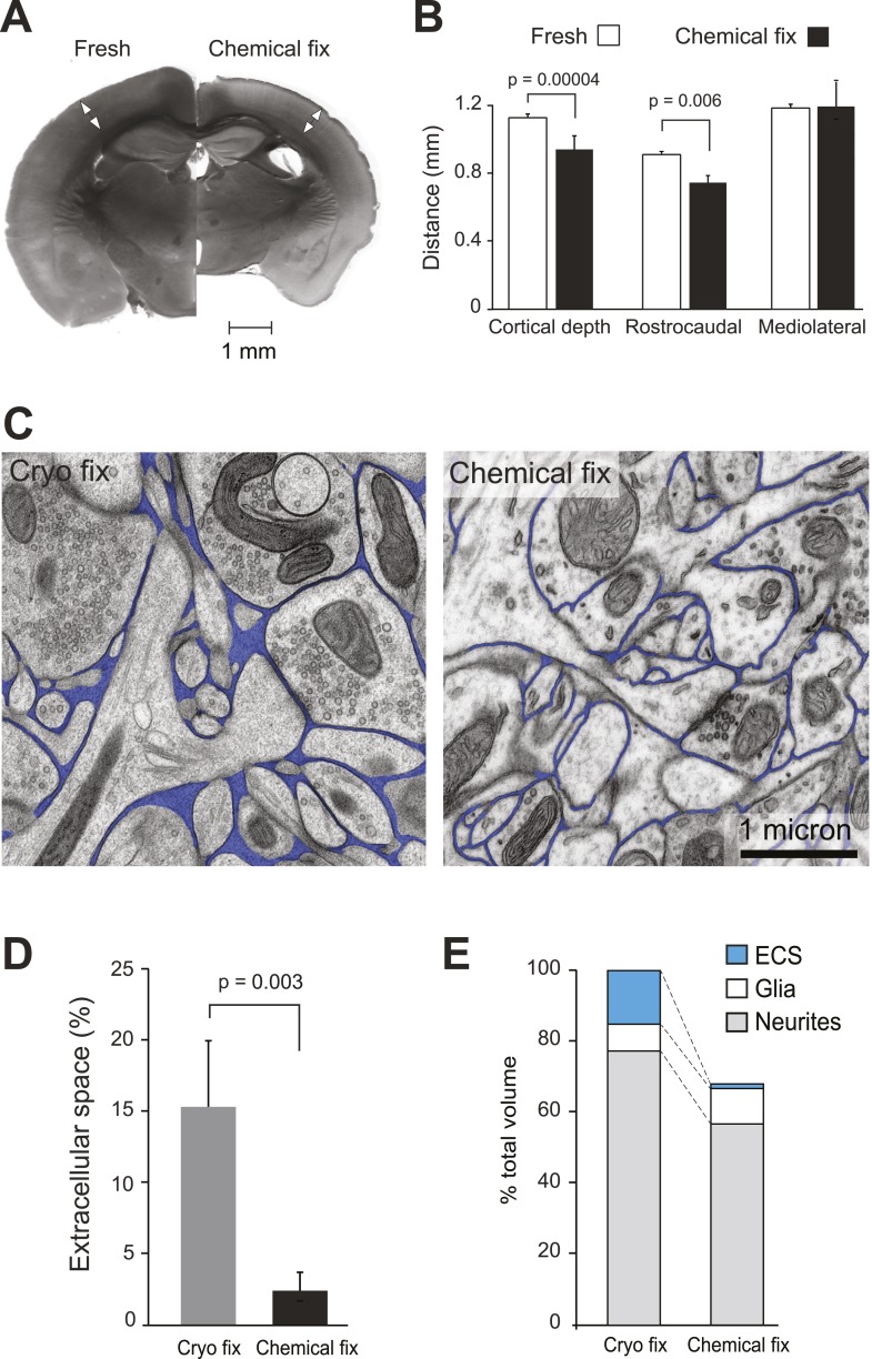
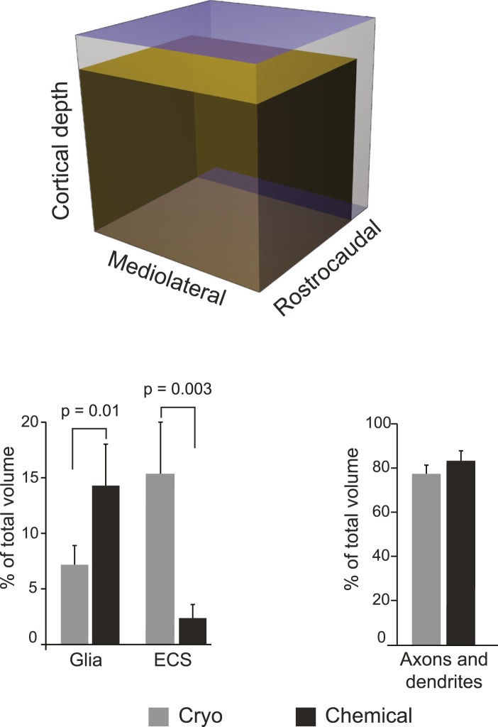
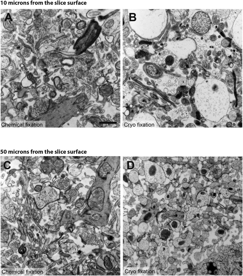
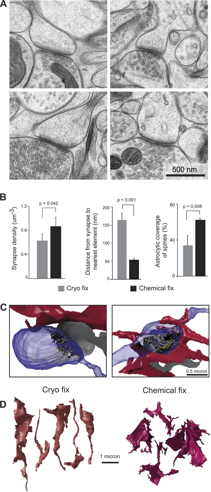
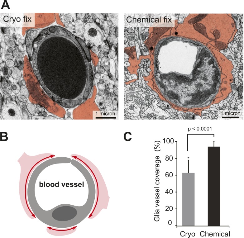
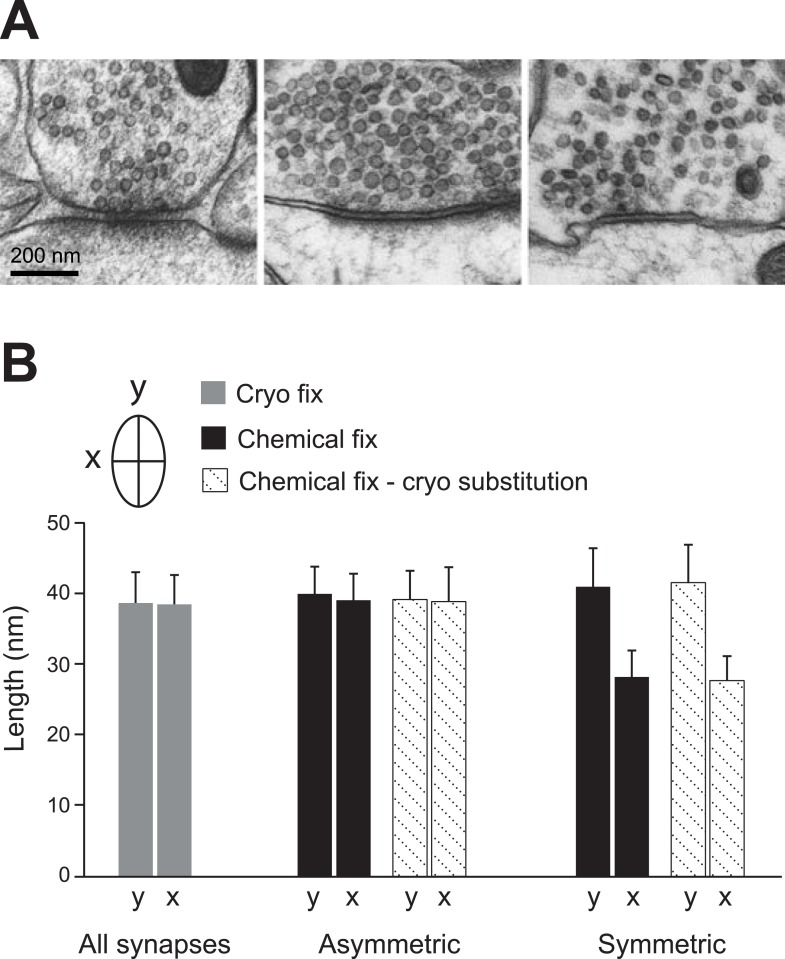
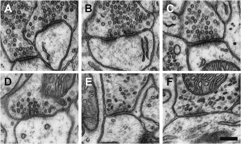
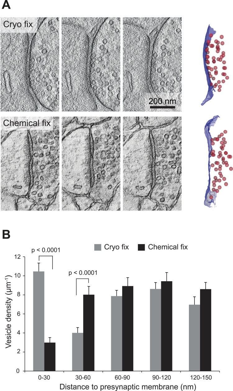
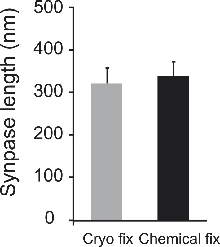
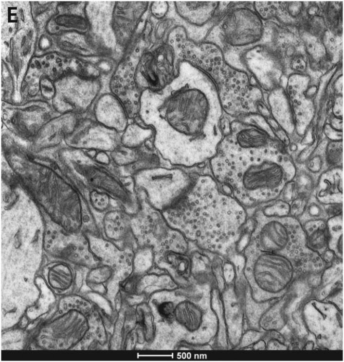
Analysis of brain ultrastructure using electron microscopy typically relies on chemical fixation. However, this is known to cause significant tissue distortion including a reduction in the extracellular space. Cryo fixation is thought to give a truer representation of biological structures, and here we use rapid, high-pressure freezing on adult mouse neocortex to quantify the extent to which these two fixation methods differ in terms of their preservation of the different cellular compartments, and the arrangement of membranes at the synapse and around blood vessels. As well as preserving a physiological extracellular space, cryo fixation reveals larger numbers of docked synaptic vesicles, a smaller glial volume, and a less intimate glial coverage of synapses and blood vessels compared to chemical fixation. The ultrastructure of mouse neocortex therefore differs significantly comparing cryo and chemical fixation conditions.
Overview
The claims and statistics of this paper haven’t been checked yet.
Run the check — free, no account needed. It reads the open-access text and checks the sources it cites.
Integrity signals
Registry
Cites
Statistics
Figures
Verification
Claims the paper makes
The claims and statistics of this paper haven’t been checked yet.
Run the check — free, no account needed. It reads the open-access text and checks the sources it cites.
Claims other papers make about this one
Each is checked against this paper’s own text: does it say what the citing paper says it does?
In the citing paper
“however, recent data with high-pressure cryopreservation show much less astrocytic coverage of both capillaries and synapses than seen with aldehyde fixation, which shrinks the extracellular space (Korogod et al., 2015).”
From Fueling thought: Management of glycolysis and oxidative phosphorylation in neuronal metabolism (2018) · cites this paper as Korogod et al., 2015
Self-check
Percentages, sums and reported differences re-computed from the paper’s own numbers.
The self-check needs the paper’s full text.
Citations
Cites
Calamita, E., Schmid, M., Kunz, M., Ndebele-Murisa, M. R., Magadza, C. H. D., Nyambe, I., & Wehrli, B. (2019) · Figshare · doi:10.26181/6153922fe919c · Cited by 4,239
Glial and neuronal control of brain blood flow
Attwell, D., Buchan, A. M., Charpak, S., Lauritzen, M., MacVicar, B. A., & Newman, E. A. (2010) · Nature · doi:10.1038/nature09613 · Cited by 2,511
Long-term in vivo imaging of experience-dependent synaptic plasticity in adult cortex
Trachtenberg, J. T., Chen, B. E., Knott, G. W., Feng, G., Sanes, J. R., Welker, E., & Svoboda, K. (2002) · Nature · doi:10.1038/nature01273 · Cited by 1,927
Self-citationAxo-somatic and axo-dendritic synapses of the cerebral cortex: an electron microscope study.
Eg, G. (1959) · PubMed · Cited by 1,768
Diffusion in Brain Extracellular Space
Syková, E., & Nicholson, C. (2008) · Physiological Reviews · doi:10.1152/physrev.00027.2007 · Cited by 1,481
Harris, K., & Stevens, J. (1989) · The Journal of Neuroscience · doi:10.1523/jneurosci.09-08-02982.1989 · Cited by 1,073
TrakEM2 Software for Neural Circuit Reconstruction
Cardona, A., Saalfeld, S., Schindelin, J., Arganda‐Carreras, I., Preibisch, S., Longair, M., Tomančák, P., Hartenstein, V., & Douglas, R. J. (2012) · PLoS ONE · doi:10.1371/journal.pone.0038011 · Cited by 1,009
Connectomic reconstruction of the inner plexiform layer in the mouse retina
Helmstaedter, M., Briggman, K. L., Turaga, S. C., Jain, V., Seung, H. S., & Denk, W. (2013) · Nature · doi:10.1038/nature12346 · Cited by 997
Colonnier, M. (1968) · Brain Research · doi:10.1016/0006-8993(68)90234-5 · Cited by 946
Characteristics of Excitatory and Inhibitory Synapses in the Central Nervous System of the Cat
UCHIZONO, K. (1965) · Nature · doi:10.1038/207642a0 · Cited by 917
Cited by
Sorted by citations
ilastik: interactive machine learning for (bio)image analysis
Berg, S., Kutra, D., Kroeger, T., Straehle, C., Kausler, B. X., Haubold, C., Schiegg, M., Aleš, J., Beier, T., Rudy, M., et al. (2019) · Nature Methods · doi:10.1038/s41592-019-0582-9 · Cited by 3,422
Verkhratsky, A., & Nedergaard, M. (2018) · Physiological Reviews · doi:10.1152/physrev.00042.2016 · Cited by 1,678
Vascular Cognitive Impairment and Dementia
Iadecola, C., Duering, M., Hachinski, V., Joutel, A., Pendlebury, S. T., Schneider, J. A., & Dichgans, M. (2019) · Journal of the American College of Cardiology · doi:10.1016/j.jacc.2019.04.034 · Cited by 865
Rasmussen, M. K., Mestre, H., & Nedergaard, M. (2021) · Physiological Reviews · doi:10.1152/physrev.00031.2020 · Cited by 642
The role of brain barriers in fluid movement in the CNS: is there a ‘glymphatic’ system?
Abbott, N. J., Pizzo, M. E., Preston, J. E., Janigro, D., & Thorne, R. G. (2018) · Acta Neuropathologica · doi:10.1007/s00401-018-1812-4 · Cited by 593
The Glymphatic System: A Novel Component of Fundamental Neurobiology
Hablitz, L. M., & Nedergaard, M. (2021) · Journal of Neuroscience · doi:10.1523/jneurosci.0619-21.2021 · Cited by 503
Perivascular spaces, glymphatic dysfunction, and small vessel disease
Mestre, H., Kostrikov, S., Mehta, R. I., & Nedergaard, M. (2017) · Clinical Science · doi:10.1042/cs20160381 · Cited by 403
Cell Densities in the Mouse Brain: A Systematic Review
Keller, D., Erö, C., & Markram, H. (2018) · Frontiers in Neuroanatomy · doi:10.3389/fnana.2018.00083 · Cited by 380
The glymphatic system: Current understanding and modeling
Bohr, T., Hjorth, P. G., Holst, S. C., Hrabětová, S., Kiviniemi, V. J., Lilius, T. O., Lundgaard, I., Mardal, K., Martens, E. A., Mori, Y., et al. (2022) · iScience · doi:10.1016/j.isci.2022.104987 · Cited by 401
Fueling thought: Management of glycolysis and oxidative phosphorylation in neuronal metabolism
Yellen, G. (2018) · Journal of Cell Biology · doi:10.1083/jcb.201803152 · Cited by 371
“For instance, astrocytes with pericapillary endfeet are ideally positioned to take up glucose from the vasculature; however, recent data with high-pressure cryopreservation show much less astrocytic coverage of both capillaries and synapses than seen with aldehyde fixation, which shrinks the extracellular space (Korog…”
BackgroundSmith, A. J., Yao, X., Dix, J. A., Jin, B.-J., & Verkman, A. S. (2017) · eLife · doi:10.7554/elife.27679 · Cited by 362
The anatomy and immunology of vasculature in the central nervous system
Mastorakos, P., & McGavern, D. (2019) · Science Immunology · doi:10.1126/sciimmunol.aav0492 · Cited by 333
eC-CLEM: flexible multidimensional registration software for correlative microscopies
Paul‐Gilloteaux, P., Heiligenstein, X., Belle, M., Domart, M., Larijani, B., Collinson, L., Raposo, G., & Salamero, J. (2017) · Nature Methods · doi:10.1038/nmeth.4170 · Cited by 327
Brain Extracellular Space: The Final Frontier of Neuroscience
Nicholson, C., & Hrabětová, S. (2017) · Biophysical Journal · doi:10.1016/j.bpj.2017.06.052 · Cited by 321
Super-Resolution Imaging of the Extracellular Space in Living Brain Tissue
Tønnesen, J., Inavalli, V. V. G. K., & Nägerl, U. V. (2018) · Cell · doi:10.1016/j.cell.2018.02.007 · Cited by 303
Improved tools to study astrocytes
Yu, X., Nagai, J., & Khakh, B. S. (2020) · Nature Reviews Neuroscience · doi:10.1038/s41583-020-0264-8 · Cited by 298
Volume scanning electron microscopy for imaging biological ultrastructure
Titze, B., & Genoud, C. (2016) · Biology of the Cell · doi:10.1111/boc.201600024 · Cited by 287
LTP Induction Boosts Glutamate Spillover by Driving Withdrawal of Perisynaptic Astroglia
Henneberger, C., Bard, L., Panatier, A., Reynolds, J. R., Kopach, O., Medvedev, N. I., Minge, D., Herde, M. K., Anders, S., Kraev, I., et al. (2020) · Neuron · doi:10.1016/j.neuron.2020.08.030 · Cited by 274
Godin, A. G., Varela, J. A., Gao, Z., Danné, N., Dupuis, J. P., Lounis, B., Groc, L., & Cognet, L. (2017) · Nature Nanotechnology · doi:10.1038/nnano.2016.248 · Cited by 253
Lactate Shuttles in Neuroenergetics—Homeostasis, Allostasis and Beyond
Mason, S. (2017) · Frontiers in Neuroscience · doi:10.3389/fnins.2017.00043 · Cited by 240
Design and Validation of Diffusion MRI Models of White Matter
Jelescu, I. O., & Budde, M. D. (2017) · Frontiers in Physics · doi:10.3389/fphy.2017.00061 · Cited by 240
Peddie, C. J., Genoud, C., Kreshuk, A., Meechan, K., Micheva, K. D., Narayan, K., Pape, C., Parton, R. G., Schieber, N. L., Schwab, Y., et al. (2022) · Nature Reviews Methods Primers · doi:10.1038/s43586-022-00131-9 · Cited by 250
Structural basis of astrocytic Ca2+ signals at tripartite synapses
Arizono, M., Inavalli, V. V. G. K., Panatier, A., Pfeiffer, T., Angibaud, J., Levet, F., ter Veer, M. J. T., Stobart, J. L., Bellocchio, L., Mikoshiba, K., et al. (2020) · Nature Communications · doi:10.1038/s41467-020-15648-4 · Cited by 221
Gao, Y., Ma, Y., Zhang, Q., Winder, A. T., Liang, Z., Antinori, L., Drew, P. J., & Zhang, N. (2016) · NeuroImage · doi:10.1016/j.neuroimage.2016.11.069 · Cited by 226
A Historical Review of Brain Drug Delivery
Pardridge, W. M. (2022) · Pharmaceutics · doi:10.3390/pharmaceutics14061283 · Cited by 219
Neuronal vs glial glutamate uptake: Resolving the conundrum
Danbolt, N. C., Furness, D. N., & Zhou, Y. (2016) · Neurochemistry International · doi:10.1016/j.neuint.2016.05.009 · Cited by 224
Tao, C.-L., Liu, Y.-T., Sun, R., Zhang, B., Qi, L., Shivakoti, S., Tian, C.-L., Zhang, P., Lau, P.-M., Zhou, Z. H., et al. (2018) · The Journal of Neuroscience · doi:10.1523/jneurosci.1548-17.2017 · Cited by 207
Hladky, S. B., & Barrand, M. A. (2018) · Fluids and Barriers of the CNS · doi:10.1186/s12987-018-0113-6 · Cited by 206
A serotonergic axon-cilium synapse drives nuclear signaling to alter chromatin accessibility
Sheu, S.-H., Upadhyayula, S., Dupuy, V., Pang, S., Deng, F., Wan, J., Walpita, D., Pasolli, H. A., Houser, J., Sanchez-Martinez, S., et al. (2022) · Cell · doi:10.1016/j.cell.2022.07.026 · Cited by 162
Jurisch‐Yaksi, N., Yaksi, E., & Kızıl, Ç. (2020) · Glia · doi:10.1002/glia.23849 · Cited by 174
Analysis of convective and diffusive transport in the brain interstitium
Ray, L., Iliff, J. J., & Heys, J. J. (2019) · Fluids and Barriers of the CNS · doi:10.1186/s12987-019-0126-9 · Cited by 167
Smith, A. J., & Verkman, A. S. (2017) · The FASEB Journal · doi:10.1096/fj.201700999 · Cited by 162
A Developmental Analysis of Juxtavascular Microglia Dynamics and Interactions with the Vasculature
Mondo, E., Becker, S. C., Kautzman, A. G., Schifferer, M., Baer, C. E., Chen, J., Huang, E. J., Simons, M., & Schafer, D. P. (2020) · Journal of Neuroscience · doi:10.1523/jneurosci.3006-19.2020 · Cited by 153
Physiology and molecular biology of barrier mechanisms in the fetal and neonatal brain
Saunders, N. R., Dzięgielewska, K. M., Møllgård, K., & Habgood, M. D. (2018) · The Journal of Physiology · doi:10.1113/jp275376 · Cited by 159
The role of astrocyte structural plasticity in regulating neural circuit function and behavior
Lawal, O., Ulloa Severino, F. P., & Eroglu, C. (2022) · Glia · doi:10.1002/glia.24191 · Cited by 142
Saunders, N. R., Habgood, M. D., Møllgård, K., & Dziegielewska, K. M. (2016) · F1000Research · doi:10.12688/f1000research.7378.1 · Cited by 143
PDGF-B Is Required for Development of the Glymphatic System
Munk, A. S. F., Wang, W., Bèchet, N. B., Eltanahy, A. M., Cheng, A. X., Sigurðsson, B., Benraiss, A., Mäe, M. A., Kress, B. T., Kelley, D. H., et al. (2019) · Cell Reports · doi:10.1016/j.celrep.2019.02.050 · Cited by 147
Aten, S., Kiyoshi, C. M., Arzola, E. P., Patterson, J., Taylor, A. T., Du, Y., Guiher, A. M., Philip, M., Camacho, E. G., Mediratta, D., et al. (2020) · Progress in Neurobiology · doi:10.1016/j.pneurobio.2022.102264 · Cited by 136
Klostranec, J. M., Vucevic, D., Bhatia, K. D., Kortman, H. G. J., Krings, T., Murphy, K., terBrugge, K. G., & Mikulis, D. J. (2021) · Radiology · doi:10.1148/radiol.2021202043 · Cited by 130
Wiring variations that enable and constrain neural computation in a sensory microcircuit
Tobin, W. F., Wilson, R. I., & Lee, W.-C. A. (2017) · eLife · doi:10.7554/elife.24838 · Cited by 123
Soria, F. N., Paviolo, C., Doudnikoff, É., Arotçarena, M.-L., Lee, A., Danné, N., Mandal, A. K., Gosset, P., Dehay, B., Groc, L., et al. (2020) · Nature Communications · doi:10.1038/s41467-020-17328-9 · Cited by 124
Post mortem mapping of connectional anatomy for the validation of diffusion MRI
Yendiki, A., Aggarwal, M., Axer, M., Howard, A. F. D., van Walsum, A. V. C., & Haber, S. N. (2022) · NeuroImage · doi:10.1016/j.neuroimage.2022.119146 · Cited by 116
Direct vascular contact is a hallmark of cerebral astrocytes
Hösli, L., Zuend, M., Bredell, G., Zanker, H. S., de Oliveira, C. E. P., Saab, A. S., & Weber, B. (2022) · Cell Reports · doi:10.1016/j.celrep.2022.110599 · Cited by 113
Towards correlative super‐resolution fluorescence and electron cryo‐microscopy
Wolff, G., Hagen, C., Grünewald, K., & Kaufmann, R. (2016) · Biology of the Cell · doi:10.1111/boc.201600008 · Cited by 122
Mitochondrial Ultrastructure Is Coupled to Synaptic Performance at Axonal Release Sites
Cserép, C., Pósfai, B., Schwarcz, A. D., & Dénes, Á. (2018) · eNeuro · doi:10.1523/eneuro.0390-17.2018 · Cited by 106
Brain Ultrastructure: Putting the Pieces Together
Nahirney, P. C., & Tremblay, M. (2021) · Frontiers in Cell and Developmental Biology · doi:10.3389/fcell.2021.629503 · Cited by 111
Aging drives cerebrovascular network remodeling and functional changes in the mouse brain
Bennett, H. C., Zhang, Q., Wu, Y.-T., Manjila, S. B., Chon, U., Shin, D., Vanselow, D. J., Pi, H.-J., Drew, P. J., & Kim, Y. (2024) · Nature Communications · doi:10.1038/s41467-024-50559-8 · Cited by 115
Battefeld, A., Klooster, J., & Kole, M. H. P. (2016) · Nature Communications · doi:10.1038/ncomms11298 · Cited by 110
McCafferty, C. L., Klumpe, S., Amaro, R. E., Kukulski, W., Collinson, L., & Engel, B. D. (2024) · Cell · doi:10.1016/j.cell.2024.01.005 · Cited by 113
Fast and precise targeting of single tumor cells in vivo by multimodal correlative microscopy
Karreman, M. A., Mercier, L., Schieber, N. L., Solecki, G. M., Allio, G., Winkler, F., Ruthensteiner, B., Goetz, J. G., & Schwab, Y. (2015) · Journal of Cell Science · doi:10.1242/jcs.181842 · Cited by 104
Schulz, J., Hartz, A. M. S., & Bauer, B. (2023) · Pharmacological Reviews · doi:10.1124/pharmrev.120.000025 · Cited by 118
The glymphatic system and meningeal lymphatics of the brain: new understanding of brain clearance
Yankova, G., Bogomyakova, O. B., & Тулупов, А. А. (2021) · Reviews in the Neurosciences · doi:10.1515/revneuro-2020-0106 · Cited by 110
Unveiling the Extracellular Space of the Brain: From Super-resolved Microstructure toIn VivoFunction
Hrabětová, S., Cognet, L., Rusakov, D. A., & Nägerl, U. V. (2018) · Journal of Neuroscience · doi:10.1523/jneurosci.1664-18.2018 · Cited by 104
Calì, C., Agus, M., Kare, K., Boges, D. J., Lehväslaiho, H., Hadwiger, M., & Magistretti, P. J. (2019) · Progress in Neurobiology · doi:10.1016/j.pneurobio.2019.101696 · Cited by 99
Astrocytes in the regulation of cerebrovascular functions
Cohen‐Salmon, M., Slaoui, L., Mazaré, N., Gilbert, A., Oudart, M., Alvear‐Perez, R., Elorza‐Vidal, X., Chever, O., & Boulay, A. (2021) · Glia · doi:10.1002/glia.23924 · Cited by 93
Игоревич, Г. Н., Golyagina, I., Brazhe, A., Scimemi, A., Турлапов, В., & Semyanov, A. V. (2018) · Frontiers in Cellular Neuroscience · doi:10.3389/fncel.2018.00248 · Cited by 91
Andersson, M., Kjer, H. M., Rafael‐Patiño, J., Pacureanu, A., Pakkenberg, B., Thiran, J., Ptito, M., Bech, M., Dahl, A. B., Dahl, V. A., et al. (2020) · Proceedings of the National Academy of Sciences · doi:10.1073/pnas.2012533117 · Cited by 104
Savage, J. C., Picard, K., Ibáñez, F. G., & Tremblay, M. (2018) · Frontiers in Immunology · doi:10.3389/fimmu.2018.00803 · Cited by 96
Zhang, D., Ruan, J., Peng, S., Li, J., Hu, X., Zhang, Y., Zhang, T., Ge, Y., Zhu, Z., Xiao, X., et al. (2024) · Nature Neuroscience · doi:10.1038/s41593-023-01515-0 · Cited by 104
Extracellular space preservation aids the connectomic analysis of neural circuits
Pallotto, M., Watkins, P. V., Fubara, B. N., Singer, J. H., & Briggman, K. L. (2015) · eLife · doi:10.7554/elife.08206 · Cited by 94
Disentangling astroglial physiology with a realistic cell model in silico
Savtchenko, L. P., Bard, L., Jensen, T. P., Reynolds, J. R., Kraev, I., Medvedev, N. I., Stewart, M. G., Henneberger, C., & Rusakov, D. A. (2018) · Nature Communications · doi:10.1038/s41467-018-05896-w · Cited by 87
A roadmap to integrate astrocytes into Systems Neuroscience
Kastanenka, K. V., Moreno‐Bote, R., Pittà, M. D., Perea, G., Eraso‐Pichot, A., Masgrau, R., Poskanzer, K. E., & Galea, E. (2019) · Glia · doi:10.1002/glia.23632 · Cited by 88
Light-microscopy-based connectomic reconstruction of mammalian brain tissue
Tavakoli, M. R., Lyudchik, J., Januszewski, M., Vistunou, V., Dueñas, N. A., Vorlaufer, J., Sommer, C., Kreuzinger, C., Oliveira, B., Cenameri, A., et al. (2025) · Nature · doi:10.1038/s41586-025-08985-1 · Cited by 91
Intravital Correlative Microscopy: Imaging Life at the Nanoscale
Karreman, M. A., Hyenne, V., Schwab, Y., & Goetz, J. G. (2016) · Trends in Cell Biology · doi:10.1016/j.tcb.2016.07.003 · Cited by 91
Whitening of odor representations by the wiring diagram of the olfactory bulb
Wanner, A. A., & Friedrich, R. W. (2020) · Nature Neuroscience · doi:10.1038/s41593-019-0576-z · Cited by 79
Computational geometry analysis of dendritic spines by structured illumination microscopy
Kashiwagi, Y., Higashi, T., Obashi, K., Sato, Y., Komiyama, N. H., Grant, S. G. N., & Okabe, S. (2019) · Nature Communications · doi:10.1038/s41467-019-09337-0 · Cited by 82
Iliff, J. J., & Simon, M. J. (2019) · The Journal of Physiology · doi:10.1113/jp277635 · Cited by 81
Neural anatomy and optical microscopy (NAOMi) simulation for evaluating calcium imaging methods
Song, A. K., Gauthier, J. L., Pillow, J. W., Tank, D. W., & Charles, A. S. (2021) · Journal of Neuroscience Methods · doi:10.1016/j.jneumeth.2021.109173 · Cited by 78
Sherpa, A. D., Xiao, F., Joseph, N., Aoki, C. J., & Hrabětová, S. (2016) · Synapse · doi:10.1002/syn.21908 · Cited by 76
Fluid Flow and Mass Transport in Brain Tissue
Ray, L., & Heys, J. J. (2019) · Fluids · doi:10.3390/fluids4040196 · Cited by 78
Local Efficacy of Glutamate Uptake Decreases with Synapse Size
Herde, M. K., Bohmbach, K., Domingos, C., Vana, N., Komorowska‐Müller, J. A., Passlick, S., Schwarz, I., Jackson, C. J., Dietrich, D., Schwarz, M. K., et al. (2020) · Cell Reports · doi:10.1016/j.celrep.2020.108182 · Cited by 71
Tsang, T. K., Bushong, E. A., Boassa, D., Hu, J., Romoli, B., Phan, S., Dulcis, D., Su, C.-Y., & Ellisman, M. H. (2018) · eLife · doi:10.7554/elife.35524 · Cited by 71
Functional hyperemia drives fluid exchange in the paravascular space
Kedarasetti, R. T., Turner, K. L., Echagarruga, C., Gluckman, B. J., Drew, P. J., & Costanzo, F. (2020) · Fluids and Barriers of the CNS · doi:10.1186/s12987-020-00214-3 · Cited by 72
A network model of glymphatic flow under different experimentally-motivated parametric scenarios
Tithof, J., Boster, K. A. S., Bork, P. A. R., Nedergaard, M., Thomas, J. H., & Kelley, D. H. (2022) · iScience · doi:10.1016/j.isci.2022.104258 · Cited by 70
Calì, C., Wawrzyniak, M., Becker, C., Maco, B., Cantoni, M., Jorstad, A., Nigro, B., Grillo, F. W., Paola, V. D., Fua, P., et al. (2018) · PLoS ONE · doi:10.1371/journal.pone.0198131 · Cited by 68
Ultrastructural Correlates of Presynaptic Functional Heterogeneity in Hippocampal Synapses
Maus, L., Lee, C., Altas, B., Sertel, S. M., Weyand, K., Rizzoli, S. O., Rhee, J., Brose, N., Imig, C., & Cooper, B. H. (2020) · Cell Reports · doi:10.1016/j.celrep.2020.02.083 · Cited by 68
Arterial vasodilation drives convective fluid flow in the brain: a poroelastic model
Kedarasetti, R. T., Drew, P. J., & Costanzo, F. (2022) · Fluids and Barriers of the CNS · doi:10.1186/s12987-022-00326-y · Cited by 71
Paviolo, C., Soria, F. N., Ferreira, J. S., Lee, A., Groc, L., Bézard, E., & Cognet, L. (2019) · Methods · doi:10.1016/j.ymeth.2019.03.005 · Cited by 67
Imig, C., López-Murcia, F. J., Maus, L., García-Plaza, I. H., Mortensen, L. S., Schwark, M., Schwarze, V., Angibaud, J., Nägerl, U. V., Taschenberger, H., et al. (2020) · Neuron · doi:10.1016/j.neuron.2020.09.004 · Cited by 66
Organizing principles of astrocytic nanoarchitecture in the mouse cerebral cortex
Salmon, C. K., Syed, T. A., Kacerovsky, J. B., Alivodej, N., Schober, A. L., Sloan, T. F. W., Pratte, M. T., Rosen, M. P., Green, M., Chirgwin-Dasgupta, A., et al. (2023) · Current Biology · doi:10.1016/j.cub.2023.01.043 · Cited by 70
Current Progress in Expansion Microscopy: Chemical Strategies and Applications
Wen, G., Leen, V., Rohand, T., Sauer, M., & Hofkens, J. (2023) · Chemical Reviews · doi:10.1021/acs.chemrev.2c00711 · Cited by 71
Local diffusion in the extracellular space of the brain
Tønnesen, J., Hrabětová, S., & Soria, F. N. (2022) · Neurobiology of Disease · doi:10.1016/j.nbd.2022.105981 · Cited by 66
Astrocytic IP3Rs: Beyond IP3R2
Sherwood, M. W., Arizono, M., Panatier, A., Mikoshiba, K., & Oliet, S. H. R. (2021) · Frontiers in Cellular Neuroscience · doi:10.3389/fncel.2021.695817 · Cited by 65
Borges-Merjane, C., Kim, O., & Jonas, P. (2020) · Neuron · doi:10.1016/j.neuron.2019.12.022 · Cited by 60
Tripathy, R., Leca, I., van Dijk, T., Weiss, M. M., van Bon, B. W. M., Sergaki, M. C., Gstrein, T., Breuss, M. W., Tian, G., Bahi‐Buisson, N., et al. (2018) · Neuron · doi:10.1016/j.neuron.2018.10.044 · Cited by 61
Dynamism of an Astrocyte In Vivo: Perspectives on Identity and Function
Poskanzer, K. E., & Molofsky, A. V. (2017) · Annual Review of Physiology · doi:10.1146/annurev-physiol-021317-121125 · Cited by 62
How does neurovascular unit dysfunction contribute to multiple sclerosis?
Cashion, J. M., Young, K. M., & Sutherland, B. A. (2023) · Neurobiology of Disease · doi:10.1016/j.nbd.2023.106028 · Cited by 58
Control of astrocyte morphology by Rho GTPases
Zeug, A., Müller, F. E., Anders, S., Herde, M. K., Minge, D., Ponimaskin, E. G., & Henneberger, C. (2017) · Brain Research Bulletin · doi:10.1016/j.brainresbull.2017.05.003 · Cited by 59
Current Techniques for Investigating the Brain Extracellular Space
Soria, F. N., Miguélez, C., Peñagarikano, O., & Tønnesen, J. (2020) · Frontiers in Neuroscience · doi:10.3389/fnins.2020.570750 · Cited by 53
Digital Reconstruction of the Neuro-Glia-Vascular Architecture
Zisis, E., Keller, D. X., Kanari, L., Arnaudon, A., Gevaert, M., Delemontex, T., Coste, B., Foni, A., Abdellah, M., Calì, C., et al. (2021) · Cerebral Cortex · doi:10.1093/cercor/bhab254 · Cited by 56
Munc13- and SNAP25-dependent molecular bridges play a key role in synaptic vesicle priming
Papantoniou, C., Laugks, U., Betzin, J., Capitanio, C., Ferrero, J. J., Sánchez‐Prieto, J., Schoch, S., Brose, N., Baumeister, W. P., Cooper, B. H., et al. (2023) · Science Advances · doi:10.1126/sciadv.adf6222 · Cited by 55
Transmission Electron Microscopy of Oligodendrocytes and Myelin
Weil, M., Ruhwedel, T., Meschkat, M., Sadowski, B., & Möbius, W. (2019) · Methods in molecular biology · doi:10.1007/978-1-4939-9072-6_20 · Cited by 55
Super-resolution STED microscopy in live brain tissue
Calovi, S., Soria, F. N., & Tønnesen, J. (2021) · Neurobiology of Disease · doi:10.1016/j.nbd.2021.105420 · Cited by 54
Testen, A., Sepulveda-Orengo, M. T., Gaines, C. H., & Reissner, K. J. (2018) · Frontiers in Cellular Neuroscience · doi:10.3389/fncel.2018.00246 · Cited by 57
Arizono, M., Inavalli, V. V. G. K., Bancelin, S., Fernández‐Monreal, M., & Nägerl, U. V. (2021) · Glia · doi:10.1002/glia.23995 · Cited by 51
Physical determinants of vesicle mobility and supply at a central synapse
Rothman, J. S., Kocsis, L., Herzog, É., Nusser, Z., & Silver, R. A. (2016) · eLife · doi:10.7554/elife.15133 · Cited by 53
Bączyńska, E., Pels, K. K., Basu, S., Włodarczyk, J., & Ruszczycki, B. (2021) · International Journal of Molecular Sciences · doi:10.3390/ijms22084053 · Cited by 49
Yakoubi, R., Rollenhagen, A., von Lehe, M., Miller, D., Walkenfort, B., Hasenberg, M., Sätzler, K., & Lübke, J. H. R. (2019) · eLife · doi:10.7554/elife.48373 · Cited by 54
Sauerbeck, A. D., Gangolli, M., Reitz, S. J., Salyards, M. H., Kim, S., Hemingway, C., Gratuze, M., Makkapati, T., Kerschensteiner, M., Holtzman, D. M., et al. (2020) · Neuron · doi:10.1016/j.neuron.2020.04.012 · Cited by 53
Hoffmann, P. C., Giandomenico, S. L., Ganeva, I., Wozny, M. R., Sutcliffe, M., Lancaster, M. A., & Kukulski, W. (2021) · eLife · doi:10.7554/elife.70269 · Cited by 53
Metadata
- Authors
- Korogod, N., Petersen, C. C., & Knott, G. W.
- Affiliations
- École Polytechnique Fédérale de Lausanne (CH)
- Corresponding author
- Petersen, Carl CH
- Knott, Graham W
- Venue
- eLife
- Publisher
- eLife Sciences Publications, Ltd
- Published
- 2015-08-11
- Volume
- 4
- ISSN
- 2050-084X (ISSN-L)
- PMC
- PMC4530226
- OpenAlex
- W1926373965
- Cited by
- 408
- References
- 63
- Citations by year
- 2026: 22 · 2025: 38 · 2024: 55 · 2023: 54 · 2022: 38 · 2021: 48 · 2020: 41 · 2019: 32 · 2018: 28 · 2017: 27 · 2016: 20 · 2015: 5
- FWCI
- 12.38
- Type
- journal article
- Language
- en
- Open access
- gold
- doi.org
- Licence
- Locations
- eLife (publishedVersion, cc-by)
- PubMed Central (publishedVersion, cc-by)
- PubMed (publishedVersion)
- DOAJ (DOAJ: Directory of Open Access Journals) (submittedVersion, cc-by-sa)
- Infoscience (Ecole Polytechnique Fédérale de Lausanne) (submittedVersion)
- Infoscience (Ecole Polytechnique Fédérale de Lausanne) (submittedVersion, cc-by-nc-nd)
- Funders
- Schweizerische Nationalfonds zur Förderung der Wissenschaftlichen Forschung
- Human Frontier Science Program (HFSP)
- European Research Council (ERC)
- Topics
- Life Sciences › Neuroscience › Cellular and Molecular Neuroscience › Neuroscience and Neuropharmacology Research
- Life Sciences › Biochemistry, Genetics and Molecular Biology › Structural Biology › Advanced Electron Microscopy Techniques and Applications
- Health Sciences › Medicine › Anatomy › Classical Anatomy and Histology
- Keywords
- neocortex, cryofixation, chemical fixation, high-pressure freezing, extracellular space, synaptic vesicles, synapses, blood vessels, tissue deformation
- MeSH terms
- Aldehydes, Animals, Fixatives, Freezing, Microscopy, Electron, Perfusion, Tissue Fixation, Neocortex, Mice
About these checks
Registry, retraction, Cites and statistics results are computed, not judged, with one exception: a printed reference that no index lists is read once by a model before it counts as a citation error. Claims are extracted by a model and kept only when their quote appears verbatim in the text; each cited source is then checked, with a model’s help, for whether it says what the claim relies on. Those verdicts can be wrong — read the claim’s quote and the cited source before relying on one.
Statistics and arithmetic are recomputed, not judged.
Confidence bands are shown only for fields calibrated on our annotated test set. None is calibrated yet, so no band is shown.
What counts as a claim ›How we measure confidence ›How we check this ›
Are you an author, or do you see a problem with this page? Request a review
