Cited by 111

The DOI matches its Crossref record; 1 of the 1 references it cites were found in a registry or an index.

Lysosomal membrane glycoproteins bind cholesterol and contribute to lysosomal cholesterol export

Li, Jian, Pfeffer, Suzanne R·2016·eLife

Abstract

LAMP1 and LAMP2 proteins are highly abundant, ubiquitous, mammalian proteins that line the lysosome limiting membrane, and protect it from lysosomal hydrolase action. LAMP2 deficiency causes Danon’s disease, an X-linked hypertrophic cardiomyopathy. LAMP2 is needed for chaperone-mediated autophagy, and its expression improves tissue function in models of aging. We show here that human LAMP1 and LAMP2 bind cholesterol in a manner that buries the cholesterol 3β-hydroxyl group; they also bind tightly to NPC1 and NPC2 proteins that export cholesterol from lysosomes. Quantitation of cellular LAMP2 and NPC1 protein levels suggest that LAMP proteins represent a significant cholesterol binding site at the lysosome limiting membrane, and may signal cholesterol availability. Functional rescue experiments show that the ability of human LAMP2 to facilitate cholesterol export from lysosomes relies on its ability to bind cholesterol directly.

Overview

The claims and statistics of this paper haven’t been checked yet.

Run the check — free, no account needed. It reads the open-access text and checks the sources it cites.

A few free checks a day · papers already verified open instantly · results are public

Integrity signals

Registry

Crossref · 10.7554/elife.21635

Cites

1 of 1 references verified0 citation errors · 0 not checkable

Statistics

Figures

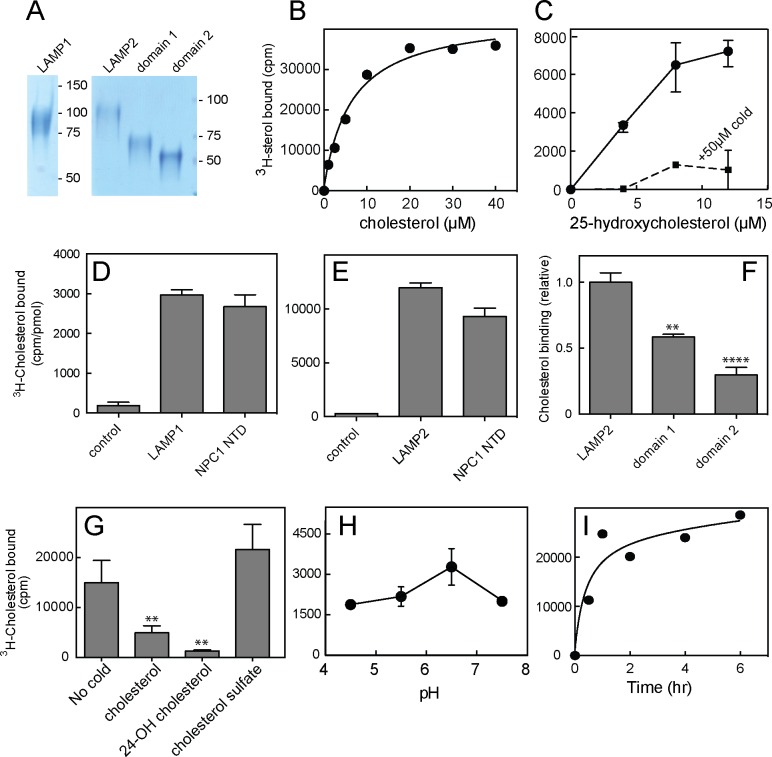
Figure 1. Cholesterol binding to LAMP proteins. (A) Coomassie-stained SDS-PAGE of purified, secreted human LAMP1, LAMP2 or LAMP domains from LAMP2. B,C. 3H-cholesterol or 3H-25 hydroxycholesterol binding to soluble LAMP2 (full length protein). Also shown in C is binding in the presence of 50 µM cold hydroxycholesterol. D,E, 3H-cholesterol binding to indicated, soluble proteins compared with the soluble NPC1 N-terminal domain. (F), Cholesterol binding to LAMP2 domains 1 and 2 compared with full length, soluble LAMP2 protein. P values were determined relative to the full length soluble LAMP2 protein. (G) Sterol competition (30 µM) for 3H-cholesterol binding to soluble LAMP2. P values were determined relative to no cold addition. H, I. pH dependence and kinetics of 3H-cholesterol binding to LAMP2. In B,C,E,H and I, a representative experiment is shown; C and E show the average of duplicates. In C, the background counts in reactions containing the control protein, GFP-binding protein, were subtracted; in D and E, the control was TIP47 protein. D,F, and G show the combined results of two experiments in duplicate. Numbers at right (A) indicate mass in kD for this and all subsequent figures. Reactions contained D,E,G,H,I, 500 nM total cholesterol; F, 5 µM total cholesterol; B and C were carried out using increasing concentrations of the indicated sterol. DOI: http://dx.doi.org/10.7554/eLife.21635.003
Figure 1. Cholesterol binding to LAMP proteins. (A) Coomassie-stained SDS-PAGE of purified, secreted human LAMP1, LAMP2 or LAMP domains from LAMP2. B,C. 3H-cholesterol or 3H-25 hydroxycholesterol binding to soluble LAMP2 (full length protein). Also shown in C is binding in the presence of 50 µM cold hydroxycholesterol. D,E, 3H-cholesterol binding to indicated, soluble proteins compared with the soluble NPC1 N-terminal domain. (F), Cholesterol binding to LAMP2 domains 1 and 2 compared with full length, soluble LAMP2 protein. P values were determined relative to the full length soluble LAMP2 protein. (G) Sterol competition (30 µM) for 3H-cholesterol binding to soluble LAMP2. P values were determined relative to no cold addition. H, I. pH dependence and kinetics of 3H-cholesterol binding to LAMP2. In B,C,E,H and I, a representative experiment is shown; C and E show the average of duplicates. In C, the background counts in reactions containing the control protein, GFP-binding protein, were subtracted; in D and E, the control was TIP47 protein. D,F, and G show the combined results of two experiments in duplicate. Numbers at right (A) indicate mass in kD for this and all subsequent figures. Reactions contained D,E,G,H,I, 500 nM total cholesterol; F, 5 µM total cholesterol; B and C were carried out using increasing concentrations of the indicated sterol. DOI: http://dx.doi.org/10.7554/eLife.21635.003CC BY · PubMed Central
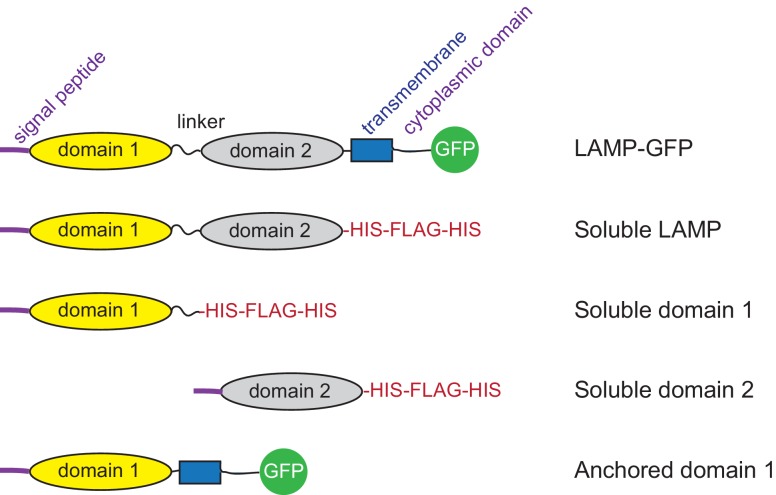
Figure 1—figure supplement 1. Diagram of constructs used. DOI: http://dx.doi.org/10.7554/eLife.21635.004
Figure 1—figure supplement 1. Diagram of constructs used. DOI: http://dx.doi.org/10.7554/eLife.21635.004CC BY · PubMed Central
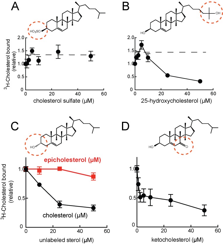
Figure 2. Cholesterol binding to LAMP2 is competed by 25-hydroxycholesterol (B), cholesterol (C), 7-ketocholesterol (D) but not cholesterol sulfate (A) or epicholesterol (C). The structures above indicate the regions of the sterol that differ from cholesterol. In C, the background obtained in reactions containing GFP was subtracted. All panels used 50nM 3H-cholesterol and the indicated amounts of competitors. DOI: http://dx.doi.org/10.7554/eLife.21635.005
Figure 2. Cholesterol binding to LAMP2 is competed by 25-hydroxycholesterol (B), cholesterol (C), 7-ketocholesterol (D) but not cholesterol sulfate (A) or epicholesterol (C). The structures above indicate the regions of the sterol that differ from cholesterol. In C, the background obtained in reactions containing GFP was subtracted. All panels used 50nM 3H-cholesterol and the indicated amounts of competitors. DOI: http://dx.doi.org/10.7554/eLife.21635.005CC BY · PubMed Central
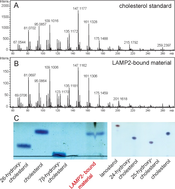
Figure 3. Mass spectrometry identification of small molecules released from LAMP2 after chloroform:methanol (2:1) extraction. (A) masses from cholesterol standard; (B) masses of LAMP2-bound material; (C) Copper sulfate/phosphoric acid detection of indicated markers after thin layer chromatography compared with material eluted from soluble LAMP2 (50 µg). Shown are the results of a representative experiment carried out twice. DOI: http://dx.doi.org/10.7554/eLife.21635.006
Figure 3. Mass spectrometry identification of small molecules released from LAMP2 after chloroform:methanol (2:1) extraction. (A) masses from cholesterol standard; (B) masses of LAMP2-bound material; (C) Copper sulfate/phosphoric acid detection of indicated markers after thin layer chromatography compared with material eluted from soluble LAMP2 (50 µg). Shown are the results of a representative experiment carried out twice. DOI: http://dx.doi.org/10.7554/eLife.21635.006CC BY · PubMed Central
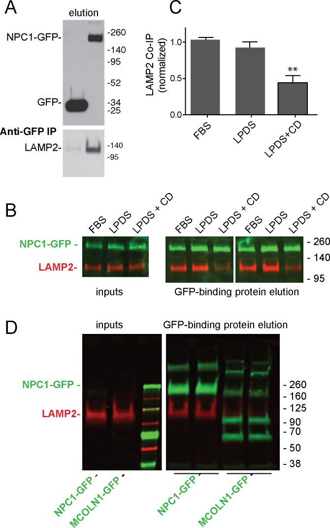
Figure 4. LAMP2 interacts with NPC1 protein. (A) Anti-GFP immunoprecipitation from HEK293T cells grown in FBS containing medium expressing GFP or mouse NPC1-GFP. The blot was developed with ECL. Upper panel, anti-GFP immunoblot (100% elution); lower panel, anti-LAMP2 immunoblot to detect endogenous, full length protein (100% elution). B,C, co-immunoprecipitation of LAMP2 and NPC1-GFP in cells grown in FBS, LPDS (5%) or LPDS + cyclodextrin (1 mM) for 24 hr. Left panels, 1% inputs; right panels, 50% elutions. Error bars represent SEM for two combined experiments carried out in duplicate; P value is from comparison with LPDS by two-tailed Student’s t-test. B shows duplicate reactions to document reproducibility. D, Anti-GFP immunoprecipitation of HEK293T cells grown in FBS expressing GFP-MCOLN1 or mouse NPC1-GFP. GFP-MCOLN1 occurs as a ~90 kD form, a proteolytically processed form, and as higher oligomers (Vergarajauregui et al., 2011). Left panels, inputs (2%); right panels, total elution carried out in duplicate. GFP proteins are green; LAMP2 is presented in red. Shown is a representative experiment carried out twice in duplicate. DOI: http://dx.doi.org/10.7554/eLife.21635.007
Figure 4. LAMP2 interacts with NPC1 protein. (A) Anti-GFP immunoprecipitation from HEK293T cells grown in FBS containing medium expressing GFP or mouse NPC1-GFP. The blot was developed with ECL. Upper panel, anti-GFP immunoblot (100% elution); lower panel, anti-LAMP2 immunoblot to detect endogenous, full length protein (100% elution). B,C, co-immunoprecipitation of LAMP2 and NPC1-GFP in cells grown in FBS, LPDS (5%) or LPDS + cyclodextrin (1 mM) for 24 hr. Left panels, 1% inputs; right panels, 50% elutions. Error bars represent SEM for two combined experiments carried out in duplicate; P value is from comparison with LPDS by two-tailed Student’s t-test. B shows duplicate reactions to document reproducibility. D, Anti-GFP immunoprecipitation of HEK293T cells grown in FBS expressing GFP-MCOLN1 or mouse NPC1-GFP. GFP-MCOLN1 occurs as a ~90 kD form, a proteolytically processed form, and as higher oligomers (Vergarajauregui et al., 2011). Left panels, inputs (2%); right panels, total elution carried out in duplicate. GFP proteins are green; LAMP2 is presented in red. Shown is a representative experiment carried out twice in duplicate. DOI: http://dx.doi.org/10.7554/eLife.21635.007CC BY · PubMed Central
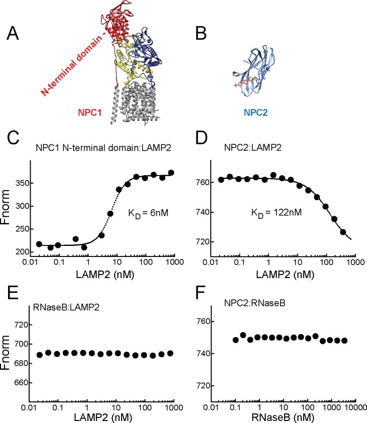
Figure 5. LAMP2 binds NPC1 and NPC2 proteins. A,B, Structures of NPC1 (Gong et al., 2016; pdb 3jD8) and NPC2 (Xu et al., 2007; pdb 2 hka) proteins; C,E, Microscale thermophoresis (E) or fluorescence (C) obtained with mixtures of soluble, AF647 labeled-NPC1 N-terminal domain or AF647-RNase B with increasing concentrations of soluble LAMP2 in 1 µM cholesterol. D,F, Microscale thermophoresis of AF647 labeled-NPC2 with increasing concentrations of soluble LAMP2 or RNase B in 1 µM cholesterol sulfate. For C–F, a representative experiment carried out at least twice is shown. DOI: http://dx.doi.org/10.7554/eLife.21635.008
Figure 5. LAMP2 binds NPC1 and NPC2 proteins. A,B, Structures of NPC1 (Gong et al., 2016; pdb 3jD8) and NPC2 (Xu et al., 2007; pdb 2 hka) proteins; C,E, Microscale thermophoresis (E) or fluorescence (C) obtained with mixtures of soluble, AF647 labeled-NPC1 N-terminal domain or AF647-RNase B with increasing concentrations of soluble LAMP2 in 1 µM cholesterol. D,F, Microscale thermophoresis of AF647 labeled-NPC2 with increasing concentrations of soluble LAMP2 or RNase B in 1 µM cholesterol sulfate. For C–F, a representative experiment carried out at least twice is shown. DOI: http://dx.doi.org/10.7554/eLife.21635.008CC BY · PubMed Central
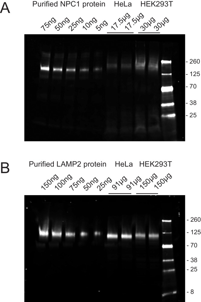
Figure 5—figure supplement 1. Quantitation of NPC1 (A) and LAMP2 (B) proteins in HeLa and HEK293T cells. The values at the top of each gel represent the amount of purified protein or total cell extract analyzed, determined by BCA protein assay. The immunoblot was developed using rabbit anti-NPC1 or mouse anti-LAMP2 antibodies followed by detection with IRDye800CW goat anti-mouse or donkey anti-rabbit antibodies. Shown is an example of an experiment carried out in duplicate; subconfluent cultures were analyzed. Purified NPC1 is slightly smaller than that in the extract because it was deglycosylated (see Materials and methods). DOI: http://dx.doi.org/10.7554/eLife.21635.009
Figure 5—figure supplement 1. Quantitation of NPC1 (A) and LAMP2 (B) proteins in HeLa and HEK293T cells. The values at the top of each gel represent the amount of purified protein or total cell extract analyzed, determined by BCA protein assay. The immunoblot was developed using rabbit anti-NPC1 or mouse anti-LAMP2 antibodies followed by detection with IRDye800CW goat anti-mouse or donkey anti-rabbit antibodies. Shown is an example of an experiment carried out in duplicate; subconfluent cultures were analyzed. Purified NPC1 is slightly smaller than that in the extract because it was deglycosylated (see Materials and methods). DOI: http://dx.doi.org/10.7554/eLife.21635.009CC BY · PubMed Central
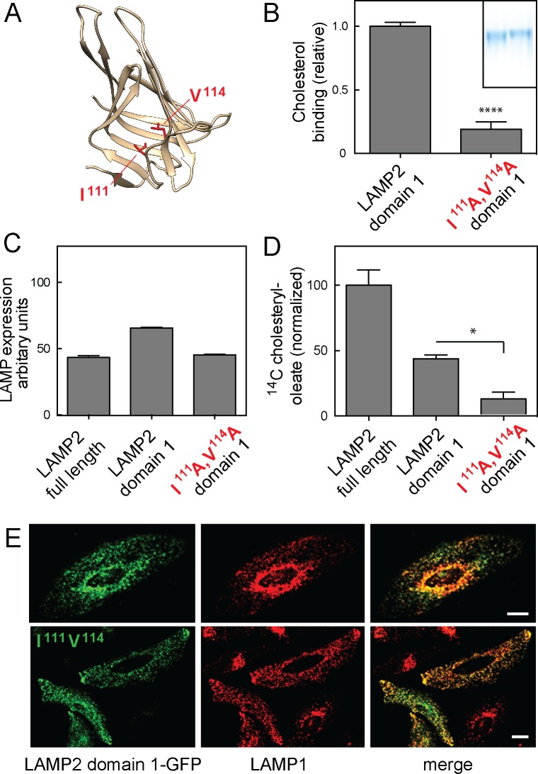
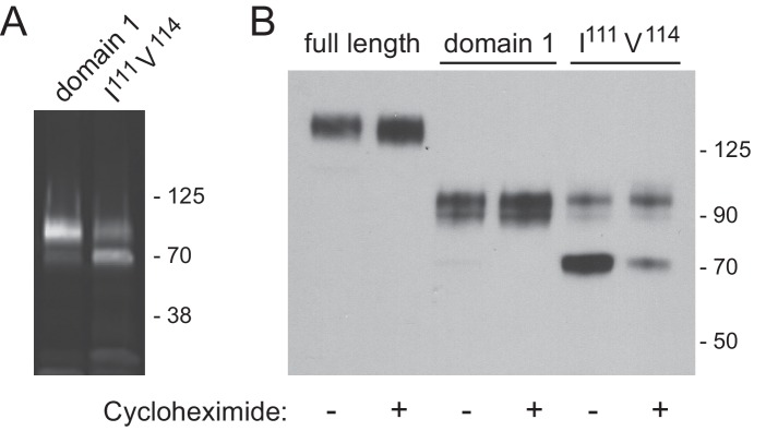
Figure 6. Cholesterol binding to LAMP2 domain 1 is required for its ability to rescue cholesterol export from LAMP-deficient lysosomes. (A), predicted structure model of LAMP2 domain 1; residues I111 and V114 are highlighted in red. (B) Relative 3H-cholesterol binding to soluble LAMP 2 domain 1 or LAMP2 domain 1-I111A/V114A. Shown is combined data from 5 independent experiments carried out in duplicate in the presence of 50 nM 3H-cholesterol. Inset, SDS-PAGE analysis of wild type (left) and domain 1-I111A/V114A (right) proteins analyzed. P value was determined by two-tailed Student’s t-test. (C) flow cytometry analysis of mean fluorescence of GFP rescue constructs in lentivirus-tranduced cells (>20,000 cells analyzed). (D) Cholesteryl oleate synthesis in MEF cells lacking LAMP1 and LAMP2 after rescue with either full length, membrane anchored LAMP2, membrane anchored LAMP2 domain 1, or membrane anchored LAMP2 domain 1-I111A/V114A. C-terminally GFP-tagged, rescue proteins were stably expressed using lentivirus transduction; shown is the combined result of 2 independent experiments, normalized for the amount of mature protein in each sample (Figure 6—figure supplement 1) relative to the amount of rescue seen with full length LAMP2 protein. P-values are in relation to full length for domain 1, or to domain 1 for the mutant protein, and were determined by one way ANOVA. E, confocal light microscopic analysis of GFP rescue construct localization (green) and endogenous LAMP1 protein (red) in transiently transfected HeLa cells; bars represent 20 µm. DOI: http://dx.doi.org/10.7554/eLife.21635.010
Figure 6. Cholesterol binding to LAMP2 domain 1 is required for its ability to rescue cholesterol export from LAMP-deficient lysosomes. (A), predicted structure model of LAMP2 domain 1; residues I111 and V114 are highlighted in red. (B) Relative 3H-cholesterol binding to soluble LAMP 2 domain 1 or LAMP2 domain 1-I111A/V114A. Shown is combined data from 5 independent experiments carried out in duplicate in the presence of 50 nM 3H-cholesterol. Inset, SDS-PAGE analysis of wild type (left) and domain 1-I111A/V114A (right) proteins analyzed. P value was determined by two-tailed Student’s t-test. (C) flow cytometry analysis of mean fluorescence of GFP rescue constructs in lentivirus-tranduced cells (>20,000 cells analyzed). (D) Cholesteryl oleate synthesis in MEF cells lacking LAMP1 and LAMP2 after rescue with either full length, membrane anchored LAMP2, membrane anchored LAMP2 domain 1, or membrane anchored LAMP2 domain 1-I111A/V114A. C-terminally GFP-tagged, rescue proteins were stably expressed using lentivirus transduction; shown is the combined result of 2 independent experiments, normalized for the amount of mature protein in each sample (Figure 6—figure supplement 1) relative to the amount of rescue seen with full length LAMP2 protein. P-values are in relation to full length for domain 1, or to domain 1 for the mutant protein, and were determined by one way ANOVA. E, confocal light microscopic analysis of GFP rescue construct localization (green) and endogenous LAMP1 protein (red) in transiently transfected HeLa cells; bars represent 20 µm. DOI: http://dx.doi.org/10.7554/eLife.21635.010CC BY · PubMed Central
Figure 6—figure supplement 1. Immunoblot analysis of LAMP2 constructs from lentivirus transduced LAMP1/LAMP2-knock out MEF cells. (A) total cell extract from cells expressing membrane anchored, GFP-LAMP2 domain 1 or GFP-LAMP2 domain 1-I111A/V114A; (B) migration of the constructs indicated, before or after 4 hr cycloheximide treatment (50 µg/ml). Panel A was developed as in Figure 4B; panel B was developed as in Figure 4A. DOI: http://dx.doi.org/10.7554/eLife.21635.011
Figure 6—figure supplement 1. Immunoblot analysis of LAMP2 constructs from lentivirus transduced LAMP1/LAMP2-knock out MEF cells. (A) total cell extract from cells expressing membrane anchored, GFP-LAMP2 domain 1 or GFP-LAMP2 domain 1-I111A/V114A; (B) migration of the constructs indicated, before or after 4 hr cycloheximide treatment (50 µg/ml). Panel A was developed as in Figure 4B; panel B was developed as in Figure 4A. DOI: http://dx.doi.org/10.7554/eLife.21635.011CC BY · PubMed Central

Verification

Claims the paper makes

The claims and statistics of this paper haven’t been checked yet.

Run the check — free, no account needed. It reads the open-access text and checks the sources it cites.

A few free checks a day · papers already verified open instantly · results are public

Claims other papers make about this one

Each is checked against this paper’s own text: does it say what the citing paper says it does?

Self-check

Percentages, sums and reported differences re-computed from the paper’s own numbers.

The self-check needs the paper’s full text.

Citations

Cites

Show all 1 references

Research works printed but not linked: 32

  1. Abi-Mosleh, 2009, Cyclodextrin overcomes deficient lysosome-to-endoplasmic reticulum transport of cholesterol in Niemann-Pick type C cells, PNAS, 106, 19316, 10.1073/pnas.0910916106 · DOI · Paper page
  2. Bandyopadhyay, 2008, The chaperone-mediated autophagy receptor organizes in dynamic protein complexes at the lysosomal membrane, Molecular and Cellular Biology, 28, 5747, 10.1128/MCB.02070-07 · DOI
  3. Cuervo, 1996, A receptor for the selective uptake and degradation of proteins by lysosomes, Science, 273, 501, 10.1126/science.273.5274.501 · DOI · Paper page
  4. Cuervo, 2000, Unique properties of lamp2a compared to other lamp2 isoforms, Journal of Cell Science, 113, 4441, 10.1242/jcs.113.24.4441 · DOI
  5. Eskelinen, 2004, Disturbed cholesterol traffic but normal proteolytic function in LAMP-1/LAMP-2 double-deficient fibroblasts, Molecular Biology of the Cell, 15, 3132, 10.1091/mbc.E04-02-0103 · DOI
  6. Goldstein, 1983, Receptor-mediated endocytosis of low-density lipoprotein in cultured cells, Methods in Enzymology, 98, 241, 10.1016/0076-6879(83)98152-1 · DOI
  7. Gong, 2016, Structural insights into the Niemann-Pick C1 (NPC1)-mediated cholesterol transfer and ebola infection, Cell, 165, 1467, 10.1016/j.cell.2016.05.022 · DOI
  8. Granger, 1990, Characterization and cloning of lgp110, a lysosomal membrane glycoprotein from mouse and rat cells, The Journal of Biological Chemistry, 265, 12036, 10.1016/S0021-9258(19)38504-7 · DOI
  9. Griffiths, 1989, A quantitative analysis of the endocytic pathway in baby hamster kidney cells, The Journal of Cell Biology, 109, 2703, 10.1083/jcb.109.6.2703 · DOI
  10. Hulce, 2013, Proteome-wide mapping of cholesterol-interacting proteins in mammalian cells, Nature Methods, 10, 259, 10.1038/nmeth.2368 · DOI
  11. Huynh, 2007, LAMP proteins are required for fusion of lysosomes with phagosomes, The EMBO Journal, 26, 313, 10.1038/sj.emboj.7601511 · DOI · Paper page
  12. Infante, 2008, Purified NPC1 protein. I. Binding of cholesterol and oxysterols to a 1278-amino acid membrane protein, The Journal of Biological Chemistry, 283, 1052, 10.1074/jbc.M707943200 · DOI
  13. Itzhak, 2016, Global, quantitative and dynamic mapping of protein subcellular localization, eLife, 5, e16950, 10.7554/eLife.16950 · DOI · Paper page
  14. Kundra, 1999, Asparagine-linked oligosaccharides protect Lamp-1 and Lamp-2 from intracellular proteolysis, Journal of Biological Chemistry, 274, 31039, 10.1074/jbc.274.43.31039 · DOI
  15. Kwon, 2009, Structure of N-terminal domain of NPC1 reveals distinct subdomains for binding and transfer of cholesterol, Cell, 137, 1213, 10.1016/j.cell.2009.03.049 · DOI · Paper page
  16. Li, 2015, Glycosylation inhibition reduces cholesterol accumulation in NPC1 protein-deficient cells, PNAS, 112, 14876, 10.1073/pnas.1520490112 · DOI
  17. Li, 2016, Clues to the mechanism of cholesterol transfer from the structure of NPC1 middle lumenal domain bound to NPC2, PNAS, 113, 10079, 10.1073/pnas.1611956113 · DOI
  18. Li, 2016, Structure of human Niemann-Pick C1 protein, PNAS, 113, 8212, 10.1073/pnas.1607795113 · DOI
  19. Pettersen, 2004, UCSF Chimera--a visualization system for exploratory research and analysis, Journal of Computational Chemistry, 25, 1605, 10.1002/jcc.20084 · DOI
  20. Quinn, 1984, Density of newly synthesized plasma membrane proteins in intracellular membranes II. Biochemical studies, The Journal of Cell Biology, 98, 2142, 10.1083/jcb.98.6.2142 · DOI
  21. Rosenbaum, 2011, Niemann-Pick type C disease: molecular mechanisms and potential therapeutic approaches, Journal of Neurochemistry, 116, 789, 10.1111/j.1471-4159.2010.06976.x · DOI
  22. Rosenbaum, 2010, Endocytosis of beta-cyclodextrins is responsible for cholesterol reduction in Niemann-Pick type C mutant cells, PNAS, 107, 5477, 10.1073/pnas.0914309107 · DOI
  23. Saftig, 2009, Lysosome biogenesis and lysosomal membrane proteins: trafficking meets function, Nature Reviews Molecular Cell Biology, 10, 623, 10.1038/nrm2745 · DOI · Paper page
  24. Schneede, 2011, Role for LAMP-2 in endosomal cholesterol transport, Journal of Cellular and Molecular Medicine, 15, 280, 10.1111/j.1582-4934.2009.00973.x · DOI
  25. Schwake, 2013, Lysosomal membrane proteins and their central role in physiology, Traffic, 14, 739, 10.1111/tra.12056 · DOI
  26. Settembre, 2013, Signals from the lysosome: a control centre for cellular clearance and energy metabolism, Nature Reviews Molecular Cell Biology, 14, 283, 10.1038/nrm3565 · DOI
  27. Tanaka, 2000, Accumulation of autophagic vacuoles and cardiomyopathy in LAMP-2-deficient mice, Nature, 406, 902, 10.1038/35022595 · DOI · Paper page
  28. Ulloth, 2007, Characterization of methyl-beta-cyclodextrin toxicity in NGF-differentiated PC12 cell death, NeuroToxicology, 28, 613, 10.1016/j.neuro.2007.01.001 · DOI
  29. Vergarajauregui, 2011, LAPTMs regulate lysosomal function and interact with mucolipin 1: new clues for understanding mucolipidosis type IV, Journal of Cell Science, 124, 459, 10.1242/jcs.076240 · DOI
  30. Wilke, 2012, Crystal structure of the conserved domain of the DC lysosomal associated membrane protein: implications for the lysosomal glycocalyx, BMC Biology, 10, 62, 10.1186/1741-7007-10-62 · DOI
  31. Xu, 2015, Lysosomal physiology, Annual Review of Physiology, 77, 57, 10.1146/annurev-physiol-021014-071649 · DOI
  32. Xu, 2007, Structural basis of sterol binding by NPC2, a lysosomal protein deficient in Niemann-Pick type C2 disease, Journal of Biological Chemistry, 282, 23525, 10.1074/jbc.M703848200 · DOI

Cited by

Sorted by citations

Metadata

Authors
Li, J., & Pfeffer, S. R.
Affiliations
Department of Biochemistry, Stanford University School of Medicine, Stanford, United States
Venue
eLife
Published
2016-09-24
Volume
5
ISSN
2050-084X
Cited by
111
References
1
Language
en
Funders
  • Ara Parseghian Medical Research Foundation
  • National Institute of Diabetes and Digestive and Kidney Diseases (DK37332)
  • National Institutes of Health (P30 CA124435)

About these checks

Registry, retraction, Cites and statistics results are computed, not judged, with one exception: a printed reference that no index lists is read once by a model before it counts as a citation error. Claims are extracted by a model and kept only when their quote appears verbatim in the text; each cited source is then checked, with a model’s help, for whether it says what the claim relies on. Those verdicts can be wrong — read the claim’s quote and the cited source before relying on one.

Statistics and arithmetic are recomputed, not judged.

Confidence bands are shown only for fields calibrated on our annotated test set. None is calibrated yet, so no band is shown.

What counts as a claim ›How we measure confidence ›How we check this ›

Are you an author, or do you see a problem with this page? Request a review