

The DOI matches its Crossref record; 32 of the 32 references it cites were found in a registry or an index.
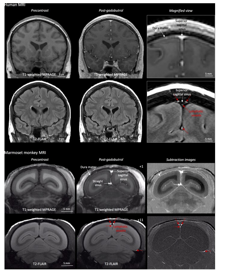
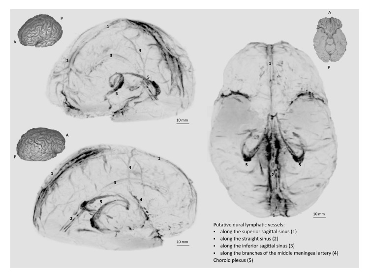
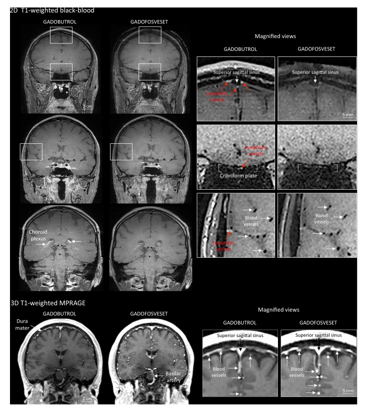
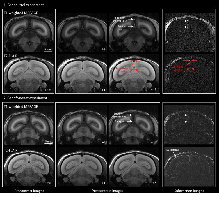
Human and nonhuman primate meninges harbor lymphatic vessels that can be visualized noninvasively by MRI.
Open accessAbstract
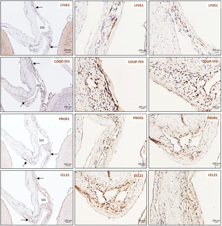
Here, we report the existence of meningeal lymphatic vessels in human and nonhuman primates (common marmoset monkeys) and the feasibility of noninvasively imaging and mapping them in vivo with high-resolution, clinical MRI. On T2-FLAIR and T1-weighted black-blood imaging, lymphatic vessels enhance with gadobutrol, a gadolinium-based contrast agent with high propensity to extravasate across a permeable capillary endothelial barrier, but not with gadofosveset, a blood-pool contrast agent. The topography of these vessels, running alongside dural venous sinuses, recapitulates the meningeal lymphatic system of rodents. In primates, meningeal lymphatics display a typical panel of lymphatic endothelial markers by immunohistochemistry. This discovery holds promise for better understanding the normal physiology of lymphatic drainage from the central nervous system and potential aberrations in neurological diseases.
Overview
The claims and statistics of this paper haven’t been checked yet.
Run the check — free, no account needed. It reads the open-access text and checks the sources it cites.
Integrity signals
Registry
Cites
Statistics
Figures
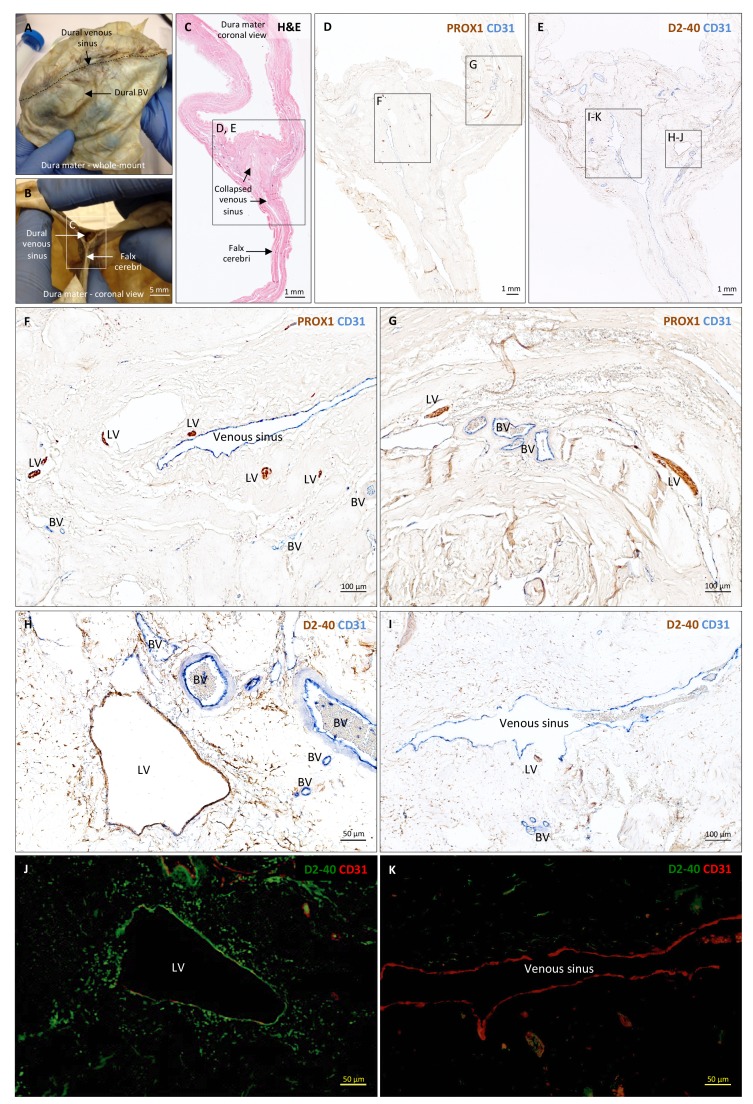
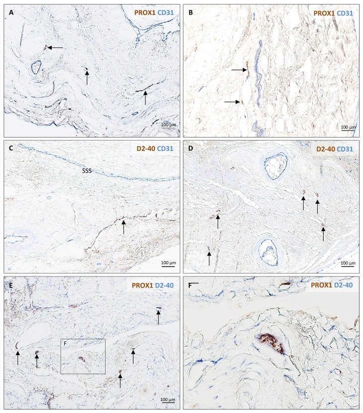
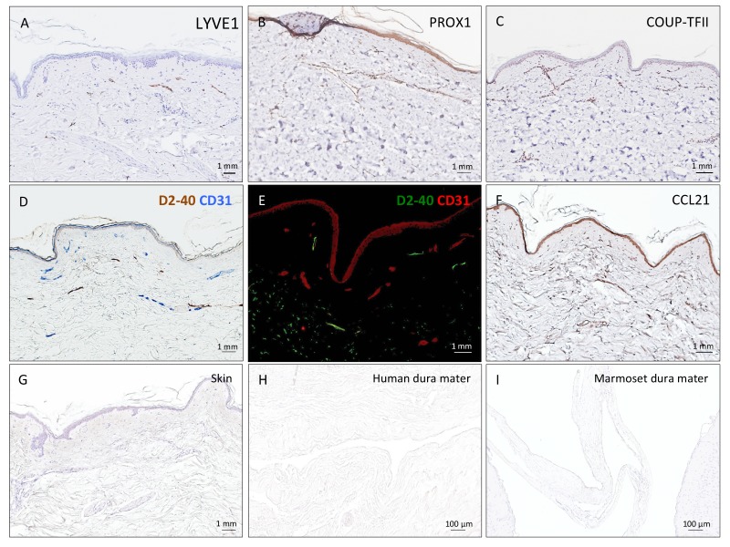
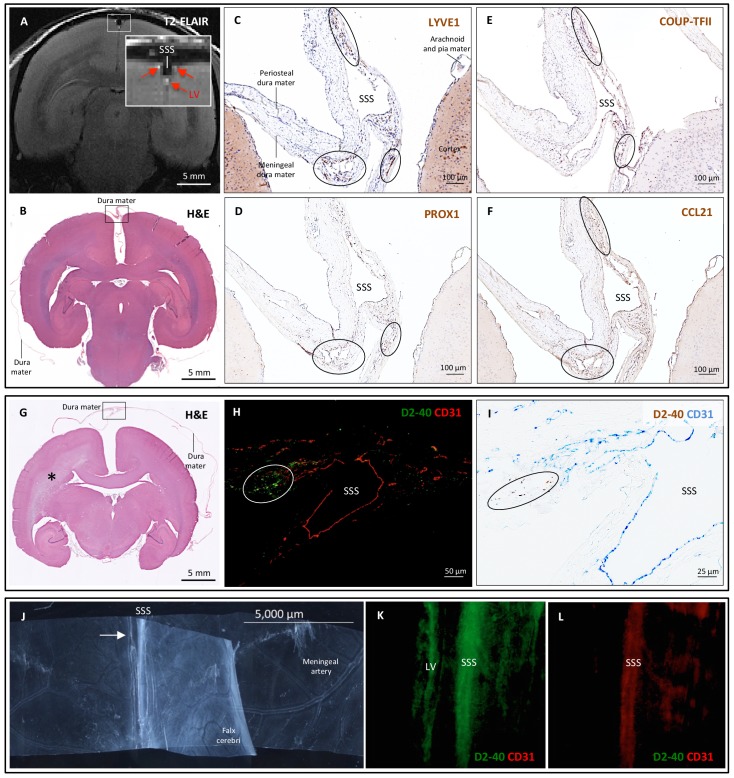
Verification
Claims the paper makes
The claims and statistics of this paper haven’t been checked yet.
Run the check — free, no account needed. It reads the open-access text and checks the sources it cites.
Claims other papers make about this one
Each is checked against this paper’s own text: does it say what the citing paper says it does?
The source backs part of the claim, not all of it — or backs it for a different population, condition or comparison.
In the citing paper
“Recent magnetic resonance imaging (MRI) studies in the living human brain and marmosets utilized a combination of gadolinium-based contrast agent (Gadovist) and blood-pool contrast agent (Vasovist) to demonstrate existence of the meningeal lymphatic system (4).”
From Blood-Brain Barrier: From Physiology to Disease and Back (2019) · cites this paper as [4]
Experimental — accuracy not yet measuredIn the citing paper
“The dural lymphatics are now recognized as a critical transport system of macromolecules, interstitial fluid, and CSF from the CNS into the cervical lymph nodes (Aspelund et al., 2015; Absinta et al., 2017; Louveau et al., 2018; Ahn et al., 2019).”
From Living on the Edge of the CNS: Meninges Cell Diversity in Health and Disease (2021) · cites this paper as Absinta et al., 2017
In the citing paper
“The extent of enhancing tissue within parasagittal dura does not resemble the previously reported appearance of true dural lymphatic vessels, which are tubular-shaped structures of sub-millimeter cross-sectional diameter near the venous sinus wall3,4,11, and also far from the classical MRI appearance of arachnoid granulations bulging into venous sinuses12.”
From Cerebrospinal fluid tracer efflux to parasagittal dura in humans (2020) · cites this paper as [11]
Self-check
Percentages, sums and reported differences re-computed from the paper’s own numbers.
The self-check needs the paper’s full text.
Citations
Cites
Iliff, J. J., Wang, M., Liao, Y., Plogg, B. A., Peng, W., Gundersen, G. A., Benveniste, H., Vates, G. E., Deane, R., Goldman, S. A., et al. (2012) · Science Translational Medicine · doi:10.1126/scitranslmed.3003748 · Cited by 5,824
Sleep Drives Metabolite Clearance from the Adult Brain
Xie, L., Kang, H., Xu, Q., Chen, M. J., Liao, Y., Thiyagarajan, M., O’Donnell, J., Christensen, D. J., Nicholson, C., Iliff, J. J., et al. (2013) · Science · doi:10.1126/science.1241224 · Cited by 5,411
Structural and functional features of central nervous system lymphatic vessels
Louveau, A., Smirnov, I., Keyes, T. J., Eccles, J. D., Rouhani, S. J., Peske, J. D., Derecki, N. C., Castle, D., Mandell, J. W., Lee, K. S., et al. (2015) · Nature · doi:10.1038/nature14432 · Cited by 4,413
A dural lymphatic vascular system that drains brain interstitial fluid and macromolecules
Aspelund, A., Antila, S., Proulx, S. T., Karlsen, T. V., Karaman, S., Detmar, M., Wiig, H., & Alitalo, K. (2015) · Journal of Experimental Medicine · doi:10.1084/jem.20142290 · Cited by 2,198
Lymphangiogenesis in development and human disease
Alitalo, K. K., Tammela, T., & Petrova, T. V. (2005) · Nature · doi:10.1038/nature04480 · Cited by 1,287
Brain-wide pathway for waste clearance captured by contrast-enhanced MRI
Iliff, J. J., Lee, H., Yu, M., Feng, T., Logan, J., Nedergaard, M., & Benveniste, H. (2013) · Journal of Clinical Investigation · doi:10.1172/jci67677 · Cited by 1,148
Lymphangiogenesis and lymphatic vessel remodelling in cancer
Stacker, S. A., Williams, S. P., Karnezis, T., Shayan, R., Fox, S. B., & Achen, M. G. (2014) · Nature reviews. Cancer · doi:10.1038/nrc3677 · Cited by 837
Mandell, D. M., Mossa‐Basha, M., Qiao, Y., Hess, C. P., Hui, F. K., Matouk, C. C., Johnson, M. H., Daemen, M. J. A. P., Vossough, A., Edjlali, M., et al. (2016) · American Journal of Neuroradiology · doi:10.3174/ajnr.a4893 · Cited by 694
Revisiting the Mechanisms of CNS Immune Privilege
Louveau, A., Harris, T. H., & Kipnis, J. (2015) · Trends in Immunology · doi:10.1016/j.it.2015.08.006 · Cited by 682
Self-citationLineage tracing demonstrates the venous origin of the mammalian lymphatic vasculaturePreprint
Srinivasan, R. S., Dillard, M. E., Lagutin, O. V., Lin, F., Tsai, S. Y., Tsai, M., Samokhvalov, I. M., & Oliver, G. C. (2007) · Genes & Development · doi:10.1101/gad.1588407 · Cited by 583
Research works printed but not linked: 1
- Mascagni, 1816, Istoria Completa Dei Vasi Linfatici, Vol. II, 195
Cited by
Sorted by citations
Items 401–500 of 643
The glymphatic system in Huntington's disease
Duan, W., Zhou, Y., & Liu, H. (2025) · Journal of Huntington s Disease · doi:10.1177/18796397251331436 · Cited by 8
Pisoni, L., Donini, L., Gagni, P., Pennuto, M., Ratti, A., Verde, F., Ticozzi, N., Mandrioli, J., Calvo, A., & Basso, M. (2025) · Cells · doi:10.3390/cells14110848 · Cited by 7
Physiological brain clearance architecture revealed by neuronal protein tracing
Chayama, Y., Rao, N. R., Perla, D., Zhang, Z., Reid, M. M., Nelson, S., Wen, X., Ding, B., Blumenfeld, J. R., Apolonio, A., et al. (2026) · Cell · doi:10.1016/j.cell.2026.04.048 · Cited by 11
Hoang, T. A., Cao, E., Gracia, G., Nicolazzo, J. A., & Trevaskis, N. L. (2023) · Frontiers in Pharmacology · doi:10.3389/fphar.2023.1111617 · Cited by 7
Borrelli, S., Leclercq, S., Pasi, M., & Maggi, P. (2024) · Multiple Sclerosis and Related Disorders · doi:10.1016/j.msard.2024.105878 · Cited by 10
Blanchette, M., & Daneman, R. (2017) · The Journal of Experimental Medicine · doi:10.1084/jem.20172031 · Cited by 7
Role of Transcriptional and Epigenetic Regulation in Lymphatic Endothelial Cell Development
La, H., Yoo, H., Park, Y. B., Thang, N. X., Park, C., Yoo, S., Lee, H., Choi, Y., Song, H., Tae, J., et al. (2022) · Cells · doi:10.3390/cells11101692 · Cited by 7
Jakimovski, D., Topolski, M., Kimura, K., Marr, K., Gandhi, S., Ramasamy, D. P., Bergsland, N., Hagemeier, J., Weinstock‐Guttman, B., & Zivadinov, R. (2018) · Clinical Autonomic Research · doi:10.1007/s10286-018-0555-6 · Cited by 7
He, X., Xiong, D., Zhao, L., Fu, J., & Luo, L. (2024) · Nature Communications · doi:10.1038/s41467-024-49818-5 · Cited by 6
Advances in Research on Meningeal Lymphatic Vessels in Central Nervous System Diseases
Shen, H., Zhu, B., Qian, Y., Jin, J., Zhou, J., Peng, G., & Mo, J. (2024) · Journal of Craniofacial Surgery · doi:10.1097/scs.0000000000010872 · Cited by 7
Back, D. B., Choi, B., Kwon, K. J., Choi, D., Shin, C. Y., Lee, J., & Kim, H. Y. (2025) · Frontiers in Molecular Neuroscience · doi:10.3389/fnmol.2025.1516265 · Cited by 6
Emerging Roles of Meningeal Lymphatic Vessels in Ischemic Stroke
Machado, R. S., Mathias, K., Joaquim, L. D. S., da Costa, M. D. A., Tiscoski, A. D. B., Gonçalves, C. L., Rezin, G. T., & Petronilho, F. C. (2025) · Molecular Neurobiology · doi:10.1007/s12035-025-04983-6 · Cited by 6
The parasagittal dural space of the human brain
Donahue, M. J., McKnight, C. D., Claassen, D. O., & Hett, K. (2025) · Brain · doi:10.1093/brain/awaf204 · Cited by 6
Han, M., Han, Y., Jiang, Z., Gao, Y., Cao, G., Zhang, H., Wang, T., & Li, Z. (2025) · Proceedings of the National Academy of Sciences · doi:10.1073/pnas.2503434122 · Cited by 8
Kamouh, M.-R. E., Spajer, M., Singhabahu, R., Marolany, A.-L. J., Sailor, K. A., Mouton, L., Doukhi, D., Koundal, S., Metcalfe, T., Langui, D., et al. (2026) · Nature Neuroscience · doi:10.1038/s41593-026-02358-1 · Cited by 10
Liu, C., Wang, X., Huang, W., Meng, W., Su, Z., Xing, Q., Shi, H., Zhang, D., Zhou, M., Zhao, Y., et al. (2021) · Stem Cell Research & Therapy · doi:10.1186/s13287-021-02427-1 · Cited by 6
Comparative study of 3D-T2WI vs. 3D-T2-FLAIR MRI in displaying human meningeal lymphatics vessels
Fang, Y., Sun, Y., Lai, T., Song, X., Hu, T., Zhao, Y., Lin, Y., & Bao, Q. (2024) · Clinical Radiology · doi:10.1016/j.crad.2024.09.006 · Cited by 6
The glymphatic system and its involvement in disorders of the nervous system
Toriello, M., González‐Quintanilla, V., & Pascual, J. (2021) · Medicina Clínica (English Edition) · doi:10.1016/j.medcle.2021.03.003 · Cited by 7
Charting the meningeal lymphatic network
Mesquita, S. D. (2022) · The Journal of Experimental Medicine · doi:10.1084/jem.20220891 · Cited by 6
Lin, J., Sun, H., Jiang, H., Xue, R., Qin, B., Lü, J., Zhu, Z., Cai, C., Han, J., Zhang, J., et al. (2023) · Chemical Engineering Journal · doi:10.1016/j.cej.2023.144686 · Cited by 6
Central Nervous System Lymphatics and Impact on Neurologic Disease
Nistal, D. A., & Mocco, J. (2017) · World Neurosurgery · doi:10.1016/j.wneu.2017.11.028 · Cited by 6
Complex Autoantibody Responses Occur Following Moderate to Severe Traumatic Brain InjuryPreprint
Needham, E., Stoevesandt, O., Thelin, E. P., Zetterberg, H., Zanier, E. R., Nimer, F. A., Ashton, N. J., Outtrim, J. G., Newcombe, V., Mousa, H. S., et al. (2020) · medRxiv · doi:10.1101/2020.07.24.20161786 · Cited by 6
An integrated single cell and spatial omics atlas of human prenatal developmentPreprint
Webb, S., Rose, A., Xu, C., Steele, L., Kuri, M. A., Stephenson, E., İnecik, K., Jafree, D. J., Foster, A. R., Basurto-Lozada, D., et al. (2026) · bioRxiv (Cold Spring Harbor Laboratory) · doi:10.64898/2026.03.30.714220 · Cited by 7
Regulation of autoimmune‐mediated neuroinflammation by endothelial cells
Kallal, N., Hugues, S., & Garnier, L. (2024) · European Journal of Immunology · doi:10.1002/eji.202350482 · Cited by 8
Khabibov, M., Гарифуллин, А., Sadreeva, A., Fernandez, M. F., Шамилевич, К. М., Topchu, I., Kharin, L. V., & Boumber, Y. (2025) · Current Neuropharmacology · doi:10.2174/011570159x359861250224051857 · Cited by 5
Zeng, L., Yin, Z., Li, W., Wang, X., Zhang, Y., Xie, M., & Zhao, L. (2025) · Frontiers in Neurology · doi:10.3389/fneur.2025.1607723 · Cited by 6
Meningeal lymphatic architecture and drainage dynamics surrounding the human middle meningeal artery
Albayram, S., Richmond, S. B., Yağmurlu, K., Tuna, İ. S., Karakaya, E., Ravichandran, H., Tufan, F., Lesha, E., Mut, M., Bunyak, F., et al. (2025) · iScience · doi:10.1016/j.isci.2025.113693 · Cited by 5
Moore, M. E., Karakaya, E., Altınbaş, Ö., Bartlett, M. J., Adilbay, D., Ergul, A., Yağmurlu, K., Albayram, M., & Albayram, Ö. (2026) · Neurobiology of Aging · doi:10.1016/j.neurobiolaging.2026.02.002 · Cited by 7
The meninges as a neuroimmune interface: structure, barriers and roles in CNS disease
Joost, S., Kaddatz, H., Vankriekelsvenne, E., & Brandenburg, L. (2026) · Fluids and Barriers of the CNS · doi:10.1186/s12987-026-00795-5 · Cited by 7
Albumin-based nanoparticle for dual-modality imaging of the lymphatic system
Li, M., Zhang, Y., Ma, J., & Du, J. (2023) · RSC Advances · doi:10.1039/d2ra07414a · Cited by 5
From blood vessels to brain cells: Connecting the circulatory system and Parkinson's disease
Olaoye, O. J., Farrow, S. L., Nyaga, D. M., Cooper, A. A., & O'Sullivan, J. M. (2025) · Journal of Parkinson s Disease · doi:10.1177/1877718x241308168 · Cited by 6
Lymphatic dysfunction in critical illness
Burke, E., & Datar, S. A. (2018) · Current Opinion in Pediatrics · doi:10.1097/mop.0000000000000623 · Cited by 5
Glymphatic Magnetic Resonance Imaging: Part I—Methodologies for Evaluation of the Glymphatic System
Lee, H., Yoo, R., & Choi, S. H. (2023) · Investigative Magnetic Resonance Imaging · doi:10.13104/imri.2023.0031 · Cited by 6
Brain lymphatic drainage system – visualization opportunities and current state of the art
Yankova, G., & Bogomyakova, O. B. (2020) · Complex Issues of Cardiovascular Diseases · doi:10.17802/2306-1278-2020-9-3-81-89 · Cited by 5
Wild-type Yellow fever virus in cerebrospinal fluid from fatal cases in Brazil, 2018
de Rezende, I. M., Cenachi, A. R. C., Costa, T. A., Oliveira, G. F. G., Rabelo, L., Menezes, L. M., Penido, I., Pereira, L. S., Arruda, M. S., Gonçalves, A. P., et al. (2022) · Frontiers in Virology · doi:10.3389/fviro.2022.936191 · Cited by 5
Kollarik, S., Humer, S. K., Zurfluh, C. E., Gousopoulos, E., & Lindenblatt, N. (2025) · Cellular and Molecular Neurobiology · doi:10.1007/s10571-025-01611-8 · Cited by 4
Tamadon, A., Afshar, A., Mussin, N. M., Zhilisbayeva, K. R., Kurmanalina, M. A., Baspakova, A. M., Aringazina, R. A., Mahdipour, M., Tanideh, N., Khoradmehr, A., et al. (2025) · ACS Chemical Neuroscience · doi:10.1021/acschemneuro.5c00533 · Cited by 4
Fabi, A., Alves, A. S., Neutzner, A., Frank, S., Lariu, A., Muller, L., Ismail, T., Gkotsoulias, D. G., Guzman, R. G., Schaefer, D. J., et al. (2025) · JPRAS Open · doi:10.1016/j.jpra.2025.12.013 · Cited by 6
The Glymphatic System and Meningeal Lymphatics: Current Understandings and Future Perspectives
Sun, H., Fan, H., Zhou, Y., Zhu, H., Chen, Y., Zhang, R., Wang, K., Pan, Y., & Zhang, A. (2026) · MedComm · doi:10.1002/mco2.70691 · Cited by 6
Brain Lymphatic Dysfunction in Subarachnoid Hemorrhage: Pathophysiology and Clinical Implications
Guo, S., Zhang, J. H., Boling, W., & Huang, L. (2026) · Biomolecules · doi:10.3390/biom16040616 · Cited by 7
Tertiary Lymphoid Structures in Central Nervous System Disorders
Vaccaro, A., Pereira, B. D. A., van de Walle, T., & Dimberg, A. (2024) · Methods in molecular biology · doi:10.1007/978-1-0716-4184-2_2 · Cited by 4
Morphology of the glymphatic and meningeal lymphatic structures of the bottlenose dolphin
Keenan, T. F., Jackson, O. N., Nelson-Maney, N., Rommel, S. A., McLellan, W. A., Pabst, D. A., Costidis, A. M., Caron, K. M., Kernagis, D., Rotstein, D. S., et al. (2025) · Scientific Reports · doi:10.1038/s41598-025-14840-0 · Cited by 4
Kwon, W., Yoo, C., Kim, J. H., Kim, T., Kim, A., Hwang, M. H., & Choi, H. (2023) · Bioengineering & Translational Medicine · doi:10.1002/btm2.10589 · Cited by 4
Major Challenges and Limitations of Big Data Analytics
Cao, B., & Reilly, J. P. (2019) · Springer eBooks · doi:10.1007/978-3-030-03553-2_2 · Cited by 5
Meningeal lymphatic network: The middleman of neuroinflammation
Pal, S., Rao, S. C., & Louveau, A. (2020) · Clinical and Experimental Neuroimmunology · doi:10.1111/cen3.12563 · Cited by 4
Evolving trends in anatomy- A global perspective
Sharma, A., & Kumar, A. (2021) · Indian Journal of Clinical Anatomy and Physiology · doi:10.18231/j.ijcap.2021.037 · Cited by 4
Lendrum, J., Seebach, B. S., Klein, B. A., & Liu, S. (2017) · bioRxiv (Cold Spring Harbor Laboratory) · doi:10.1101/199075 · Cited by 4
Zhong, L. X., Lu, A., Lu, X., Liu, X., Cao, L., Zhu, S., Diao, S., Cheng, X., Wu, H., & Chen, J. (2025) · Technology in Cancer Research & Treatment · doi:10.1177/15330338251319394 · Cited by 3
Zhou, X. Y., Cervera, S. G. D. B., Sun, Y., Li, Y., Li, W., Pereira, L. P., Sair, H. I., Paez, A. G., Knutsson, L., van Zijl, P. C. M., et al. (2025) · Fluids and Barriers of the CNS · doi:10.1186/s12987-025-00710-4 · Cited by 3
Zhang, Y., Jin, S., Guo, P., Yin, Y., Hua, Y., Ding, X., Zhang, Z. Q., Chen, S., Han, X., Li, B., et al. (2026) · Advanced Science · doi:10.1002/advs.202516988 · Cited by 3
Functional and Structural Evidence of Neurofluid Circuit Aberrations in Huntington Disease
Hett, K., Dubois, A., Leguizamon, M., Song, A. K., Trujillo, P., McKnight, C. D., Considine, C. M., Donahue, M. J., & Claassen, D. O. (2026) · Annals of Clinical and Translational Neurology · doi:10.1002/acn3.70328 · Cited by 4
Perivascular fibroblasts at the nexus of CNS immunity
Verdon, M., Skipness, V., Häusle, L., & Pikor, N. B. (2026) · The Journal of Immunology · doi:10.1093/jimmun/vkag003 · Cited by 3
Li, Z., Wang, S., Yang, J., Hu, H., Zheng, Y., Li, C., & Ding, Z. (2026) · Neuroscience · doi:10.1016/j.neuroscience.2026.04.021 · Cited by 3
The Glymphatic System in Idiopathic Intracranial Hypertension
Bouffard, M. A., Ford, J. N., & Grech, O. (2026) · Journal of Neuro-Ophthalmology · doi:10.1097/wno.0000000000002483 · Cited by 3
Quantifying the Lymphatic Transport of Model Therapeutics from the Brain in Rats
Hoang, T. A., Gracia, G., Cao, E., Nicolazzo, J. A., & Trevaskis, N. L. (2024) · Molecular Pharmaceutics · doi:10.1021/acs.molpharmaceut.4c00026 · Cited by 3
Imaging evaluation and volumetric measurement of the space surrounding the diploic veins
Nakamichi, R., Taoka, T., Ito, R., Yoshida, T., Sone, M., & Naganawa, S. (2024) · Japanese Journal of Radiology · doi:10.1007/s11604-024-01572-w · Cited by 3
Zhou, Z., Zhou, Y., Wang, R., Yan, S., Xiao, Z., Luo, Z., Ke, H., Zhang, K., Fang, M., Sun, J., et al. (2024) · Aging and Disease · doi:10.14336/ad.2024.0415 · Cited by 3
Xu, H., Zhao, X., & Luo, J. (2024) · Cellular and Molecular Life Sciences · doi:10.1007/s00018-024-05300-5 · Cited by 5
Brain lymphatic vessels accompanying meningeal arteries in the human
Çavdar, S., Altınöz, D., Demir, T. D., Bayraktaroğlu, A., Gürses, İ. A., & Özcan, G. (2024) · Translational Research in Anatomy · doi:10.1016/j.tria.2024.100321 · Cited by 3
Disorders of Lymphatic Architecture and Flow in Critical Illness
Itkin, M. G., Horák, J., L., J. L. P., Chang, C. W. J., Lile, D. J., Tomita, B., Bass, G. A., Kovach, S. J., & Kaplan, L. J. (2025) · Critical Care Medicine · doi:10.1097/ccm.0000000000006561 · Cited by 3
MR Imaging of Neurofluids in the Developing Brain
Agarwal, N., Klein, W. M., & Tuura, R. (2025) · Neuroimaging Clinics of North America · doi:10.1016/j.nic.2024.12.005 · Cited by 2
Meningeal Lymphatic and Glymphatic Structures in a Pelagic Delphinid (Delphinus delphis)
Jackson, O. N., Keenan, T. F., Nelson-Maney, N., Rommel, S. A., McLellan, W. A., Pabst, D. A., Costidis, A. M., Caron, K. M., Kernagis, D., Rotstein, D. S., et al. (2025) · Animals · doi:10.3390/ani15050729 · Cited by 4
Neurodegenerative Dementias: Improving Brain Health to Decrease Risk
Bateman, J. R., Hurley, R. A., & Taber, K. H. (2019) · Journal of Neuropsychiatry · doi:10.1176/appi.neuropsych.18110287 · Cited by 3
Bair, M. M., & Munakomi, S. (2019) · StatPearls · Cited by 3
Gasser, T. C. (2021) · Springer eBooks · doi:10.1007/978-3-030-70966-2_2 · Cited by 3
The New Anatomy of Neuroimmunology
Solaro, C. M., Barratt, D., & Vaccarezza, M. (2022) · Immuno · doi:10.3390/immuno2010016 · Cited by 4
Malik, P., & Shroff, M. M. (2022) · Neuroradiology · doi:10.1007/s00234-022-03100-x · Cited by 3
A pilot study of lymphoscintigraphy with tracer injection into the human brain
Coxon, A. T., Desai, R. R., Patel, P. R., Vellimana, A. K., Willie, J. T., Dowling, J. L., Leuthardt, E. C., Kim, A. H., Johanns, T. M., Siegel, B. A., et al. (2023) · Journal of Cerebral Blood Flow & Metabolism · doi:10.1177/0271678x231160891 · Cited by 4
LYMPHATIC DRAINAGE SYSTEM OF THE BRAIN: A NEW PLAYER IN NEUROSCIENCE
Semyachkina-Glushkovskaya, O. V., Postnov, D. E., Khorovodov, A. P., Наволокин, Н. А., & Kurthz, Y. G. G. (2023) · Журнал эволюционной биохимии и физиологии · doi:10.31857/s0044452923010084 · Cited by 3
Wallis, Z. K., Midkiff, C. C., Zhao, M., Autissier, P., Kim, S., Amadeck, A. Q., Wang, Y., Jakubowski, M., Burdo, T. H., Miller, A. D., et al. (2025) · PLoS Pathogens · doi:10.1371/journal.ppat.1013180 · Cited by 3
Physiologic constraints of using exosomes in vivo as systemic delivery vehicles
Lee, D. S., Suh, M., Kang, S. Y., & Hwang, D. W. (2019) · Precision Nanomedicine · doi:10.33218/prnano2(3)070819.1 · Cited by 3
Parasagittal Cystic Lesions May Arise from the Pial Sheath around the Cortical Venous Wall
Naganawa, S., Ito, R., Taoka, T., Yoshida, T., & Sone, M. (2021) · Magnetic Resonance in Medical Sciences · doi:10.2463/mrms.bc.2021-0141 · Cited by 3
Idiopathic intracranial hypertension in patients with cerebral small vessel disease: A case report
Liu, W., Jia, L., Xu, L., Yang, F., Cheng, H., Li, H., Hou, J., Zhang, D., & Liu, Y. (2023) · Medicine · doi:10.1097/md.0000000000032639 · Cited by 3
MRI Parameters of 3D FLAIR T2 Imaging Technique for Visualization of the Brain Lymphatic SignalsPreprint
Albayram, S., & Albayram, Ö. (2023) · Research Square · doi:10.21203/rs.3.pex-2228/v1 · Cited by 3
Cerebral Venous Blood Flow Regulates Brain Fluid Clearance via Dural LymphaticsPreprint
Kamouh, M.-R. E., Spajer, M., Singhabahu, R., Sailor, K. A., Bourrienne, M., Mouton, L., Koundal, S., Doukhi, D., Grine, A., Ninnemann, J., et al. (2024) · bioRxiv (Cold Spring Harbor Laboratory) · doi:10.1101/2024.12.12.628127 · Cited by 3
He, J., Meng, Q., Miao, C., Hao, J., & Dai, M. (2024) · Postgraduate Medical Journal · doi:10.1093/postmj/qgae125 · Cited by 2
RetractedMeningeal Immunity: Anatomy, Function, and Neonatal Unique Features
Lorga, I., & Andrade, E. B. (2025) · European Journal of Immunology · doi:10.1002/eji.202451618 · Cited by 3
Mishra, J., Dickinson, J. D., & Maurya, S. K. (2025) · Frontiers in Aging Neuroscience · doi:10.3389/fnagi.2025.1666837 · Cited by 2
Umehana, M., Fushimi, Y., Okuchi, S., Otani, S., Yamamoto, T., Sakata, A., Nakajima, S., Ikeda, S., Ito, S., Grinstead, J. W., et al. (2025) · Japanese Journal of Radiology · doi:10.1007/s11604-025-01908-0 · Cited by 3
A cellular and molecular perspective on organotypic lymphatic (dys)function
Kumar, S. S., Uphoff, K., Hötte, S., Prokosch, V., Schulte‐Merker, S., & Schulte-Ostermann, D. (2025) · Seminars in Cell and Developmental Biology · doi:10.1016/j.semcdb.2025.103665 · Cited by 3
Lymph‐based liquid biopsy: Current progress and future potential
Liu, D., Cheng, L., Hu, X., Bernardini, S., Zhang, H., Zhu, L., & Luo, Y. (2026) · Interdisciplinary medicine · doi:10.1002/inmd.70092 · Cited by 2
Wu, Y., Wang, K., Ju, L., Li, A., Qin, Q., Xu, F., Lin, D. D. M., Kleinberg, L. R., & Xu, J. (2026) · Magnetic Resonance in Medicine · doi:10.1002/mrm.70298 · Cited by 4
Meningeal lymphatic-associated brain swelling in acute stroke
Patil, R. M., Back, D. B., Hamanaka, G., Desai, R., Hayakawa, A., Chan, S. J., Ahn, B. J., Pignataro, G., Hayakawa, K., & Esposito, E. (2026) · PLoS ONE · doi:10.1371/journal.pone.0342643 · Cited by 2
Breaking barriers: how cytomegaloviruses manipulate myeloid cells to invade tissues
Dey, A., Gres, V., Nelipovich, A., Kolb, P., Immler, R., Ruzsics, Z., Kierdorf, K., Henneke, P., & Baasch, S. (2026) · EMBO Reports · doi:10.1038/s44319-026-00763-y · Cited by 2
Zhang, J., Zhang, J., Ding, J., Zhang, X., Wang, Y., Wei, W., Chai, Y., Zhang, S., & Chen, X. (2026) · Alzheimer s & Dementia · doi:10.1002/alz.71451 · Cited by 3
Ultrafast venous and sagittal sinus constrictions in the brain driven by abdominal pressure
Zhang, Q., Garborg, C. S., Frank, N., Salehi, F., Turner, K. L., & Drew, P. J. (2026) · Proceedings of the National Academy of Sciences · doi:10.1073/pnas.2604172123 · Cited by 3
Kolotilov, N. (2021) · Radiation Diagnostics Radiation Therapy · doi:10.37336/2707-0700-2021-2-3 · Cited by 2
Achievements of PVS (Primo Vascular System) Research from a Historical Perspective
Kim, H.-G. (2022) · Journal of Acupuncture and Meridian Studies · doi:10.51507/j.jams.2022.15.1.50 · Cited by 2
Intranasally Applied Human Olfactory Mucosa Neural Progenitor Cells Migrate to Damaged Brain Regions
Kronner, J. M., Folbe, A. J., Meythaler, J. M., Nelson, J. O., Borisov, A. B., & Peduzzi, J. D. (2022) · Future Science OA · doi:10.2144/fsoa-2022-0012 · Cited by 2
Magnetic Resonance Imaging of the Glymphatic System in the Human Brain
Lee, H., & Park, K. M. (2022) · Journal of Sleep Medicine · doi:10.13078/jsm.220025 · Cited by 2
Immunity in the brain and surrounding tissues
Kanamori, M., & Ito, M. (2023) · The Journal of Biochemistry · doi:10.1093/jb/mvad010 · Cited by 2
Cerebrospinal and Interstitial Fluids: Production, Outflow, and Circulation
Hladky, S. B., & Barrand, M. A. (2023) · Springer eBooks · doi:10.1007/978-3-031-39558-1_7 · Cited by 2
Association between the Presence of the Parasagittal Cyst-like Structures and Cognitive Function
Ohashi, T., Ito, R., Yamamoto, R., Ukai, K., & Naganawa, S. (2024) · Magnetic Resonance in Medical Sciences · doi:10.2463/mrms.mp.2024-0138 · Cited by 2
Beyond plaques and tangles: The role of immune cell dysfunction in Alzheimer's disease
Hussain, N., Khan, M. M., Sharma, A., Singh, R. K., & Khan, R. H. (2025) · Neurochemistry International · doi:10.1016/j.neuint.2025.105947 · Cited by 2
Gadolinium-Based Imaging and the Study of Neurofluid Dynamics
Ringstad, G. A., Eide, P. K., Naganawa, S., & Agarwal, N. (2025) · Neuroimaging Clinics of North America · doi:10.1016/j.nic.2024.12.008 · Cited by 3
Vascular Contribution to Cerebral Waste Clearance Affected by Aging or Diabetes
Shen, Y., Zhang, L., Ding, G., Boyd, E., Kaur, J., Li, Q., Haacke, E. M., Hu, J., & Jiang, Q. (2025) · Diagnostics · doi:10.3390/diagnostics15081019 · Cited by 2
Traumatic meningeal injury and repair mechanisms
Loane, D. J., & Faden, A. I. (2018) · Nature Immunology · doi:10.1038/s41590-018-0093-3 · Cited by 2
CLEANING THE BRAIN THROUGH TURBULENT GLYMPHATIC FLOW: THE WASHING MACHINE HYPOTHESIS
Kuo, P. H. (2022) · Lymphology · doi:10.2458/lymph.4787 · Cited by 2
Leptomeningeal Disease and the Role of Intrathecal Therapy
Saadeh, F. S., & Boire, A. A. (2020) · Springer eBooks · doi:10.1007/978-3-030-42958-4_12 · Cited by 2
Klibanov, A. L. (2021) · Radiology · doi:10.1148/radiol.2021204271 · Cited by 2
Metadata
- Authors
- Absinta, M., Ha, S.-K., Nair, G., Sati, P., Luciano, N. J., Palisoc, M., Louveau, A., Zaghloul, K. A., Pittaluga, S., Kipnis, J., & Reich, D. S.
- Affiliations
- National Institutes of Health (US)
- University of Virginia (US)
- Corresponding author
- Reich, Daniel S
- ORCID
- Venue
- eLife
- Publisher
- eLife Sciences Publications, Ltd
- Published
- 2017-10-03
- Volume
- 6
- ISSN
- 2050-084X (ISSN-L)
- PMC
- PMC5626482
- OpenAlex
- W2760815599
- Cited by
- 643
- References
- 32
- Citations by year
- 2026: 61 · 2025: 88 · 2024: 73 · 2023: 77 · 2022: 79 · 2021: 78 · 2020: 70 · 2019: 60 · 2018: 51 · 2017: 4
- FWCI
- 22.31
- Type
- journal article
- Language
- en
- Open access
- gold
- doi.org
- Licence
- Locations
- eLife (publishedVersion, cc-by)
- Europe PMC (PubMed Central) (publishedVersion, public-domain)
- PubMed Central (publishedVersion, public-domain)
- PubMed (publishedVersion)
- DOAJ (DOAJ: Directory of Open Access Journals) (submittedVersion, cc-by-sa)
- Funders
- National Institute of Neurological Disorders and Stroke (Intramural Research Program)
- National Multiple Sclerosis Society (Postdoctoral Fellowship)
- Conrad N. Hilton Foundation (Marilyn Hilton Award for Innovation in Multiple Sclerosis Research)
- National Cancer Institute (Intramural Research Program)
- National Institute on Aging (R01AG034113)
- Lymphatic Education and Research Network (Postdoctoral fellowship)
- Topics
- Health Sciences › Medicine › Oncology › Lymphatic System and Diseases
- Health Sciences › Medicine › Clinical Neurology › Cerebrospinal fluid and hydrocephalus
- Health Sciences › Medicine › Surgery › Lymphatic Disorders and Treatments
- Keywords
- meningeal lymphatic vessels, meningeal lymphatic system, non-human primates, Callithrix jacchus, human brain, gadobutrol, gadofosveset, contrast extravasation, lymphatic endothelial markers, dural venous sinuses, black-blood imaging
- MeSH terms
- Animals, Callithrix, Humans, Magnetic Resonance Imaging, Meninges, Lymphatic Vessels
About these checks
Registry, retraction, Cites and statistics results are computed, not judged, with one exception: a printed reference that no index lists is read once by a model before it counts as a citation error. Claims are extracted by a model and kept only when their quote appears verbatim in the text; each cited source is then checked, with a model’s help, for whether it says what the claim relies on. Those verdicts can be wrong — read the claim’s quote and the cited source before relying on one.
Statistics and arithmetic are recomputed, not judged.
Confidence bands are shown only for fields calibrated on our annotated test set. None is calibrated yet, so no band is shown.
What counts as a claim ›How we measure confidence ›How we check this ›
Are you an author, or do you see a problem with this page? Request a review
