

The DOI matches its Crossref record; 32 of the 32 references it cites were found in a registry or an index.
Human and nonhuman primate meninges harbor lymphatic vessels that can be visualized noninvasively by MRI.
Open accessAbstract
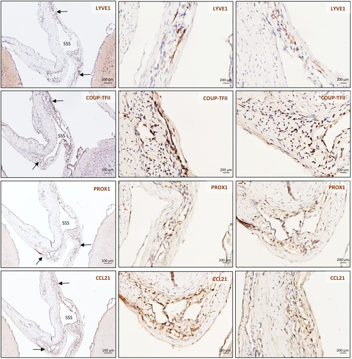
Here, we report the existence of meningeal lymphatic vessels in human and nonhuman primates (common marmoset monkeys) and the feasibility of noninvasively imaging and mapping them in vivo with high-resolution, clinical MRI. On T2-FLAIR and T1-weighted black-blood imaging, lymphatic vessels enhance with gadobutrol, a gadolinium-based contrast agent with high propensity to extravasate across a permeable capillary endothelial barrier, but not with gadofosveset, a blood-pool contrast agent. The topography of these vessels, running alongside dural venous sinuses, recapitulates the meningeal lymphatic system of rodents. In primates, meningeal lymphatics display a typical panel of lymphatic endothelial markers by immunohistochemistry. This discovery holds promise for better understanding the normal physiology of lymphatic drainage from the central nervous system and potential aberrations in neurological diseases.
Overview
The claims and statistics of this paper haven’t been checked yet.
Run the check — free, no account needed. It reads the open-access text and checks the sources it cites.
Integrity signals
Registry
Cites
Statistics
Figures
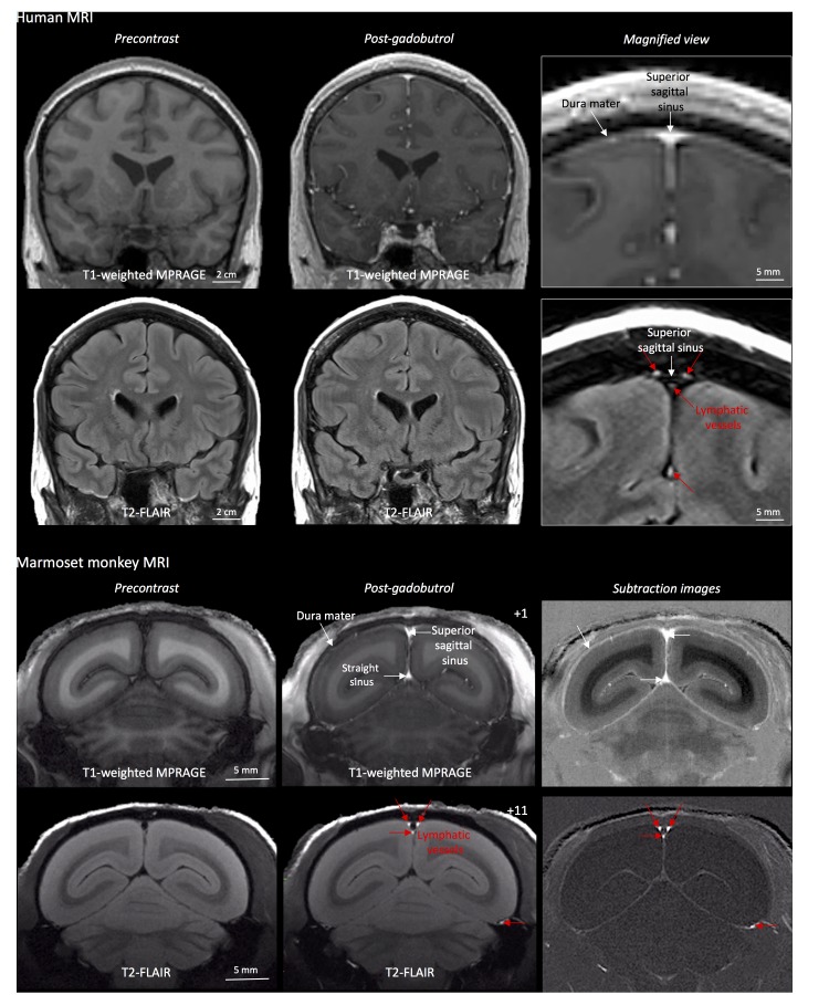
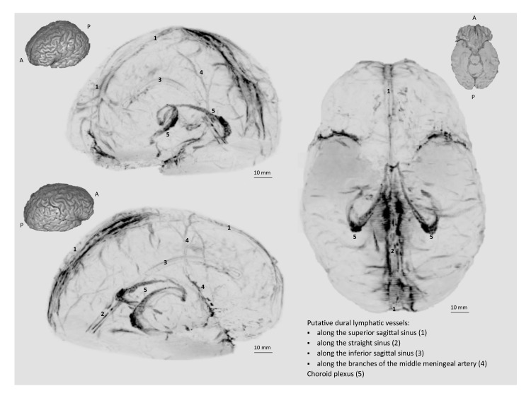
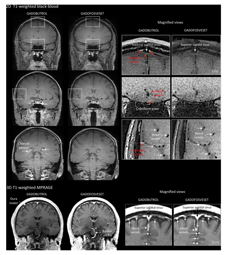
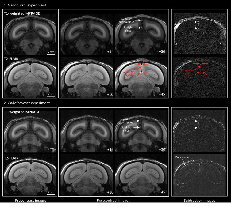
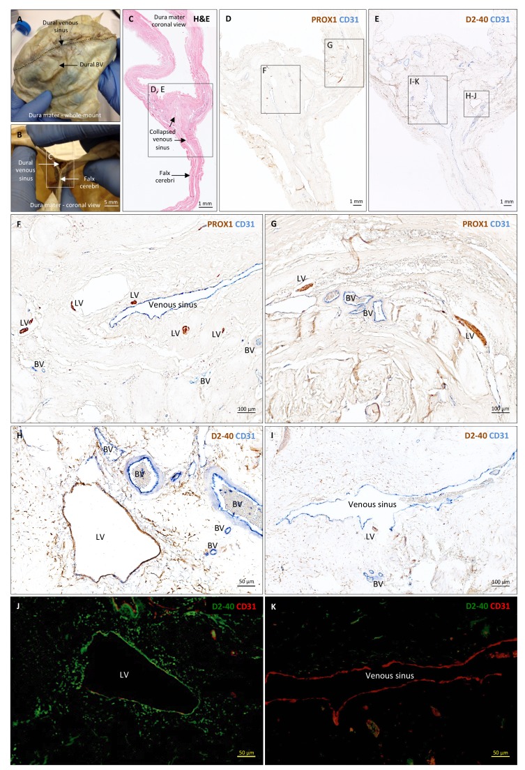
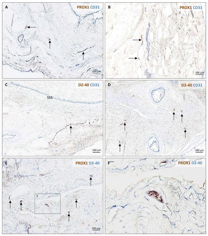
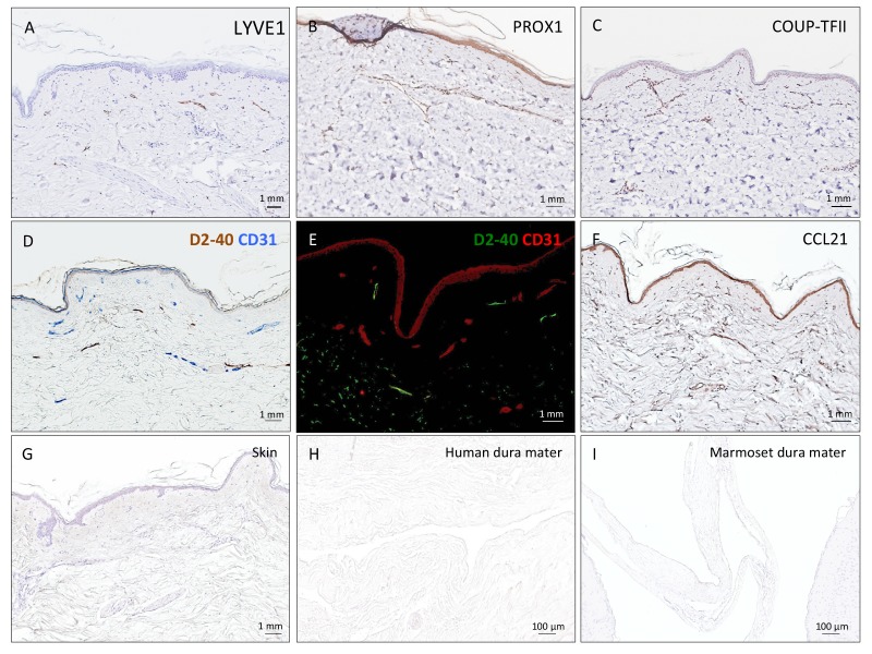
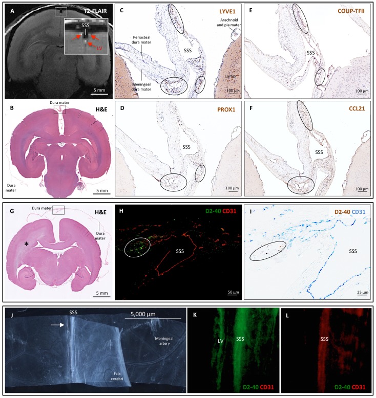
Verification
Claims the paper makes
The claims and statistics of this paper haven’t been checked yet.
Run the check — free, no account needed. It reads the open-access text and checks the sources it cites.
Claims other papers make about this one
Each is checked against this paper’s own text: does it say what the citing paper says it does?
The source backs part of the claim, not all of it — or backs it for a different population, condition or comparison.
In the citing paper
“Recent magnetic resonance imaging (MRI) studies in the living human brain and marmosets utilized a combination of gadolinium-based contrast agent (Gadovist) and blood-pool contrast agent (Vasovist) to demonstrate existence of the meningeal lymphatic system (4).”
From Blood-Brain Barrier: From Physiology to Disease and Back (2019) · cites this paper as [4]
Experimental — accuracy not yet measuredIn the citing paper
“The dural lymphatics are now recognized as a critical transport system of macromolecules, interstitial fluid, and CSF from the CNS into the cervical lymph nodes (Aspelund et al., 2015; Absinta et al., 2017; Louveau et al., 2018; Ahn et al., 2019).”
From Living on the Edge of the CNS: Meninges Cell Diversity in Health and Disease (2021) · cites this paper as Absinta et al., 2017
In the citing paper
“The extent of enhancing tissue within parasagittal dura does not resemble the previously reported appearance of true dural lymphatic vessels, which are tubular-shaped structures of sub-millimeter cross-sectional diameter near the venous sinus wall3,4,11, and also far from the classical MRI appearance of arachnoid granulations bulging into venous sinuses12.”
From Cerebrospinal fluid tracer efflux to parasagittal dura in humans (2020) · cites this paper as [11]
Self-check
Percentages, sums and reported differences re-computed from the paper’s own numbers.
The self-check needs the paper’s full text.
Citations
Cites
Iliff, J. J., Wang, M., Liao, Y., Plogg, B. A., Peng, W., Gundersen, G. A., Benveniste, H., Vates, G. E., Deane, R., Goldman, S. A., et al. (2012) · Science Translational Medicine · doi:10.1126/scitranslmed.3003748 · Cited by 5,824
Sleep Drives Metabolite Clearance from the Adult Brain
Xie, L., Kang, H., Xu, Q., Chen, M. J., Liao, Y., Thiyagarajan, M., O’Donnell, J., Christensen, D. J., Nicholson, C., Iliff, J. J., et al. (2013) · Science · doi:10.1126/science.1241224 · Cited by 5,411
Structural and functional features of central nervous system lymphatic vessels
Louveau, A., Smirnov, I., Keyes, T. J., Eccles, J. D., Rouhani, S. J., Peske, J. D., Derecki, N. C., Castle, D., Mandell, J. W., Lee, K. S., et al. (2015) · Nature · doi:10.1038/nature14432 · Cited by 4,413
A dural lymphatic vascular system that drains brain interstitial fluid and macromolecules
Aspelund, A., Antila, S., Proulx, S. T., Karlsen, T. V., Karaman, S., Detmar, M., Wiig, H., & Alitalo, K. (2015) · Journal of Experimental Medicine · doi:10.1084/jem.20142290 · Cited by 2,198
Lymphangiogenesis in development and human disease
Alitalo, K. K., Tammela, T., & Petrova, T. V. (2005) · Nature · doi:10.1038/nature04480 · Cited by 1,287
Brain-wide pathway for waste clearance captured by contrast-enhanced MRI
Iliff, J. J., Lee, H., Yu, M., Feng, T., Logan, J., Nedergaard, M., & Benveniste, H. (2013) · Journal of Clinical Investigation · doi:10.1172/jci67677 · Cited by 1,148
Lymphangiogenesis and lymphatic vessel remodelling in cancer
Stacker, S. A., Williams, S. P., Karnezis, T., Shayan, R., Fox, S. B., & Achen, M. G. (2014) · Nature reviews. Cancer · doi:10.1038/nrc3677 · Cited by 837
Mandell, D. M., Mossa‐Basha, M., Qiao, Y., Hess, C. P., Hui, F. K., Matouk, C. C., Johnson, M. H., Daemen, M. J. A. P., Vossough, A., Edjlali, M., et al. (2016) · American Journal of Neuroradiology · doi:10.3174/ajnr.a4893 · Cited by 694
Revisiting the Mechanisms of CNS Immune Privilege
Louveau, A., Harris, T. H., & Kipnis, J. (2015) · Trends in Immunology · doi:10.1016/j.it.2015.08.006 · Cited by 682
Self-citationLineage tracing demonstrates the venous origin of the mammalian lymphatic vasculaturePreprint
Srinivasan, R. S., Dillard, M. E., Lagutin, O. V., Lin, F., Tsai, S. Y., Tsai, M., Samokhvalov, I. M., & Oliver, G. C. (2007) · Genes & Development · doi:10.1101/gad.1588407 · Cited by 583
Kida, S., Pantazis, A., & Weller, R. O. (1993) · Neuropathology and Applied Neurobiology · doi:10.1111/j.1365-2990.1993.tb00476.x · Cited by 520
Patterns of Contrast Enhancement in the Brain and Meninges
Smirniotopoulos, J. G., Murphy, F. M., Rushing, E. J., Rees, J., & Schroeder, J. W. (2007) · Radiographics · doi:10.1148/rg.272065155 · Cited by 503
Lymphatic drainage of the brain and the pathophysiology of neurological disease
Weller, R. O., Djuanda, E., Yow, H.-Y., & Carare, R. O. (2009) · Acta Neuropathologica · doi:10.1007/s00401-008-0457-0 · Cited by 488
Live imaging of lymphatic development in the zebrafish
Yaniv, K., Isogai, S., Castranova, D., Dye, L., Hitomi, J., & Weinstein, B. M. (2006) · Nature Medicine · doi:10.1038/nm1427 · Cited by 486
MS-325: albumin-targeted contrast agent for MR angiography.
Lauffer, R. B., Parmelee, D. J., Dunham, S. U., Ouellet, H. S., Dolan, R. P., Witte, S., McMurry, T. J., & Walovitch, R. C. (1998) · Radiology · doi:10.1148/radiology.207.2.9577506 · Cited by 457
Multifaceted interactions between adaptive immunity and the central nervous system
Kipnis, J. (2016) · Science · doi:10.1126/science.aag2638 · Cited by 376
Self-citationGadolinium-based MRI characterization of leptomeningeal inflammation in multiple sclerosis
Absinta, M., Vuolo, L., Rao, A., Nair, G., Sati, P., Cortese, I. C. M., Ohayon, J., Fenton, K., Reyes-Mantilla, M. I., Maric, D., et al. (2015) · Neurology · doi:10.1212/wnl.0000000000001587 · Cited by 296 · cited 6×
“They provide an alternate conduit for drainage of immune cells and cerebrospinal fluid (CSF) from the brain, beyond previously described pathways of flow: via arachnoid granulations into the dural venous sinuses, and via the cribriform plate into the ethmoid region (Weller et al., 2009).”
BackgroundMethodInfluentialSelf-citationThe Schlemm’s canal is a VEGF-C/VEGFR-3–responsive lymphatic-like vessel
Aspelund, A., Tammela, T., Antila, S., Nurmi, H. J., Leppänen, V., Zarkada, G., Stanczuk, L., François, M., Mäkinen, T., Saharinen, P., et al. (2014) · Journal of Clinical Investigation · doi:10.1172/jci75395 · Cited by 235
A genetic Xenopus laevis tadpole model to study lymphangiogenesis
Ny, A., Koch, M., Schneider, M., Neven, E., Tong, R. T., Maity, S., Fischer, C., Plaisance, S., Lambrechts, D., Héligon, C., et al. (2005) · Nature Medicine · doi:10.1038/nm1285 · Cited by 226
Brain: Gadolinium-enhanced Fast Fluid-attenuated Inversion-Recovery MR Imaging
Mathews, V. P., Caldemeyer, K. S., Lowe, M. J., Greenspan, S. L., Weber, D. M., & Ulmer, J. L. (1999) · Radiology · doi:10.1148/radiology.211.1.r99mr25257 · Cited by 211
How Do Meningeal Lymphatic Vessels Drain the CNS?
Raper, D. M. S., Louveau, A., & Kipnis, J. (2016) · Trends in Neurosciences · doi:10.1016/j.tins.2016.07.001 · Cited by 184
New Horizons for Imaging Lymphatic Function
Sharma, R. G., Wendt, J. A., Rasmussen, J. C., Adams, K. E., Marshall, M. V., & Sevick-Muraca, E. M. (2008) · Annals of the New York Academy of Sciences · doi:10.1196/annals.1413.002 · Cited by 126
Dura mater is a potential source of Aβ seeds
Kovács, G. G., Lutz, M. I., Ricken, G., Ströbel, T., Höftberger, R., Preusser, M., Regelsberger, G., Hönigschnabl, S., Reiner, A., Fischer, P. A., et al. (2016) · Acta Neuropathologica · doi:10.1007/s00401-016-1565-x · Cited by 110
Regulation and implications of inflammatory lymphangiogenesis
Kim, H., Kataru, R. P., & Koh, G. Y. (2012) · Trends in Immunology · doi:10.1016/j.it.2012.03.006 · Cited by 99
Mamourian, A. C., Hoopes, P. J., & Lewis, L. D. (2000) · PubMed · Cited by 95
Podoplanin and CLEC-2 drive cerebrovascular patterning and integrity during development
Lowe, K. L., Finney, B. A., Deppermann, C., Hägerling, R., Gazit, S. L., Frampton, J., Buckley, C. D., Camerer, E., Nieswandt, B., Kiefer, F., et al. (2015) · Blood · doi:10.1182/blood-2014-09-603803 · Cited by 94
Postmortem Magnetic Resonance Imaging to Guide the Pathologic Cut
Absinta, M., Nair, G., Filippi, M., Ray‐Chaudhury, A., Reyes-Mantilla, M. I., Pardo, C. A., & Reich, D. S. (2014) · Journal of Neuropathology & Experimental Neurology · doi:10.1097/nen.0000000000000096 · Cited by 74
Pachymeningeal enhancement—a comprehensive review of literature
Antony, J., Hacking, C., & Jeffree, R. L. (2015) · Neurosurgical Review · doi:10.1007/s10143-015-0646-y · Cited by 58
Gaitán, M. I., Maggi, P., Wohler, J. E., Leibovitch, E. C., Sati, P., Calandri, I. L., Merkle, H., Massacesi, L., Silva, A. C., Jacobson, S., et al. (2013) · Multiple Sclerosis Journal · doi:10.1177/1352458513492244 · Cited by 31
Utilizing 3D Printing Technology to Merge MRI with Histology: A Protocol for Brain Sectioning
Luciano, N. J., Sati, P., Nair, G., Guy, J. R., Ha, S.-K., Absinta, M., Chiang, W.-Y., Leibovitch, E. C., Jacobson, S., Silva, A. C., et al. (2016) · Journal of Visualized Experiments · doi:10.3791/54780 · Cited by 30
Custom fit 3D-printed brain holders for comparison of histology with MRI in marmosets
Guy, J. R., Sati, P., Leibovitch, E. C., Jacobson, S., Silva, A. C., & Reich, D. S. (2015) · Journal of Neuroscience Methods · doi:10.1016/j.jneumeth.2015.09.002 · Cited by 29
Richardson, O., Bane, O., Scott, M. L. J., Tanner, S. F., Waterton, J. C., Sourbron, S. P., Carroll, T. J., & Buckley, D. L. (2014) · Magnetic Resonance in Medicine · doi:10.1002/mrm.25128 · Cited by 15
Research works printed but not linked: 1
- Mascagni, 1816, Istoria Completa Dei Vasi Linfatici, Vol. II, 195
Cited by
Sorted by citations
Blood-Brain Barrier: From Physiology to Disease and Back
Sweeney, M. D., Zhao, Z., Montagne, A., Nelson, A. R., & Zlokovic, B. V. (2019) · Physiological Reviews · doi:10.1152/physrev.00050.2017 · Cited by 2,312
The glymphatic pathway in neurological disorders.
Rasmussen, M. K., Mestre, H., & Nedergaard, M. (2018) · The Lancet Neurology · doi:10.1016/s1474-4422(18)30318-1 · Cited by 1,560
Functional aspects of meningeal lymphatics in ageing and Alzheimer’s disease
Da Mesquita, S., Louveau, A., Vaccari, A., Smirnov, I., Cornelison, R. C., Kingsmore, K. M., Contarino, C., Onengut-Gumuscu, S., Farber, E., Raper, D., et al. (2018) · Nature · doi:10.1038/s41586-018-0368-8 · Cited by 1,445
Generation of circadian rhythms in the suprachiasmatic nucleus
Hastings, M. H., Maywood, E. S., & Brancaccio, M. (2018) · Nature Reviews Neuroscience · doi:10.1038/s41583-018-0026-z · Cited by 1,065
CNS lymphatic drainage and neuroinflammation are regulated by meningeal lymphatic vasculature.
Louveau, A., Herz, J., Alme, M. N., Salvador, A. F., Dong, M. Q., Viar, K. E., Herod, S. G., Knopp, J., Setliff, J. C., Lupi, A. L., et al. (2018) · Nature Neuroscience · doi:10.1038/s41593-018-0227-9 · Cited by 956
Metadata
- Authors
- Absinta, M., Ha, S.-K., Nair, G., Sati, P., Luciano, N. J., Palisoc, M., Louveau, A., Zaghloul, K. A., Pittaluga, S., Kipnis, J., & Reich, D. S.
- Affiliations
- National Institutes of Health (US)
- University of Virginia (US)
- Corresponding author
- Reich, Daniel S
- ORCID
- Venue
- eLife
- Publisher
- eLife Sciences Publications, Ltd
- Published
- 2017-10-03
- Volume
- 6
- ISSN
- 2050-084X (ISSN-L)
- PMC
- PMC5626482
- OpenAlex
- W2760815599
- Cited by
- 643
- References
- 32
- Citations by year
- 2026: 61 · 2025: 88 · 2024: 73 · 2023: 77 · 2022: 79 · 2021: 78 · 2020: 70 · 2019: 60 · 2018: 51 · 2017: 4
- FWCI
- 22.31
- Type
- journal article
- Language
- en
- Open access
- gold
- doi.org
- Licence
- Locations
- eLife (publishedVersion, cc-by)
- Europe PMC (PubMed Central) (publishedVersion, public-domain)
- PubMed Central (publishedVersion, public-domain)
- PubMed (publishedVersion)
- DOAJ (DOAJ: Directory of Open Access Journals) (submittedVersion, cc-by-sa)
- Funders
- National Institute of Neurological Disorders and Stroke (Intramural Research Program)
- National Multiple Sclerosis Society (Postdoctoral Fellowship)
- Conrad N. Hilton Foundation (Marilyn Hilton Award for Innovation in Multiple Sclerosis Research)
- National Cancer Institute (Intramural Research Program)
- National Institute on Aging (R01AG034113)
- Lymphatic Education and Research Network (Postdoctoral fellowship)
- Topics
- Health Sciences › Medicine › Oncology › Lymphatic System and Diseases
- Health Sciences › Medicine › Clinical Neurology › Cerebrospinal fluid and hydrocephalus
- Health Sciences › Medicine › Surgery › Lymphatic Disorders and Treatments
- Keywords
- meningeal lymphatic vessels, meningeal lymphatic system, non-human primates, Callithrix jacchus, human brain, gadobutrol, gadofosveset, contrast extravasation, lymphatic endothelial markers, dural venous sinuses, black-blood imaging
- MeSH terms
- Animals, Callithrix, Humans, Magnetic Resonance Imaging, Meninges, Lymphatic Vessels
About these checks
Registry, retraction, Cites and statistics results are computed, not judged, with one exception: a printed reference that no index lists is read once by a model before it counts as a citation error. Claims are extracted by a model and kept only when their quote appears verbatim in the text; each cited source is then checked, with a model’s help, for whether it says what the claim relies on. Those verdicts can be wrong — read the claim’s quote and the cited source before relying on one.
Statistics and arithmetic are recomputed, not judged.
Confidence bands are shown only for fields calibrated on our annotated test set. None is calibrated yet, so no band is shown.
What counts as a claim ›How we measure confidence ›How we check this ›
Are you an author, or do you see a problem with this page? Request a review
