

The DOI matches its Crossref record; 32 of the 32 references it cites were found in a registry or an index.
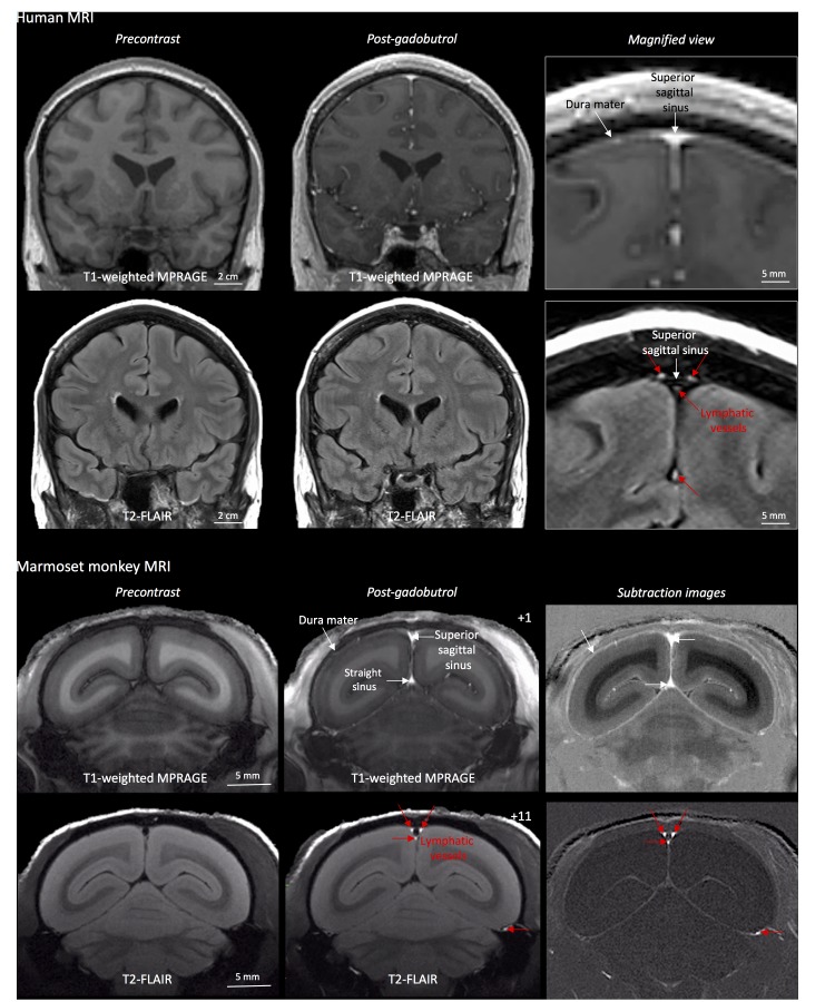
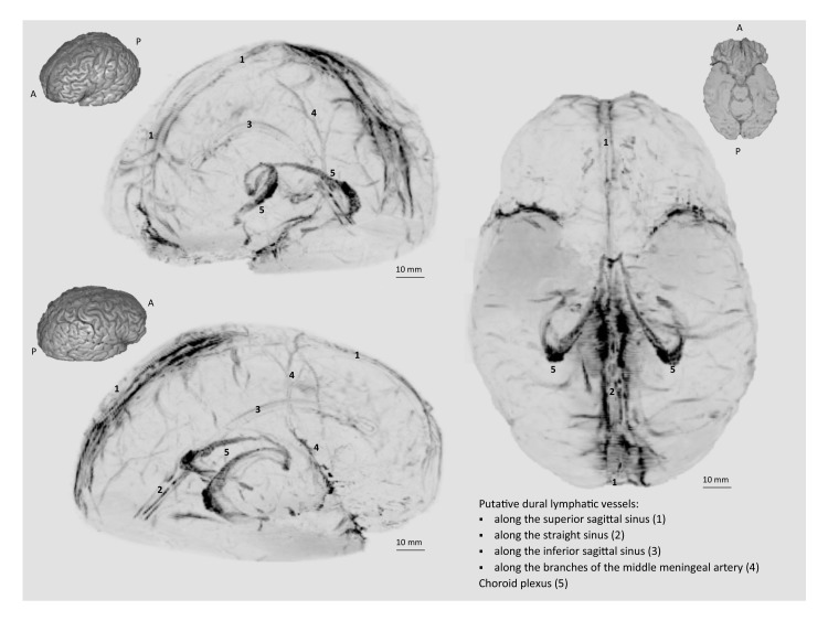
Human and nonhuman primate meninges harbor lymphatic vessels that can be visualized noninvasively by MRI.
Open accessAbstract
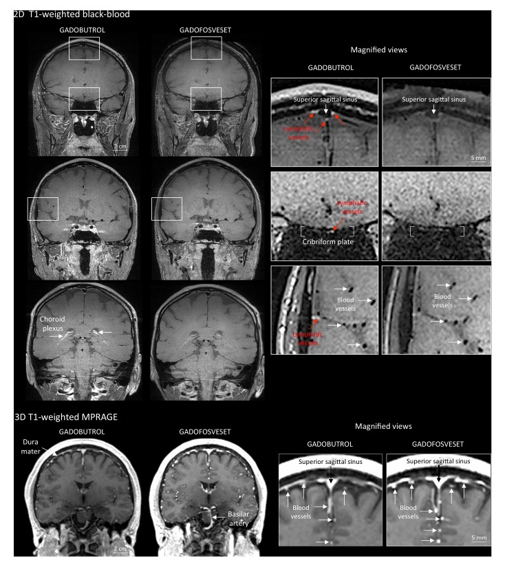
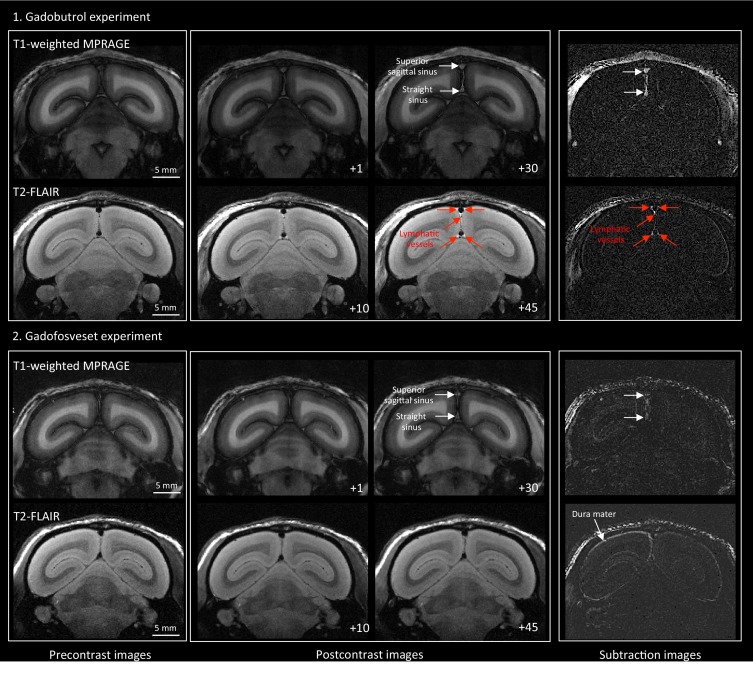
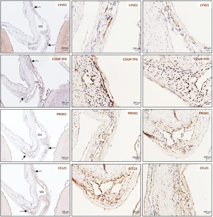
Here, we report the existence of meningeal lymphatic vessels in human and nonhuman primates (common marmoset monkeys) and the feasibility of noninvasively imaging and mapping them in vivo with high-resolution, clinical MRI. On T2-FLAIR and T1-weighted black-blood imaging, lymphatic vessels enhance with gadobutrol, a gadolinium-based contrast agent with high propensity to extravasate across a permeable capillary endothelial barrier, but not with gadofosveset, a blood-pool contrast agent. The topography of these vessels, running alongside dural venous sinuses, recapitulates the meningeal lymphatic system of rodents. In primates, meningeal lymphatics display a typical panel of lymphatic endothelial markers by immunohistochemistry. This discovery holds promise for better understanding the normal physiology of lymphatic drainage from the central nervous system and potential aberrations in neurological diseases.
Overview
The claims and statistics of this paper haven’t been checked yet.
Run the check — free, no account needed. It reads the open-access text and checks the sources it cites.
Integrity signals
Registry
Cites
Statistics
Figures
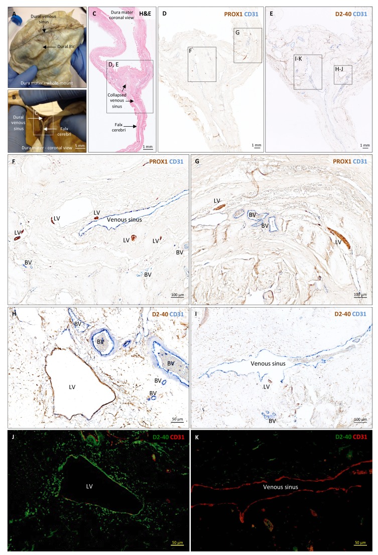
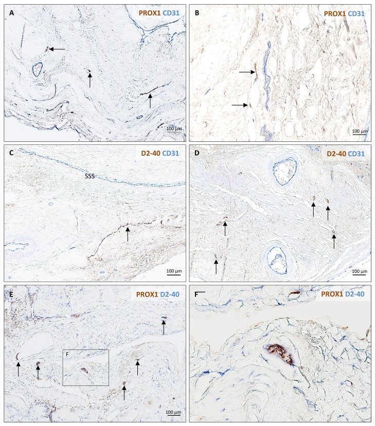
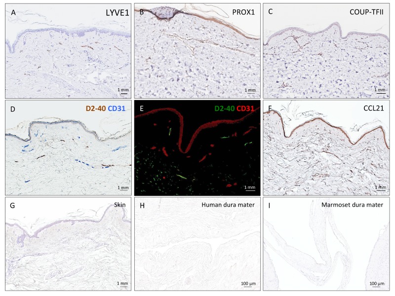
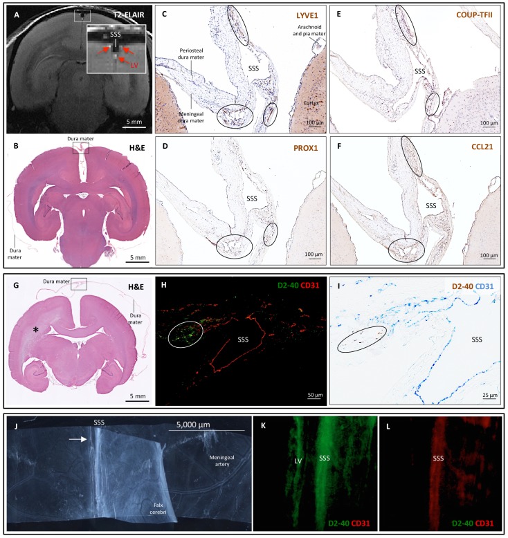
Verification
Claims the paper makes
The claims and statistics of this paper haven’t been checked yet.
Run the check — free, no account needed. It reads the open-access text and checks the sources it cites.
Claims other papers make about this one
Each is checked against this paper’s own text: does it say what the citing paper says it does?
The source backs part of the claim, not all of it — or backs it for a different population, condition or comparison.
In the citing paper
“Recent magnetic resonance imaging (MRI) studies in the living human brain and marmosets utilized a combination of gadolinium-based contrast agent (Gadovist) and blood-pool contrast agent (Vasovist) to demonstrate existence of the meningeal lymphatic system (4).”
From Blood-Brain Barrier: From Physiology to Disease and Back (2019) · cites this paper as [4]
Experimental — accuracy not yet measuredIn the citing paper
“The dural lymphatics are now recognized as a critical transport system of macromolecules, interstitial fluid, and CSF from the CNS into the cervical lymph nodes (Aspelund et al., 2015; Absinta et al., 2017; Louveau et al., 2018; Ahn et al., 2019).”
From Living on the Edge of the CNS: Meninges Cell Diversity in Health and Disease (2021) · cites this paper as Absinta et al., 2017
In the citing paper
“The extent of enhancing tissue within parasagittal dura does not resemble the previously reported appearance of true dural lymphatic vessels, which are tubular-shaped structures of sub-millimeter cross-sectional diameter near the venous sinus wall3,4,11, and also far from the classical MRI appearance of arachnoid granulations bulging into venous sinuses12.”
From Cerebrospinal fluid tracer efflux to parasagittal dura in humans (2020) · cites this paper as [11]
Self-check
Percentages, sums and reported differences re-computed from the paper’s own numbers.
The self-check needs the paper’s full text.
Citations
Cites
Iliff, J. J., Wang, M., Liao, Y., Plogg, B. A., Peng, W., Gundersen, G. A., Benveniste, H., Vates, G. E., Deane, R., Goldman, S. A., et al. (2012) · Science Translational Medicine · doi:10.1126/scitranslmed.3003748 · Cited by 5,824
Sleep Drives Metabolite Clearance from the Adult Brain
Xie, L., Kang, H., Xu, Q., Chen, M. J., Liao, Y., Thiyagarajan, M., O’Donnell, J., Christensen, D. J., Nicholson, C., Iliff, J. J., et al. (2013) · Science · doi:10.1126/science.1241224 · Cited by 5,411
Structural and functional features of central nervous system lymphatic vessels
Louveau, A., Smirnov, I., Keyes, T. J., Eccles, J. D., Rouhani, S. J., Peske, J. D., Derecki, N. C., Castle, D., Mandell, J. W., Lee, K. S., et al. (2015) · Nature · doi:10.1038/nature14432 · Cited by 4,413
A dural lymphatic vascular system that drains brain interstitial fluid and macromolecules
Aspelund, A., Antila, S., Proulx, S. T., Karlsen, T. V., Karaman, S., Detmar, M., Wiig, H., & Alitalo, K. (2015) · Journal of Experimental Medicine · doi:10.1084/jem.20142290 · Cited by 2,198
Lymphangiogenesis in development and human disease
Alitalo, K. K., Tammela, T., & Petrova, T. V. (2005) · Nature · doi:10.1038/nature04480 · Cited by 1,287
Brain-wide pathway for waste clearance captured by contrast-enhanced MRI
Iliff, J. J., Lee, H., Yu, M., Feng, T., Logan, J., Nedergaard, M., & Benveniste, H. (2013) · Journal of Clinical Investigation · doi:10.1172/jci67677 · Cited by 1,148
Lymphangiogenesis and lymphatic vessel remodelling in cancer
Stacker, S. A., Williams, S. P., Karnezis, T., Shayan, R., Fox, S. B., & Achen, M. G. (2014) · Nature reviews. Cancer · doi:10.1038/nrc3677 · Cited by 837
Mandell, D. M., Mossa‐Basha, M., Qiao, Y., Hess, C. P., Hui, F. K., Matouk, C. C., Johnson, M. H., Daemen, M. J. A. P., Vossough, A., Edjlali, M., et al. (2016) · American Journal of Neuroradiology · doi:10.3174/ajnr.a4893 · Cited by 694
Revisiting the Mechanisms of CNS Immune Privilege
Louveau, A., Harris, T. H., & Kipnis, J. (2015) · Trends in Immunology · doi:10.1016/j.it.2015.08.006 · Cited by 682
Self-citationLineage tracing demonstrates the venous origin of the mammalian lymphatic vasculaturePreprint
Srinivasan, R. S., Dillard, M. E., Lagutin, O. V., Lin, F., Tsai, S. Y., Tsai, M., Samokhvalov, I. M., & Oliver, G. C. (2007) · Genes & Development · doi:10.1101/gad.1588407 · Cited by 583
Research works printed but not linked: 1
- Mascagni, 1816, Istoria Completa Dei Vasi Linfatici, Vol. II, 195
Cited by
Sorted by citations
Blood-Brain Barrier: From Physiology to Disease and Back
Sweeney, M. D., Zhao, Z., Montagne, A., Nelson, A. R., & Zlokovic, B. V. (2019) · Physiological Reviews · doi:10.1152/physrev.00050.2017 · Cited by 2,312
The glymphatic pathway in neurological disorders.
Rasmussen, M. K., Mestre, H., & Nedergaard, M. (2018) · The Lancet Neurology · doi:10.1016/s1474-4422(18)30318-1 · Cited by 1,560
Functional aspects of meningeal lymphatics in ageing and Alzheimer’s disease
Da Mesquita, S., Louveau, A., Vaccari, A., Smirnov, I., Cornelison, R. C., Kingsmore, K. M., Contarino, C., Onengut-Gumuscu, S., Farber, E., Raper, D., et al. (2018) · Nature · doi:10.1038/s41586-018-0368-8 · Cited by 1,445
Generation of circadian rhythms in the suprachiasmatic nucleus
Hastings, M. H., Maywood, E. S., & Brancaccio, M. (2018) · Nature Reviews Neuroscience · doi:10.1038/s41583-018-0026-z · Cited by 1,065
CNS lymphatic drainage and neuroinflammation are regulated by meningeal lymphatic vasculature.
Louveau, A., Herz, J., Alme, M. N., Salvador, A. F., Dong, M. Q., Viar, K. E., Herod, S. G., Knopp, J., Setliff, J. C., Lupi, A. L., et al. (2018) · Nature Neuroscience · doi:10.1038/s41593-018-0227-9 · Cited by 956
Perivascular spaces in the brain: anatomy, physiology and pathology
Wardlaw, J. M., Benveniste, H., Nedergaard, M., Zlokovic, B. V., Mestre, H., Lee, H., Doubal, F. N., Brown, R., Ramirez, J., MacIntosh, B. J., et al. (2020) · Nature Reviews Neurology · doi:10.1038/s41582-020-0312-z · Cited by 870
Meningeal lymphatic vessels at the skull base drain cerebrospinal fluid
Ahn, J. H., Cho, H., Kim, J.-H., Kim, S. H., Ham, J.-S., Park, I., Suh, S. H., Hong, S. P., Song, J.-H., Hong, Y.-K., et al. (2019) · Nature · doi:10.1038/s41586-019-1419-5 · Cited by 811
History and progress of hypotheses and clinical trials for Alzheimer’s disease
Liu, P.-P., Xie, Y., Meng, X.-Y., & Kang, J.-S. (2019) · Signal Transduction and Targeted Therapy · doi:10.1038/s41392-019-0063-8 · Cited by 723
The Brain’s Glymphatic System: Current Controversies
Mestre, H., Mori, Y., & Nedergaard, M. (2020) · Trends in Neurosciences · doi:10.1016/j.tins.2020.04.003 · Cited by 609
Rasmussen, M. K., Mestre, H., & Nedergaard, M. (2021) · Physiological Reviews · doi:10.1152/physrev.00031.2020 · Cited by 642
The Glymphatic System and Waste Clearance with Brain Aging: A Review
Benveniste, H., Liu, X., Koundal, S., Sanggaard, S., Lee, H., & Wardlaw, J. (2019) · Gerontology · doi:10.1159/000490349 · Cited by 517
Neurovascular and Cognitive Dysfunction in Hypertension
Iadecola, C., & Gottesman, R. F. (2019) · Circulation Research · doi:10.1161/circresaha.118.313260 · Cited by 530
Vasomotion as a Driving Force for Paravascular Clearance in the Awake Mouse Brain
van Veluw, S. J., Hou, S. S., Calvo-Rodriguez, M., Arbel-Ornath, M., Snyder, A. C., Frosch, M. P., Greenberg, S. M., & Bacskai, B. J. (2020) · Neuron · doi:10.1016/j.neuron.2019.10.033 · Cited by 500
The Meningeal Lymphatic System: A New Player in Neurophysiology
Da Mesquita, S., Fu, Z., & Kipnis, J. (2018) · Neuron · doi:10.1016/j.neuron.2018.09.022 · Cited by 479
Biological functions of lymphatic vessels
Petrova, T. V., & Koh, G. Y. (2020) · Science · doi:10.1126/science.aax4063 · Cited by 474
Immunoneuropsychiatry — novel perspectives on brain disorders
Pape, K., Tamouza, R., Leboyer, M., & Zipp, F. (2019) · Nature Reviews Neurology · doi:10.1038/s41582-019-0174-4 · Cited by 461
Macrophages at CNS interfaces: Ontogeny and function in health and disease
Kierdorf, K., Masuda, T., Jordão, M. J. C., & Prinz, M. (2019) · Nature Reviews Neuroscience · doi:10.1038/s41583-019-0201-x · Cited by 416
Reassessing B cell contributions in multiple sclerosis
Li, R., Patterson, K. R., & Bar‐Or, A. (2018) · Nature Immunology · doi:10.1038/s41590-018-0135-x · Cited by 417
Zou, W., Pu, T., Feng, W., Lu, M., Zheng, Y., Du, R., Xiao, M., & Hu, G. (2019) · Translational Neurodegeneration · doi:10.1186/s40035-019-0147-y · Cited by 396
Proulx, S. T. (2021) · Cellular and Molecular Life Sciences · doi:10.1007/s00018-020-03706-5 · Cited by 412
The immunology of multiple sclerosis
Attfield, K. E., Jensen, L. T., Kaufmann, M., Friese, M. A., & Fugger, L. (2022) · Nature Reviews Immunology · doi:10.1038/s41577-022-00718-z · Cited by 380
Meningeal lymphatic vessels regulate brain tumor drainage and immunity
Hu, X., Deng, Q., Ma, L., Li, Q., Chen, Y., Liao, Y., Zhou, F., Zhang, C., Shao, L., Feng, J., et al. (2020) · Cell Research · doi:10.1038/s41422-020-0287-8 · Cited by 378
Meningeal lymphatic dysfunction exacerbates traumatic brain injury pathogenesis.
Bolte, A. C., Dutta, A. B., Hurt, M. E., Smirnov, I., Kovacs, M. A., McKee, C. A., Ennerfelt, H. E., Shapiro, D., Nguyen, B. H., Frost, E. L., et al. (2020) · Nature Communications · doi:10.1038/s41467-020-18113-4 · Cited by 357
Alves de Lima, K., Rustenhoven, J., & Kipnis, J. (2020) · Annual Review of Immunology · doi:10.1146/annurev-immunol-102319-103410 · Cited by 348
Advances in Meningeal Immunity
Rua, R., & McGavern, D. B. (2018) · Trends in Molecular Medicine · doi:10.1016/j.molmed.2018.04.003 · Cited by 326
Impairment of the Glymphatic Pathway and Putative Meningeal Lymphatic Vessels in the Aging Human
Zhou, Y., Cai, J., Zhang, W., Gong, X., Yan, S., Zhang, K., Luo, Z., Sun, J., Jiang, Q., & Lou, M. (2020) · Annals of Neurology · doi:10.1002/ana.25670 · Cited by 305
The Evolving Landscape of Brain Metastasis
Valiente, M., Ahluwalia, M. S., Boire, A. A., Brastianos, P. K., Goldberg, S. B., Lee, E. Q., Rhun, É. L., Preusser, M., Winkler, F., & Soffietti, R. (2018) · Trends in cancer · doi:10.1016/j.trecan.2018.01.003 · Cited by 315
Organ-specific lymphatic vasculature: From development to pathophysiology
Petrova, T. V., & Koh, G. Y. (2017) · The Journal of Experimental Medicine · doi:10.1084/jem.20171868 · Cited by 309
Cerebrospinal fluid tracer efflux to parasagittal dura in humans
Ringstad, G., & Eide, P. K. (2020) · Nature Communications · doi:10.1038/s41467-019-14195-x · Cited by 292
Eide, P. K., Vatnehol, S. A. S., Emblem, K. E., & Ringstad, G. (2018) · Scientific Reports · doi:10.1038/s41598-018-25666-4 · Cited by 299
Glymphatic system impairment in multiple sclerosis: relation with brain damage and disability
Carotenuto, A., Cacciaguerra, L., Pagani, E., Preziosa, P., Filippi, M., & Rocca, M. A. (2021) · Brain · doi:10.1093/brain/awab454 · Cited by 288
Identification of direct connections between the dura and the brain
Smyth, L. C. D., Xu, D., Okar, S. V., Dykstra, T., Rustenhoven, J., Papadopoulos, Z., Bhasiin, K., Kim, M. W., Drieu, A., Mamuladze, T., et al. (2024) · Nature · doi:10.1038/s41586-023-06993-7 · Cited by 264
Brain borders at the central stage of neuroimmunology
Rustenhoven, J., & Kipnis, J. (2022) · Nature · doi:10.1038/s41586-022-05474-7 · Cited by 253
Zhao, P., Wang, Y., Kang, X., Wu, A., Yin, W., Tang, Y., Wang, J., Zhang, M., Duan, Y., & Huang, Y. (2018) · Chemical Science · doi:10.1039/c7sc04853j · Cited by 249
Taoka, T., & Naganawa, S. (2019) · Journal of Magnetic Resonance Imaging · doi:10.1002/jmri.26892 · Cited by 244
An ocular glymphatic clearance system removes β-amyloid from the rodent eye
Wang, X., Lou, N., Eberhardt, A., Yang, Y., Kusk, P., Xu, Q., Förstera, B., Peng, S., Shi, M., Ladrón-de-Guevara, A., et al. (2020) · Science Translational Medicine · doi:10.1126/scitranslmed.aaw3210 · Cited by 239
Brain Microvascular Pericytes in Vascular Cognitive Impairment and Dementia
Uemura, M. T., Maki, T., Ihara, M., Lee, V. M. ‐., & Trojanowski, J. Q. (2020) · Frontiers in Aging Neuroscience · doi:10.3389/fnagi.2020.00080 · Cited by 246
The glymphatic system: implications for drugs for central nervous system diseases
Lohela, T. J., Lilius, T. O., & Nedergaard, M. (2022) · Nature Reviews Drug Discovery · doi:10.1038/s41573-022-00500-9 · Cited by 254
Xiong, Y., Mahmood, A., & Chopp, M. (2018) · Chinese Journal of Traumatology · doi:10.1016/j.cjtee.2018.02.003 · Cited by 253
Non-invasive MR imaging of human brain lymphatic networks with connections to cervical lymph nodes
Albayram, S., Smith, G., Tufan, F., Tuna, İ. S., Bostancıklıoğlu, M., Zile, M. R., & Albayram, Ö. (2022) · Nature Communications · doi:10.1038/s41467-021-27887-0 · Cited by 246
COVID-19 and chronic fatigue syndrome: Is the worst yet to come?
Wostyn, P. (2021) · Medical Hypotheses · doi:10.1016/j.mehy.2020.110469 · Cited by 236
Nasopharyngeal lymphatic plexus is a hub for cerebrospinal fluid drainage
Yoon, J., Jin, H., Kim, H. J., Hong, S. P., Yang, M. J., Ahn, J. H., Kim, Y. C., Seo, J., Lee, Y., McDonald, D. M., et al. (2024) · Nature · doi:10.1038/s41586-023-06899-4 · Cited by 210
Hladky, S. B., & Barrand, M. A. (2018) · Fluids and Barriers of the CNS · doi:10.1186/s12987-018-0113-6 · Cited by 206
Si, X., Guo, T., Wang, Z., Fang, Y., Gu, L., Cao, L., Yang, W., Gao, T., Song, Z., Tian, J., et al. (2022) · npj Parkinson s Disease · doi:10.1038/s41531-022-00316-9 · Cited by 204
Conserved meningeal lymphatic drainage circuits in mice and humans
Jacob, L., Brito, J. M., Lenck, S., Corcy, C., Benbelkacem, F., Geraldo, L. H., Xu, Y., Thomas, J.-M., Kamouh, M.-R. E., Spajer, M., et al. (2022) · The Journal of Experimental Medicine · doi:10.1084/jem.20220035 · Cited by 206
Wei, J., Chen, P., Gupta, P., Ott, M., Zamler, D. B., Kassab, C., Bhat, K., Curran, M. A., de Groot, J., & Heimberger, A. B. (2019) · Neuro-Oncology · doi:10.1093/neuonc/noz212 · Cited by 192
Cheng, Y., Tian, D., & Wang, Y.-J. (2020) · Translational Neurodegeneration · doi:10.1186/s40035-020-00195-1 · Cited by 197
Glymphatic System Function in Relation to Anesthesia and Sleep States
Benveniste, H., Heerdt, P. M., Fontes, M. L., Rothman, D. L., & Volkow, N. D. (2019) · Anesthesia & Analgesia · doi:10.1213/ane.0000000000004069 · Cited by 182
Aging Microbiota-Gut-Brain Axis in Stroke Risk and Outcome
Honarpisheh, P., Bryan, R. M., & McCullough, L. D. (2022) · Circulation Research · doi:10.1161/circresaha.122.319983 · Cited by 176
Hu, B., & Arya, A. K. (2018) · Brain Circulation · doi:10.4103/bc.bc_32_18 · Cited by 170
Gouveia-Freitas, K., & de Bastos-Leite, A. J. (2021) · Neuroradiology · doi:10.1007/s00234-021-02718-7 · Cited by 166
Advances and Challenges in Understanding Cerebral Toxoplasmosis
Schlüter, D., & Barragán, A. (2019) · Frontiers in Immunology · doi:10.3389/fimmu.2019.00242 · Cited by 162
Mohan, R., Liu, A. Y., Brown, P. D., Mahajan, A., Dinh, J., Chung, C. D., McAvoy, S., McAleer, M. F., Lin, S. H., Li, J., et al. (2020) · Neuro-Oncology · doi:10.1093/neuonc/noaa182 · Cited by 156
The immunological response to traumatic brain injury
Needham, E., Helmy, A. E., Zanier, E. R., Jones, J., Coles, A. J., & Menon, D. K. (2019) · Journal of Neuroimmunology · doi:10.1016/j.jneuroim.2019.04.005 · Cited by 161
Imaging perivascular space structure and function using brain MRI
Barisano, G., Lynch, K. M., Sibilia, F., Lan, H., Shih, N., Sepehrband, F., & Choupan, J. (2022) · NeuroImage · doi:10.1016/j.neuroimage.2022.119329 · Cited by 151
Chen, S., Shao, L., & Ma, L. (2021) · Frontiers in Cellular Neuroscience · doi:10.3389/fncel.2021.716825 · Cited by 154
Anatomy and function of the vertebral column lymphatic network in mice
Jacob, L., Boisserand, L. S. B., Geraldo, L. H. M., de Brito Neto, J., Mathivet, T., Antila, S., Barka, B., Xu, Y., Thomas, J.-M., Pestel, J., et al. (2019) · Nature Communications · doi:10.1038/s41467-019-12568-w · Cited by 144
The role of sleep deprivation and circadian rhythm disruption as risk factors of Alzheimer’s disease
Wu, H., Dunnett, S. L., Ho, J. Y. S., & Chang, R. C. (2019) · Frontiers in Neuroendocrinology · doi:10.1016/j.yfrne.2019.100764 · Cited by 154
McKnight, C. D., Trujillo, P., Lopez, A. M., Petersen, K. J., Considine, C. M., Lin, Y., Yan, Y., Kang, H., Donahue, M. J., & Claassen, D. O. (2021) · Parkinsonism & Related Disorders · doi:10.1016/j.parkreldis.2021.06.004 · Cited by 142
Pu, T., Zou, W., Feng, W., Zhang, Y., Wang, L., Wang, H., & Xiao, M. (2019) · Experimental Neurobiology · doi:10.5607/en.2019.28.1.104 · Cited by 143
Zivadinov, R., Guan, Y., Jakimovski, D., Ramanathan, M. K., & Weinstock‐Guttman, B. (2018) · Neural Regeneration Research · doi:10.4103/1673-5374.245462 · Cited by 150
The enigma and implications of brain hemispheric asymmetry in neurodegenerative diseases
Lubben, N., Ensink, E., Coetzee, G. A., & Labrie, V. (2021) · Brain Communications · doi:10.1093/braincomms/fcab211 · Cited by 139
The immune system and psychiatric disease: a basic science perspective
Bennett, F. C., & Molofsky, A. V. (2019) · Clinical & Experimental Immunology · doi:10.1111/cei.13334 · Cited by 141
Living on the Edge of the CNS: Meninges Cell Diversity in Health and Disease
Derk, J., Jones, H. E., Como, C., Pawlikowski, B., & Siegenthaler, J. A. (2021) · Frontiers in Cellular Neuroscience · doi:10.3389/fncel.2021.703944 · Cited by 120
“The dural lymphatics are now recognized as a critical transport system of macromolecules, interstitial fluid, and CSF from the CNS into the cervical lymph nodes (Aspelund et al., 2015; Absinta et al., 2017; Louveau et al., 2018; Ahn et al., 2019).”
BackgroundMicrostructural imaging of human neocortex in vivo
Edwards, L., Kirilina, E. P., Mohammadi, S., & Weiskopf, N. (2018) · NeuroImage · doi:10.1016/j.neuroimage.2018.02.055 · Cited by 122
Zheng, Z.-N., Zhang, J.-X., Jiang, J., He, Y., Zhang, W.-Y., Mo, X.-P., Kang, X.-J., Xu, Q., Wang, B., & Huang, Y.-Z. (2020) · Journal for ImmunoTherapy of Cancer · doi:10.1136/jitc-2019-000207 · Cited by 116
Current understanding of meningeal and cerebral vascular function underlying migraine headache
Levy, D., Labastida‐Ramírez, A., & MaassenVanDenBrink, A. (2018) · Cephalalgia · doi:10.1177/0333102418771350 · Cited by 130
Glymphatics visualization after focused ultrasound-induced blood-brain barrier opening in humans
Meng, Y., Abrahao, A., Heyn, C. C., Bethune, A. J., Huang, Y., Pople, C. B., Aubert, I., Hamani, C., Zinman, L., Hynynen, K., et al. (2019) · Annals of Neurology · doi:10.1002/ana.25604 · Cited by 114
Skull bone marrow channels as immune gateways to the central nervous system
Mazzitelli, J. A., Pulous, F. E., Smyth, L. C. D., Kaya, Z., Rustenhoven, J., Moskowitz, M. A., Kipnis, J., & Nahrendorf, M. (2023) · Nature Neuroscience · doi:10.1038/s41593-023-01487-1 · Cited by 116
Idiopathic intracranial hypertension
Lenck, S., Radovanovic, I., Nicholson, P. J., Hodaie, M., Krings, T., & Pereira, V. M. (2018) · Neurology · doi:10.1212/wnl.0000000000006166 · Cited by 124
The pathogenesis of cerebral small vessel disease and vascular cognitive impairment
Markus, H. S., & Joutel, A. (2025) · Physiological Reviews · doi:10.1152/physrev.00028.2024 · Cited by 140
Klostranec, J. M., Vucevic, D., Bhatia, K. D., Kortman, H. G. J., Krings, T., Murphy, K., terBrugge, K. G., & Mikulis, D. J. (2021) · Radiology · doi:10.1148/radiol.2021204088 · Cited by 121
Endothelial cell barriers: Transport of molecules between blood and tissues
Yazdani, S., Jaldín‐Fincati, J. R., Pereira, R. V. S., & Klip, A. (2019) · Traffic · doi:10.1111/tra.12645 · Cited by 126
Naganawa, S., & Taoka, T. (2020) · Magnetic Resonance in Medical Sciences · doi:10.2463/mrms.rev.2020-0122 · Cited by 122
Perivascular Spaces, Glymphatic System and MR
Yu, L., Hu, X., Li, H., & Zhao, Y. (2022) · Frontiers in Neurology · doi:10.3389/fneur.2022.844938 · Cited by 118
Microbiota–gut–brain axis: interplay between microbiota, barrier function and lymphatic system
Zhuang, M., Zhang, X., & Cai, J. (2024) · Gut Microbes · doi:10.1080/19490976.2024.2387800 · Cited by 129
The role of the immune system in Alzheimer’s disease
Wu, K., Zhang, Y.-R., Huang, Y., Dong, Q., Tan, L., & Yu, J. (2021) · Ageing Research Reviews · doi:10.1016/j.arr.2021.101409 · Cited by 119
The Glymphatic System in Humans: Investigations With Magnetic Resonance Imaging
Naganawa, S., Taoka, T., Ito, R., & Kawamura, M. (2023) · Investigative Radiology · doi:10.1097/rli.0000000000000969 · Cited by 120
Demiral, Ş. B., Tomasi, D., Sarlls, J., Lee, H., Wiers, C. E., Zehra, A., Srivastava, T., Ke, K., Shokri-Kojori, E., Freeman, C. R., et al. (2019) · NeuroImage · doi:10.1016/j.neuroimage.2018.10.043 · Cited by 110
Meningeal Lymphatics: From Anatomy to Central Nervous System Immune Surveillance
Papadopoulos, Z., Herz, J., & Kipnis, J. (2020) · The Journal of Immunology · doi:10.4049/jimmunol.1900838 · Cited by 114
Glymphatic System as a Gateway to Connect Neurodegeneration From Periphery to CNS
Natale, G., Limanaqi, F., Busceti, C. L., Mastroiacovo, F., Nicoletti, F., Puglisi‐Allegra, S., & Fornai, F. (2021) · Frontiers in Neuroscience · doi:10.3389/fnins.2021.639140 · Cited by 116
Shah, T., Leurgans, S. E., Mehta, R. I., Yang, J., Galloway, C. A., Bentley, K. L. D. M., Schneider, J. A., & Mehta, R. I. (2022) · The Journal of Experimental Medicine · doi:10.1084/jem.20220618 · Cited by 109
Age-related alterations in meningeal immunity drive impaired CNS lymphatic drainage
Rustenhoven, J., Pavlou, G., Storck, S. E., Dykstra, T., Du, S., Wan, Z., Quintero, D., Scallan, J. P., Smirnov, I., Kamm, R. D., et al. (2023) · The Journal of Experimental Medicine · doi:10.1084/jem.20221929 · Cited by 108
A (delayed) history of the brain lymphatic system
Sandrone, S., Moreno-Zambrano, D., Kipnis, J., & van Gijn, J. (2019) · Nature Medicine · doi:10.1038/s41591-019-0417-3 · Cited by 102
Christensen, J., Yamakawa, G. R., Shultz, S. R., & Mychasiuk, R. (2020) · Progress in Neurobiology · doi:10.1016/j.pneurobio.2020.101917 · Cited by 110
Kaur, J., Fahmy, L. M., Davoodi‐Bojd, E., Zhang, L., Ding, G., Hu, J., Zhang, Z., Chopp, M., & Jiang, Q. (2021) · Frontiers in Neuroanatomy · doi:10.3389/fnana.2021.665803 · Cited by 108
Group B Streptococcal Neonatal Meningitis
Tavares, T., Pinho, L., & Andrade, E. B. (2022) · Clinical Microbiology Reviews · doi:10.1128/cmr.00079-21 · Cited by 102
The glymphatic system and meningeal lymphatics of the brain: new understanding of brain clearance
Yankova, G., Bogomyakova, O. B., & Тулупов, А. А. (2021) · Reviews in the Neurosciences · doi:10.1515/revneuro-2020-0106 · Cited by 110
Transportation of Single-Domain Antibodies through the Blood–Brain Barrier
Ruiz-López, E., & Schuhmacher, A. J. (2021) · Biomolecules · doi:10.3390/biom11081131 · Cited by 104
A clinical primer for the glymphatic system
Carlstrom, L. P., Eltanahy, A. M., Perry, A., Rabinstein, A. A., Elder, B. D., Morris, J. M., Meyer, F. B., Graffeo, C. S., Lundgaard, I., & Burns, T. C. (2021) · Brain · doi:10.1093/brain/awab428 · Cited by 103
Al‐Diwani, A., Theorell, J., Damato, V., Bull, J. A., McGlashan, N., Green, E., Kienzler, A., Harrison, R., Hassanali, T., Campo, L., et al. (2022) · Brain · doi:10.1093/brain/awac088 · Cited by 97
Li, G., Cao, Y., Tang, X., Huang, J., Cai, L., & Zhou, L. (2022) · Journal of Cerebral Blood Flow & Metabolism · doi:10.1177/0271678x221098145 · Cited by 103
Piantino, J. A., Lim, M. M., Newgard, C. D., & Iliff, J. J. (2019) · Current Pain and Headache Reports · doi:10.1007/s11916-019-0799-4 · Cited by 98
Immune dynamics in the CNS and its barriers during homeostasis and disease*
Buckley, M. W., & McGavern, D. B. (2022) · Immunological Reviews · doi:10.1111/imr.13066 · Cited by 107
Microenvironmental Heterogeneity in Brain Malignancies
Perus, L. J. M., & Walsh, L. A. (2019) · Frontiers in Immunology · doi:10.3389/fimmu.2019.02294 · Cited by 96
Gut Inflammation in Association With Pathogenesis of Parkinson’s Disease
Chen, Q.-Q., Haikal, C., Li, W., & Li, J. (2019) · Frontiers in Molecular Neuroscience · doi:10.3389/fnmol.2019.00218 · Cited by 88
Wu, C., Lirng, J., Ling, Y., Wang, Y.-F., Wu, H., Fuh, J., Lin, P., Wang, S., & Chen, S. (2020) · Annals of Neurology · doi:10.1002/ana.25928 · Cited by 91
Wong-Guerra, M., Calfío, C., Maccioni, R. B., & Rojo, L. E. (2023) · Frontiers in Pharmacology · doi:10.3389/fphar.2023.1161850 · Cited by 97
Stanton, E. H., Persson, N. D. Å., Gomolka, R. S., Lilius, T. O., Sigurðsson, B., Lee, H., Xavier, A. L. R., Benveniste, H., Nedergaard, M., & Mori, Y. (2021) · Magnetic Resonance in Medicine · doi:10.1002/mrm.28645 · Cited by 86
Apolipoprotein E4 and meningeal lymphatics in Alzheimer disease: a conceptual framework
Mentis, A. A., Dardiotis, E., & Chrousos, G. P. (2020) · Molecular Psychiatry · doi:10.1038/s41380-020-0731-7 · Cited by 83
Metadata
- Authors
- Absinta, M., Ha, S.-K., Nair, G., Sati, P., Luciano, N. J., Palisoc, M., Louveau, A., Zaghloul, K. A., Pittaluga, S., Kipnis, J., & Reich, D. S.
- Affiliations
- National Institutes of Health (US)
- University of Virginia (US)
- Corresponding author
- Reich, Daniel S
- ORCID
- Venue
- eLife
- Publisher
- eLife Sciences Publications, Ltd
- Published
- 2017-10-03
- Volume
- 6
- ISSN
- 2050-084X (ISSN-L)
- PMC
- PMC5626482
- OpenAlex
- W2760815599
- Cited by
- 643
- References
- 32
- Citations by year
- 2026: 61 · 2025: 88 · 2024: 73 · 2023: 77 · 2022: 79 · 2021: 78 · 2020: 70 · 2019: 60 · 2018: 51 · 2017: 4
- FWCI
- 22.31
- Type
- journal article
- Language
- en
- Open access
- gold
- doi.org
- Licence
- Locations
- eLife (publishedVersion, cc-by)
- Europe PMC (PubMed Central) (publishedVersion, public-domain)
- PubMed Central (publishedVersion, public-domain)
- PubMed (publishedVersion)
- DOAJ (DOAJ: Directory of Open Access Journals) (submittedVersion, cc-by-sa)
- Funders
- National Institute of Neurological Disorders and Stroke (Intramural Research Program)
- National Multiple Sclerosis Society (Postdoctoral Fellowship)
- Conrad N. Hilton Foundation (Marilyn Hilton Award for Innovation in Multiple Sclerosis Research)
- National Cancer Institute (Intramural Research Program)
- National Institute on Aging (R01AG034113)
- Lymphatic Education and Research Network (Postdoctoral fellowship)
- Topics
- Health Sciences › Medicine › Oncology › Lymphatic System and Diseases
- Health Sciences › Medicine › Clinical Neurology › Cerebrospinal fluid and hydrocephalus
- Health Sciences › Medicine › Surgery › Lymphatic Disorders and Treatments
- Keywords
- meningeal lymphatic vessels, meningeal lymphatic system, non-human primates, Callithrix jacchus, human brain, gadobutrol, gadofosveset, contrast extravasation, lymphatic endothelial markers, dural venous sinuses, black-blood imaging
- MeSH terms
- Animals, Callithrix, Humans, Magnetic Resonance Imaging, Meninges, Lymphatic Vessels
About these checks
Registry, retraction, Cites and statistics results are computed, not judged, with one exception: a printed reference that no index lists is read once by a model before it counts as a citation error. Claims are extracted by a model and kept only when their quote appears verbatim in the text; each cited source is then checked, with a model’s help, for whether it says what the claim relies on. Those verdicts can be wrong — read the claim’s quote and the cited source before relying on one.
Statistics and arithmetic are recomputed, not judged.
Confidence bands are shown only for fields calibrated on our annotated test set. None is calibrated yet, so no band is shown.
What counts as a claim ›How we measure confidence ›How we check this ›
Are you an author, or do you see a problem with this page? Request a review
