

The DOI matches its Crossref record; 32 of the 32 references it cites were found in a registry or an index.
Human and nonhuman primate meninges harbor lymphatic vessels that can be visualized noninvasively by MRI.
Open accessAbstract
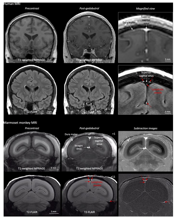
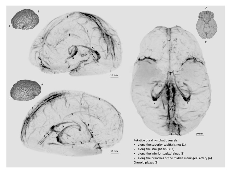
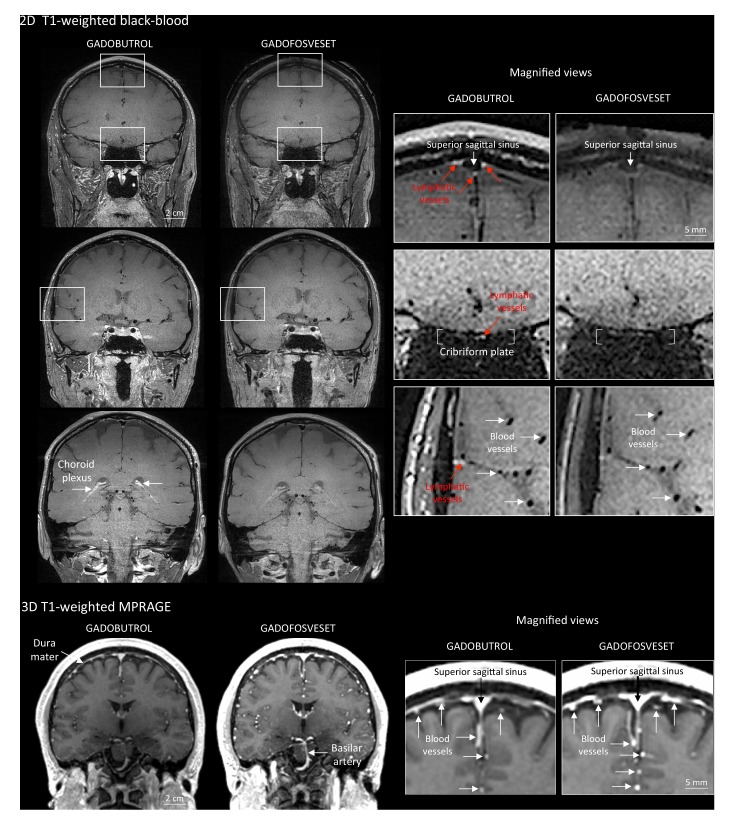
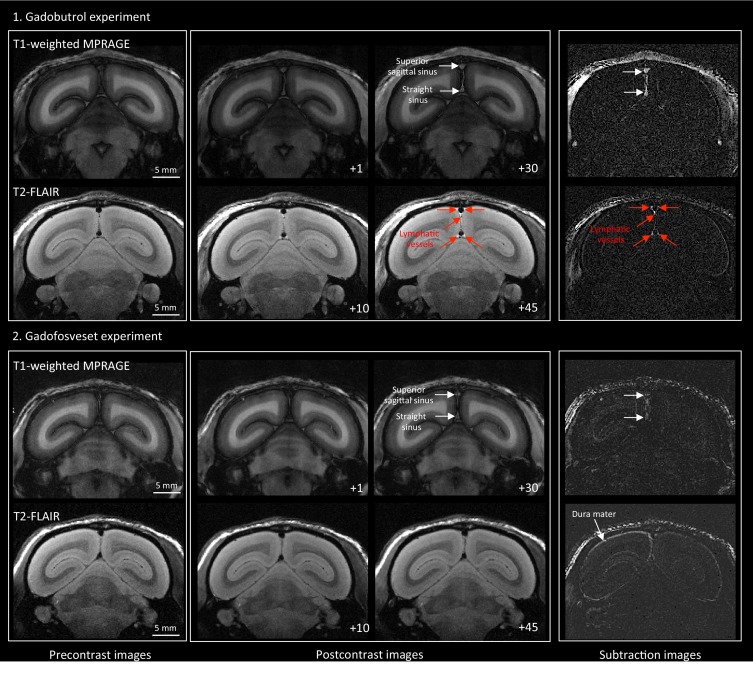
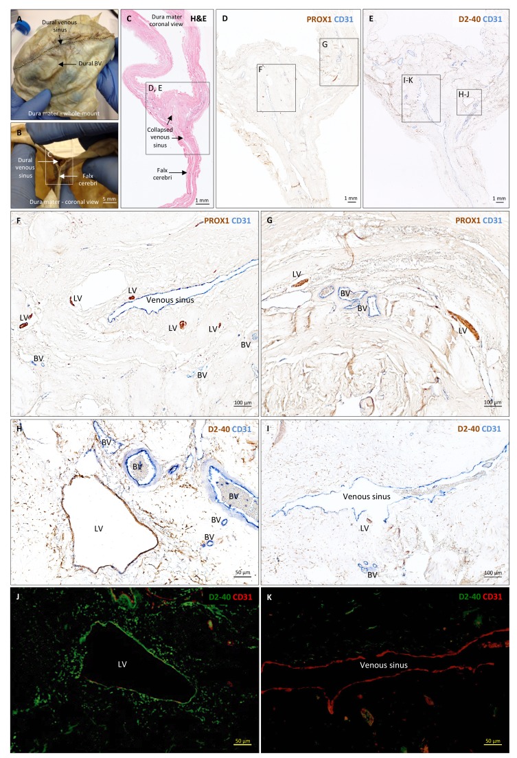
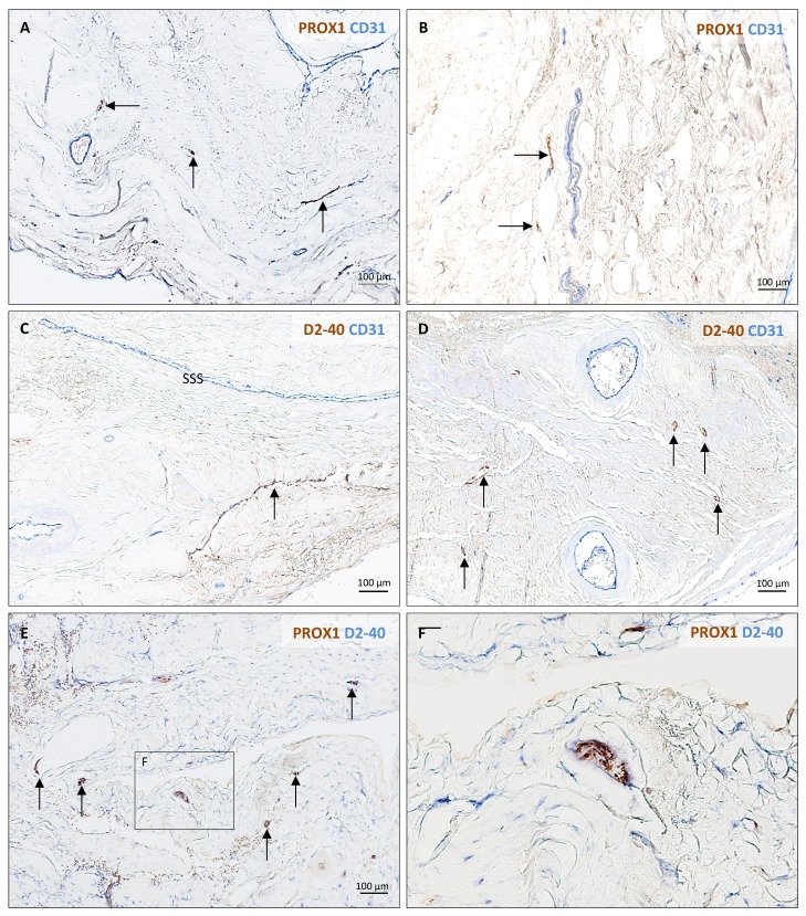
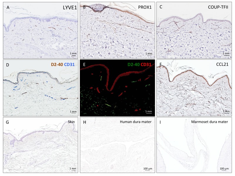
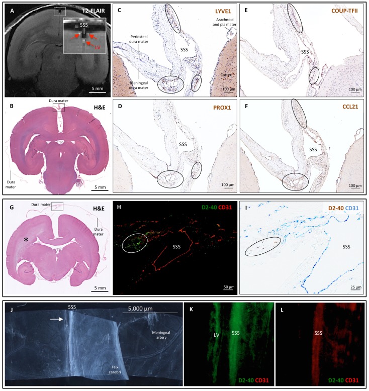
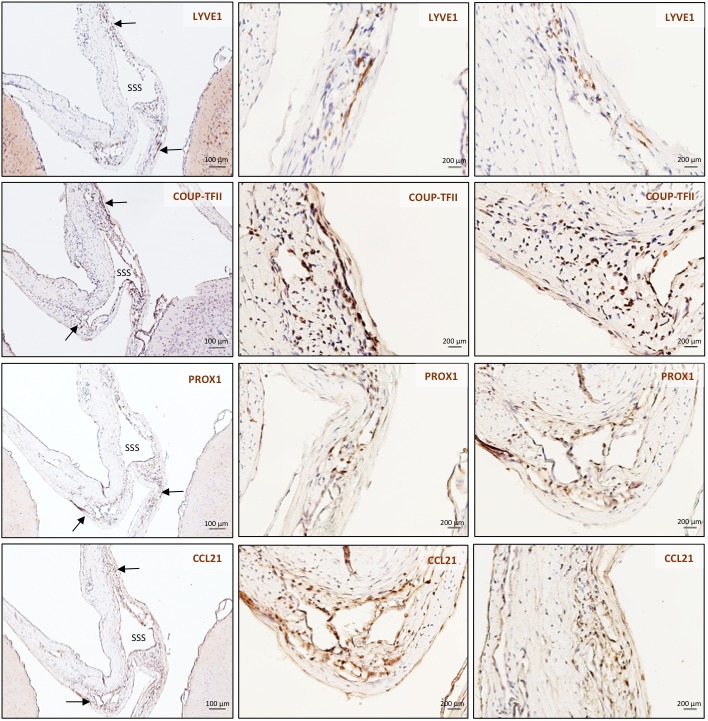
Here, we report the existence of meningeal lymphatic vessels in human and nonhuman primates (common marmoset monkeys) and the feasibility of noninvasively imaging and mapping them in vivo with high-resolution, clinical MRI. On T2-FLAIR and T1-weighted black-blood imaging, lymphatic vessels enhance with gadobutrol, a gadolinium-based contrast agent with high propensity to extravasate across a permeable capillary endothelial barrier, but not with gadofosveset, a blood-pool contrast agent. The topography of these vessels, running alongside dural venous sinuses, recapitulates the meningeal lymphatic system of rodents. In primates, meningeal lymphatics display a typical panel of lymphatic endothelial markers by immunohistochemistry. This discovery holds promise for better understanding the normal physiology of lymphatic drainage from the central nervous system and potential aberrations in neurological diseases.
Overview
The claims and statistics of this paper haven’t been checked yet.
Run the check — free, no account needed. It reads the open-access text and checks the sources it cites.
Integrity signals
Registry
Cites
Statistics
Figures
Verification
Claims the paper makes
The claims and statistics of this paper haven’t been checked yet.
Run the check — free, no account needed. It reads the open-access text and checks the sources it cites.
Claims other papers make about this one
Each is checked against this paper’s own text: does it say what the citing paper says it does?
The source backs part of the claim, not all of it — or backs it for a different population, condition or comparison.
In the citing paper
“Recent magnetic resonance imaging (MRI) studies in the living human brain and marmosets utilized a combination of gadolinium-based contrast agent (Gadovist) and blood-pool contrast agent (Vasovist) to demonstrate existence of the meningeal lymphatic system (4).”
From Blood-Brain Barrier: From Physiology to Disease and Back (2019) · cites this paper as [4]
Experimental — accuracy not yet measuredIn the citing paper
“The dural lymphatics are now recognized as a critical transport system of macromolecules, interstitial fluid, and CSF from the CNS into the cervical lymph nodes (Aspelund et al., 2015; Absinta et al., 2017; Louveau et al., 2018; Ahn et al., 2019).”
From Living on the Edge of the CNS: Meninges Cell Diversity in Health and Disease (2021) · cites this paper as Absinta et al., 2017
In the citing paper
“The extent of enhancing tissue within parasagittal dura does not resemble the previously reported appearance of true dural lymphatic vessels, which are tubular-shaped structures of sub-millimeter cross-sectional diameter near the venous sinus wall3,4,11, and also far from the classical MRI appearance of arachnoid granulations bulging into venous sinuses12.”
From Cerebrospinal fluid tracer efflux to parasagittal dura in humans (2020) · cites this paper as [11]
Self-check
Percentages, sums and reported differences re-computed from the paper’s own numbers.
The self-check needs the paper’s full text.
Citations
Cites
Iliff, J. J., Wang, M., Liao, Y., Plogg, B. A., Peng, W., Gundersen, G. A., Benveniste, H., Vates, G. E., Deane, R., Goldman, S. A., et al. (2012) · Science Translational Medicine · doi:10.1126/scitranslmed.3003748 · Cited by 5,824
Sleep Drives Metabolite Clearance from the Adult Brain
Xie, L., Kang, H., Xu, Q., Chen, M. J., Liao, Y., Thiyagarajan, M., O’Donnell, J., Christensen, D. J., Nicholson, C., Iliff, J. J., et al. (2013) · Science · doi:10.1126/science.1241224 · Cited by 5,411
Structural and functional features of central nervous system lymphatic vessels
Louveau, A., Smirnov, I., Keyes, T. J., Eccles, J. D., Rouhani, S. J., Peske, J. D., Derecki, N. C., Castle, D., Mandell, J. W., Lee, K. S., et al. (2015) · Nature · doi:10.1038/nature14432 · Cited by 4,413
A dural lymphatic vascular system that drains brain interstitial fluid and macromolecules
Aspelund, A., Antila, S., Proulx, S. T., Karlsen, T. V., Karaman, S., Detmar, M., Wiig, H., & Alitalo, K. (2015) · Journal of Experimental Medicine · doi:10.1084/jem.20142290 · Cited by 2,198
Lymphangiogenesis in development and human disease
Alitalo, K. K., Tammela, T., & Petrova, T. V. (2005) · Nature · doi:10.1038/nature04480 · Cited by 1,287
Brain-wide pathway for waste clearance captured by contrast-enhanced MRI
Iliff, J. J., Lee, H., Yu, M., Feng, T., Logan, J., Nedergaard, M., & Benveniste, H. (2013) · Journal of Clinical Investigation · doi:10.1172/jci67677 · Cited by 1,148
Lymphangiogenesis and lymphatic vessel remodelling in cancer
Stacker, S. A., Williams, S. P., Karnezis, T., Shayan, R., Fox, S. B., & Achen, M. G. (2014) · Nature reviews. Cancer · doi:10.1038/nrc3677 · Cited by 837
Mandell, D. M., Mossa‐Basha, M., Qiao, Y., Hess, C. P., Hui, F. K., Matouk, C. C., Johnson, M. H., Daemen, M. J. A. P., Vossough, A., Edjlali, M., et al. (2016) · American Journal of Neuroradiology · doi:10.3174/ajnr.a4893 · Cited by 694
Revisiting the Mechanisms of CNS Immune Privilege
Louveau, A., Harris, T. H., & Kipnis, J. (2015) · Trends in Immunology · doi:10.1016/j.it.2015.08.006 · Cited by 682
Self-citationLineage tracing demonstrates the venous origin of the mammalian lymphatic vasculaturePreprint
Srinivasan, R. S., Dillard, M. E., Lagutin, O. V., Lin, F., Tsai, S. Y., Tsai, M., Samokhvalov, I. M., & Oliver, G. C. (2007) · Genes & Development · doi:10.1101/gad.1588407 · Cited by 583
Research works printed but not linked: 1
- Mascagni, 1816, Istoria Completa Dei Vasi Linfatici, Vol. II, 195
Cited by
Sorted by citations
Items 501–600 of 643
Malis, V., Bae, W. C., Yamamoto, A., McEvoy, L. K., McDonald, M. A., & Miyazaki, M. (2022) · bioRxiv (Cold Spring Harbor Laboratory) · doi:10.1101/2022.07.14.500033 · Cited by 2
Kaur, J., Chopp, M., & Jiang, Q. (2024) · Elsevier eBooks · doi:10.1016/b978-0-323-95702-1.00243-8 · Cited by 1
Retrospective evaluation of multiplex PCR panel results from CSF samples in a university hospital
Şahin, F., Özcan, N., Özbek, E., Atmaca, S., & Temiz, H. (2025) · Northwestern Medical Journal · doi:10.54307/2025.nwmj.90 · Cited by 2
A synthesized view of the CSF-blood barrier and its surgical implications for aging disorders
Taha, B. R., McGovern, R. A., & Lam, C. H. (2025) · Frontiers in Aging Neuroscience · doi:10.3389/fnagi.2024.1492449 · Cited by 2
Hamzeh, J., Harati, H., Ayoubi, F., Saab, M., Saab, L., Ahmar, E. A., & Estephan, E. (2025) · Journal of Clinical Medicine · doi:10.3390/jcm14124387 · Cited by 1
Different Glymphatic-Lymphatic Coupling in the Nasal Mucosa and Parasagittal Dura
Urbach, H., Kavus, M., Lützen, N., Zander, C., Demerath, T., Rau, A., Wolf, K., Beck, J., & Duman, İ. E. (2025) · Investigative Radiology · doi:10.1097/rli.0000000000001220 · Cited by 2
Morphologic and functional alterations in the parasagittal dural space in mild cognitive impairment
Joo, B., Park, M., Kim, S. S., Ahn, S. J., Kim, H., Cho, H., Lyoo, C. H., & Suh, S. H. (2025) · Scientific Reports · doi:10.1038/s41598-025-07909-3 · Cited by 1
Shi, C., Zhou, Y., Zhang, Q., Jin, W., & Dong, H. (2025) · Mediators of Inflammation · doi:10.1155/mi/4288671 · Cited by 1
Incorporating meningeal immunity into vaccine development
Menon, M., Haile, C. N., & Dowling, D. J. (2025) · Nature Communications · doi:10.1038/s41467-025-64476-x · Cited by 1
Marsters, C. M., Phua, C. W. W., MacDonald, M., Boldt, G., & Climans, S. (2025) · Cancers · doi:10.3390/cancers17203392 · Cited by 1
Pediatric hydrocephalus and imaging evaluation of ventriculomegaly in the emergency setting
Elzieny, A. A., Pace-Soler, E., Thomas, A. K., & Setty, B. N. (2025) · Pediatric Radiology · doi:10.1007/s00247-025-06397-x · Cited by 1
Mechanisms of parasite‐mediated disruption of brain vessels
Loira, L., Arroz‐Madeira, S., Franco, C. A., & Pereira, S. S. (2025) · FEBS Letters · doi:10.1002/1873-3468.70255 · Cited by 1
Siow, T. Y., Huang, S., Lin, S.-N., Wong, A. M., Chen, Y., Lin, C., & Toh, C. (2025) · SLEEP · doi:10.1093/sleep/zsaf399 · Cited by 1
Possible Impact of Lymphatic Drainage on Brain Injury After Aneurysmal Subarachnoid Hemorrhage
Suzuki, H., Hakozaki, K., Aoki, K., Kawakita, F., Nakatsuka, Y., Kitano, Y., Nishikawa, H., & Yasuda, R. (2026) · International Journal of Molecular Sciences · doi:10.3390/ijms27031329 · Cited by 1
Differences in brain drainage and cognitive function between BALB/c and C57Bl/6 mice
Evsukova, A., Zlatogorskaya, D., Adushkina, V. V., Saranceva, E. I., & Дмитриевна, И. Т. (2026) · The European Physical Journal Special Topics · doi:10.1140/epjs/s11734-026-02179-0 · Cited by 1
The spinal lymphatic system: an emerging pathway bridging fluid homeostasis, immunity, and disease
Hou, Y., Wu, J., Yang, S., Hongwei, W., Wang, X., Lu, Z., Chen, Z., Feng, J., & 王宏利 (2026) · Bone Research · doi:10.1038/s41413-026-00508-6 · Cited by 2
Munir, A., Zhang, J., Hoyek, N. E., & Raghavan, P. (2026) · Seminars in Roentgenology · doi:10.1016/j.ro.2026.150986 · Cited by 1
Liu, L., Li, J.-H., Bai, Y.-D., Wang, Y., & Bu, X. (2026) · Molecular Neurodegeneration · doi:10.1186/s13024-026-00962-0 · Cited by 1
Metabolism of myeloid cells in brain tumors
Saxena, A., & Bayık, D. (2026) · Frontiers in Immunology · doi:10.3389/fimmu.2026.1894833 · Cited by 1
Mechanisms of immune suppression in glioblastoma
Ellsworth, S. G., & Grossman, S. A. (2021) · Elsevier eBooks · doi:10.1016/b978-0-12-819755-4.00016-3 · Cited by 1
Brain injury biomarkers: Proteins and autoantibodies interplay
Abdelhady, S., Issa, H., Ashekyan, O., Haidar, M. A., Outani, O., Samir, Y., Belal, E., Shakkour, Z., Mondello, S., & Kobeissy, F. H. (2022) · Elsevier eBooks · doi:10.1016/b978-0-12-823347-4.00036-1 · Cited by 1
Advances in research on glioma microenvironment and immunotherapeutic targets
Tang, A., Liang, J., Mai, Y., & Sun, H. (2022) · Journal of Cancer Discovery · doi:10.55976/jcd.1202218514-29 · Cited by 1
Venous Outflow for Brain Arteriovenous Malformations: Overview and Treatment Implications.
Osorno‐Cruz, C., Segherlou, Z. H., Peart, R. A., Dodd, W., Laurent, D., Aghili-Mehrizi, S., Lucke-Wold, B. P., & Chalouhi, N. (2022) · PubMed · Cited by 1
Hashemzadeh, M. S. (2022) · Romanian Journal of Military Medicine · doi:10.55453/rjmm.2022.125.4.19 · Cited by 1
Lenfatik Sistem Tarihçesi’ne Yolculuk: Keşifler ve Gelişmeler
Çelik, M. (2023) · Mersin Üniversitesi Tıp Fakültesi Lokman Hekim Tıp Tarihi ve Folklorik Tıp Dergisi · doi:10.31020/mutftd.1339551 · Cited by 1
Immunotherapy in the context of immune-specialized environment of brain metastases
James, F., & Lorger, M. (2023) · Discovery Immunology · doi:10.1093/discim/kyad023 · Cited by 1
Korn, T. (2024) · Elsevier eBooks · doi:10.1016/b978-0-12-823848-6.00004-x · Cited by 1
Guffart, E., & Prinz, M. (2024) · Advances in neurobiology · doi:10.1007/978-3-031-55529-9_3 · Cited by 1
Multiple sclerosis-like manifestations in systemic autoimmune and inflammatory disorders: an update
Raftopoulou, S., Kapsali, I., Evangelopoulos, M., & Mavragani, C. P. (2025) · Expert Review of Clinical Immunology · doi:10.1080/1744666x.2025.2522270 · Cited by 2
Studying the blood–brain barrier will provide new insights into neurodegeneration – Commentary
Bittner, S., & Zipp, F. (2018) · Multiple Sclerosis Journal · doi:10.1177/1352458518759430 · Cited by 1
Eide, P. K., & Ringstad, G. A. (2018) · Springer eBooks · doi:10.1007/978-3-319-97928-1_4 · Cited by 1
Lymphatic Meningeal Role in Processes of Brain Clearing: in vivo Visualization
Torres, E. N. D., Abdurashitov, A. S., Nаmykin, A. A., Shirokov, A. A., Shushunova, N. A., Saranceva, E. I., & Semyachkina-Glushkovskaya, O. V. (2018) · Izvestiya of Saratov University Chemistry Biology Ecology · doi:10.18500/1816-9775-2018-18-4-433-438 · Cited by 1
Update in Pediatric Neuro-Oncology
Khatua, S., & Pillay Smiley, N. (2019) · MDPI eBooks · doi:10.3390/books978-3-03897-540-3 · Cited by 1
Tzahor, E., & Yaniv, K. (2022) · Nature Cardiovascular Research · doi:10.1038/s44161-021-00010-8 · Cited by 1
Anatomy of the vertebral column lymphatic network in micePreprint
Jacob, L., Boisserand, L. S. B., Pestel, J., Antila, S., Thomas, J.-M., Aigrot, M., Mathivet, T., Lee, S., Alitalo, K. K., Renier, N., et al. (2019) · arXiv (Cornell University) · doi:10.48550/arxiv.1901.03864 · Cited by 1
MRI-Based Demonstration of The Normal Glymphatic System in a Human Population: A Systematic ReviewPreprint
Lee, M. K., Cho, S. J., Bae, Y. J., & Kim, J. (2021) · Research Square · doi:10.21203/rs.3.rs-604443/v1 · Cited by 1
Neurobiology and Neurophysiology of Behavioral/Psychiatric Disorders
Boorin, S. J., & Keltner, N. L. (2021) · Wiley eBooks · doi:10.1002/9781119487593.ch3 · Cited by 1
Hua, X., Ye, M., Liu, D., Li, H., Fan, C., Mai, W., Jia, W., Xiaojun, Y., Sun, M., Liu, X., et al. (2024) · Research Square · doi:10.21203/rs.3.rs-5244229/v1 · Cited by 1
Wu, Y., Wang, K., Ju, L., Li, A., Qin, Q., Xu, F., Lin, D. D. M., Kleinberg, L. R., & Xu, J. (2025) · medRxiv · doi:10.1101/2025.08.28.25334379 · Cited by 1
Neurobiology and Neurophysiology of Behavioral/Psychiatric Disorders
Scahill, L. D., & Boorin, S. J. (2012) · Wiley eBooks · doi:10.1002/9781118704660.ch3
Chronic fatigue syndrome/myalgic encephalomyelitis: diagnosis from an osteopathic perspective
Perrin, R. N. (2018) · Russian Osteopathic Journal · doi:10.32885/2220-0975-2018-1-2-19-27
Lymph Node Blood Vessels: Exit Route for Systemic Dissemination of Cancer
Skopec, R. (2018) · Current Trends On Biotechnology & Microbiology Open Access Publishers · doi:10.32474/ctbm.2018.01.000104
Optical monitoring of the meningeal lymphatic clearance after opening of blood-brain barrier
Torres, E. D., Abdurashitov, A., Nаmykin, A. A., Shirokov, A. A., Shushunova, N. A., Khorovodov, A. P., Saranceva, E. I., & Semyachkina-Glushkovskaya, O. V. (2019) · Saratov Fall Meeting: Computations and Data Analysis: from Nanoscale Tools to Brain Functions (SFM) · doi:10.1117/12.2524427
Melanoma Brain Metastases: Unique Biology and Implications for Systemic Therapy
Margolin, K. A., Davies, M. A., Kluger, H. M., & Tawbi, H. A. (2019) · Springer eBooks · doi:10.1007/978-3-319-46029-1_65-1
Immunologie de la sclérose en plaques
Defer, G. L., Debouverie, M., Bardou, I., Benamar, M., Bernard‐Valnet, R., Branger, P., Brassat, D., Benallegue, N., Calocer, F., Damotte, V., et al. (2019) · Elsevier eBooks · doi:10.1016/b978-2-294-71420-7.00005-7
Treviranus, G. R. S. (2019) · PubMed · Cited by 1
Structure and Function Study on Lymphatic System in Central Nervous System and Peripheral Organs
Yin, C. (2020) · International Conference on Biomedical Engineering and Technology (ICBET) · doi:10.1145/3397391.3397393
Norwood, J. N., Gharpure, A., Kumal, R. R., Turner, K. L., Pistone, L. F., Wal, R. L. V., & Drew, P. J. (2020) · bioRxiv (Cold Spring Harbor Laboratory) · doi:10.1101/2020.08.19.256297
Wpływ snu na funkcjonowanie układu glimfatycznego
Obara‐Michlewska, M. (2020) · Kosmos · doi:10.36921/kos.2020_2691
Isolation and in vitro culture of primary pia mater cellsPreprint
Saadeh, F. S., Remšík, J., Derderian, C., Chi, Y., & Boire, A. A. (2021) · bioRxiv (Cold Spring Harbor Laboratory) · doi:10.1101/2021.02.01.429222
El Altar de la Cruz de Eduardo Chillida en St. Peter de Colonia
Mennekes, F. (2002) · Ars sacra: Revista de patrimonio cultural, archivos, artes plásticas, arquitectura, museos y música
Immunotherapy for glioblastoma as a means to overcome resistance to standard therapy
Batich, K. A., Sampson, J. H., Khasraw, M., Friedman, H. S., & Ashley, D. M. (2021) · Elsevier eBooks · doi:10.1016/b978-0-12-821567-8.00018-x
Alshuhri, M. S. (2021) · Enlighten: Theses (The University of Glasgow) · doi:10.5525/gla.thesis.82303
Patent Highlights April–May 2021
Mucke, H. A. (2021) · Pharmaceutical Patent Analyst · doi:10.4155/ppa-2021-0011
Duque‐Wilckens, N., Sarno, E., Teis, R. E., Stölting, F., Khalid, S., Dairi, Z., Douma, A., Maradiaga, N. C., Thelen, K. M., Robison, A. J., et al. (2021) · bioRxiv (Cold Spring Harbor Laboratory) · doi:10.1101/2021.09.17.460793
High permeability of the CSF flow in the juvenile mouse brain by brain-wide DCE-MRIPreprint
Wada, Y., Imai, H., Yamawaki, Y., Parvez, M. S. A., Matsui, S., & Ohtsuki, G. (2021) · bioRxiv (Cold Spring Harbor Laboratory) · doi:10.1101/2021.09.21.461189
Audshasai, T., Coles, J. A., Panagiotou, S., Khandaker, S., Scales, H. E., Kjos, M., Baltazar, M., Vignau, J., Brewer, J. M., Kadioglu, A., et al. (2021) · bioRxiv (Cold Spring Harbor Laboratory) · doi:10.1101/2021.09.28.462246
Hett, K., McKnight, C. D., Eisma, J. J., Elenberger, J., Lindsey, J. D., Considine, C. M., Claassen, D. O., & Donahue, M. J. (2021) · Research Square · doi:10.21203/rs.3.rs-1083306/v1
The Role of Lymphatic Marker Prox-1 in Relation to Brain Tumours
Teleky, J., & Király, J. (2021) · Folia veterinaria · doi:10.2478/fv-2021-0040
Hypoproliferative human NPC xenografts survived extendedly in the brain of immunocompetent ratsPreprint
Liu, C., Wang, X., Huang, W., Meng, W., Su, Z., Xing, Q., Shi, H., Zhang, D., Zhou, M., Zhao, Y., et al. (2020) · doi:10.21203/rs.3.rs-92873/v1
Melanoma Brain Metastases: Unique Biology and Implications for Systemic Therapy
Margolin, K. A., Davies, M. A., Kluger, H. M., & Tawbi, H. A. (2020) · Springer eBooks · doi:10.1007/978-3-030-05070-2_65
Oliver, G. C., & Liu, X. (2019) · Faculty Opinions – Post-Publication Peer Review of the Biomedical Literature · doi:10.3410/f.733687657.793565950
Characterization of non-human primate dura in health and neurodegenerationPreprint
Janson, C., Hauser, K., Muller, S., Hansen, E., Lam, C. H., Kordower, J. H., & Romanova, L. G. (2022) · bioRxiv (Cold Spring Harbor Laboratory) · doi:10.1101/2022.06.03.477517
Cao, D., Sun, Y., Su, P., Pillai, J. J., Qiao, Y., Lu, H., van Zijl, P. C. M., Knutsson, L., & Hua, J. (2022) · bioRxiv (Cold Spring Harbor Laboratory) · doi:10.1101/2022.09.26.509366
Jacob, L., Brito, J. M., & Thomas, J. (2020) · Journal of Visualized Experiments · doi:10.3791/61099-v
New Drug Developments for Stroke: from Basics to Clinics
Chen, W., Wang, Y., Jiang, L., Wang, Y., & Wang, Y. (2023) · Translational Stroke Research · doi:10.1007/s12975-023-01130-y
Zhang, X., Wang, T., Bai, Y., Li, Z., Zhu, D., Cheng, A., Li, Y., & Lu, Q. (2023) · Research Square · doi:10.21203/rs.3.rs-2655218/v1
Magnetic resonance imaging-based progress in human glymphatic system research
Chen, S., Chen, Y., & Wu, R. (2023) · Radiology Science · doi:10.15212/radsci-2022-0011
Gadolinium-based imaging of the blood brain barrier and brain waste clearance pathways
(2023) · Advances in magnetic resonance technology and applications · doi:10.1016/b978-0-323-91771-1.00017-4
Anatomical and Functional Features of the Central Nervous System Lymphatic System
Donahue, M. J., Donahue, P. M. C., Crescenzi, R. L., & McKnight, C. D. (2023) · Springer eBooks · doi:10.1007/978-3-031-10909-6_59
Microglia and macrophages in diseases
Kanamori, M., Harada, Y., & Ito, M. (2023) · Folia Pharmacologica Japonica · doi:10.1254/fpj.22155
He, J., Bo, Y., Li, B., Hao, J., Miao, C., Sha, R., & Yu, H. (2023) · Research Square · doi:10.21203/rs.3.rs-3712801/v1
Jansson, D., & Iliff, J. J. (2023) · Springer eBooks · doi:10.1007/978-3-031-39558-1_8
Modulação colinérgica de células dendríticas: relevante no contexto do glioblastoma?
Cardoso, R. D. S. (2023) · Digital Library of Theses and Dissertations (Universidade de São Paulo) · doi:10.11606/d.42.2023.tde-12012024-160445
High Resolution Imaging of Superior Sagittal Lymphatic Vasculature in Dedicated Brain SPECT
Kalluri, K. S., Mathur, P., Pells, S., Auer, B., May, M., Segars, W. P., Kuo, P. H., Furenlid, L. R., & King, M. A. (2022) · IEEE Nuclear Science Symposium and Medical Imaging Conference (NSS/MIC) · doi:10.1109/nss/mic44845.2022.10398996
Guo, B., Chang, J., & Abumaria, N. (2024) · Stress and Brain · doi:10.26599/sab.2024.9060001
Bao, W., Jiang, P., Xu, P., Lin, H., Xu, J., Lai, M., Yuan, J., & Xu, J. (2024) · SSRN Electronic Journal · doi:10.2139/ssrn.4878784
A Clinical Primer on the Anatomy and Physiology of Neurofluids in the Human Brain
Agarwal, N. (2025) · Neuroimaging Clinics of North America · doi:10.1016/j.nic.2024.12.007
Magnetic Resonance Methodologies
Lu, H., & Bandettini, P. A. (2025) · Oxford University Press eBooks · doi:10.1093/med/9780197640654.003.0008
李, 越. (2025) · Advances in Clinical Medicine · doi:10.12677/acm.2025.154977
Gasser, T. C. (2025) · Springer eBooks · doi:10.1007/978-3-031-52637-4_2
Serum NR1 and NR2 concentrations in first-episode schizophrenia and clinical high-risk for psychosis
Mao, Z., Li, F., Ge, L., Hou, W., Ding, Y., Wang, F., Wen, Y., Wang, X., Cheng, Y., Hou, W., et al. (2025) · BMC Psychiatry · doi:10.1186/s12888-025-06950-w
Topbas, A. S., & Bordes, S. J. (2025) · Springer eBooks · doi:10.1007/978-3-031-78326-5_20
Zhu, D., Wang, T., Bai, Y., Zhang, X., Li, Z., Cheng, A., Li, Y., & Lu, Q. (2025) · Neuroradiology · doi:10.1007/s00234-025-03658-2
Research Progress on the Mechanism and Treatment of Post-Hemorrhagic Hydrocephalus
白, 伟. (2025) · Advances in Clinical Medicine · doi:10.12677/acm.2025.1561797
Web of Science Based Visual Analysis of Literature Related to Chronic Subdural Haematoma
郭, 徽. (2025) · Advances in Clinical Medicine · doi:10.12677/acm.2025.1561904
Feng, L., Lin, Y., Du, C., Ma, C., Ni, N., & Liu, S. (2025) · SSRN Electronic Journal · doi:10.2139/ssrn.5375486
The Evidence That Brain Cancers Could Be Effectively Treated with In-Home Radiofrequency Waves
Arendash, G. W. (2025) · Cancers · doi:10.3390/cancers17162665
Murine Model of Cerebral Venous Outflow Occlusion Through Bilateral Ligation of Jugular Veins
Kamouh, M.-R. E., Spajer, M., Bourrienne, M., Marolany, A.-L. J., Singhabahu, R., Mazighi, M., Thomas, J., & Lenck, S. (2025) · Journal of Visualized Experiments · doi:10.3791/68626
Live longitudinal imaging of meningeal cerebrovascular injury and its sequelae in adult zebrafishPreprint
Kraus, A., Prosper-Santiago, J. S., Potapova, A., Prevedel, J., Castranova, D., & Weinstein, B. M. (2025) · bioRxiv (Cold Spring Harbor Laboratory) · doi:10.1101/2025.11.14.688311
Hett, K., McKnight, C. D., Eisma, J. J., Elenberger, J., Lindsey, J. S., Considine, C. M., Claassen, D. O., & Donahue, M. J. (2022) · Figshare · doi:10.6084/m9.figshare.19395840
Jukkola, J., Kaakinen, M., Singh, A., Moradi, S., Ferdinando, H., Myllylä, T., Kiviniemi, V. V., & Eklund, L. (2024) · Figshare · doi:10.6084/m9.figshare.26673456
Sun, Y., Cao, D., Pillai, J. J., Paez, A. G., Li, Y., Gu, C., Pogson, J. M., Knutsson, L., Barker, P. B., van Zijl, P. C. M., et al. (2024) · Figshare · doi:10.6084/m9.figshare.27042294
Kim, J.-H., Yoo, R.-E., Choi, S. H., & Park, S.-H. (2024) · Figshare · doi:10.6084/m9.figshare.26592248
Corrêa‐da‐Silva, F., Kalsbeek, M. J. T., Gadella, F. S., Oppersma, J., Jiang, W., Wolff, S. E. C., Korpel, N. L., Swaab, D. F., Fliers, E., Kalsbeek, A., et al. (2024) · Figshare · doi:10.6084/m9.figshare.26601676
Jukkola, J., Kaakinen, M., Singh, A., Moradi, S., Ferdinando, H., Myllylä, T., Kiviniemi, V. V., & Eklund, L. (2024) · Figshare · doi:10.6084/m9.figshare.26673450
Jukkola, J., Kaakinen, M., Singh, A., Moradi, S., Ferdinando, H., Myllylä, T., Kiviniemi, V. V., & Eklund, L. (2024) · Figshare · doi:10.6084/m9.figshare.26673462
Jukkola, J., Kaakinen, M., Singh, A., Moradi, S., Ferdinando, H., Myllylä, T., Kiviniemi, V. V., & Eklund, L. (2024) · Figshare · doi:10.6084/m9.figshare.26673459
Jukkola, J., Kaakinen, M., Singh, A., Moradi, S., Ferdinando, H., Myllylä, T., Kiviniemi, V. V., & Eklund, L. (2024) · Figshare · doi:10.6084/m9.figshare.26673453
Corrêa‐da‐Silva, F., Kalsbeek, M. J. T., Gadella, F. S., Oppersma, J., Jiang, W., Wolff, S. E. C., Korpel, N. L., Swaab, D. F., Fliers, E., Kalsbeek, A., et al. (2024) · Figshare · doi:10.6084/m9.figshare.26601673
Metadata
- Authors
- Absinta, M., Ha, S.-K., Nair, G., Sati, P., Luciano, N. J., Palisoc, M., Louveau, A., Zaghloul, K. A., Pittaluga, S., Kipnis, J., & Reich, D. S.
- Affiliations
- National Institutes of Health (US)
- University of Virginia (US)
- Corresponding author
- Reich, Daniel S
- ORCID
- Venue
- eLife
- Publisher
- eLife Sciences Publications, Ltd
- Published
- 2017-10-03
- Volume
- 6
- ISSN
- 2050-084X (ISSN-L)
- PMC
- PMC5626482
- OpenAlex
- W2760815599
- Cited by
- 643
- References
- 32
- Citations by year
- 2026: 61 · 2025: 88 · 2024: 73 · 2023: 77 · 2022: 79 · 2021: 78 · 2020: 70 · 2019: 60 · 2018: 51 · 2017: 4
- FWCI
- 22.31
- Type
- journal article
- Language
- en
- Open access
- gold
- doi.org
- Licence
- Locations
- eLife (publishedVersion, cc-by)
- Europe PMC (PubMed Central) (publishedVersion, public-domain)
- PubMed Central (publishedVersion, public-domain)
- PubMed (publishedVersion)
- DOAJ (DOAJ: Directory of Open Access Journals) (submittedVersion, cc-by-sa)
- Funders
- National Institute of Neurological Disorders and Stroke (Intramural Research Program)
- National Multiple Sclerosis Society (Postdoctoral Fellowship)
- Conrad N. Hilton Foundation (Marilyn Hilton Award for Innovation in Multiple Sclerosis Research)
- National Cancer Institute (Intramural Research Program)
- National Institute on Aging (R01AG034113)
- Lymphatic Education and Research Network (Postdoctoral fellowship)
- Topics
- Health Sciences › Medicine › Oncology › Lymphatic System and Diseases
- Health Sciences › Medicine › Clinical Neurology › Cerebrospinal fluid and hydrocephalus
- Health Sciences › Medicine › Surgery › Lymphatic Disorders and Treatments
- Keywords
- meningeal lymphatic vessels, meningeal lymphatic system, non-human primates, Callithrix jacchus, human brain, gadobutrol, gadofosveset, contrast extravasation, lymphatic endothelial markers, dural venous sinuses, black-blood imaging
- MeSH terms
- Animals, Callithrix, Humans, Magnetic Resonance Imaging, Meninges, Lymphatic Vessels
About these checks
Registry, retraction, Cites and statistics results are computed, not judged, with one exception: a printed reference that no index lists is read once by a model before it counts as a citation error. Claims are extracted by a model and kept only when their quote appears verbatim in the text; each cited source is then checked, with a model’s help, for whether it says what the claim relies on. Those verdicts can be wrong — read the claim’s quote and the cited source before relying on one.
Statistics and arithmetic are recomputed, not judged.
Confidence bands are shown only for fields calibrated on our annotated test set. None is calibrated yet, so no band is shown.
What counts as a claim ›How we measure confidence ›How we check this ›
Are you an author, or do you see a problem with this page? Request a review
