

The DOI matches its Crossref record; 32 of the 32 references it cites were found in a registry or an index.
Human and nonhuman primate meninges harbor lymphatic vessels that can be visualized noninvasively by MRI.
Open accessAbstract
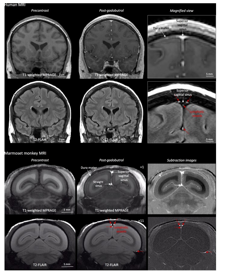
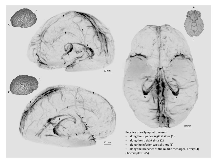
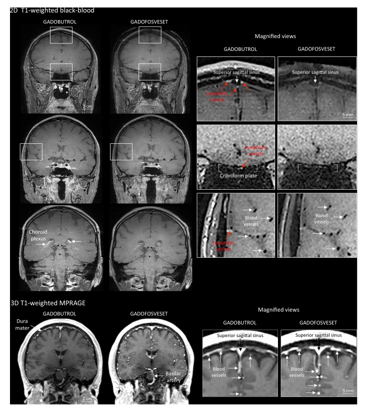
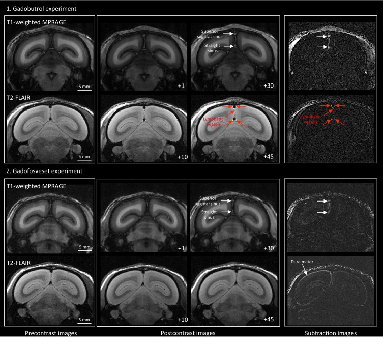
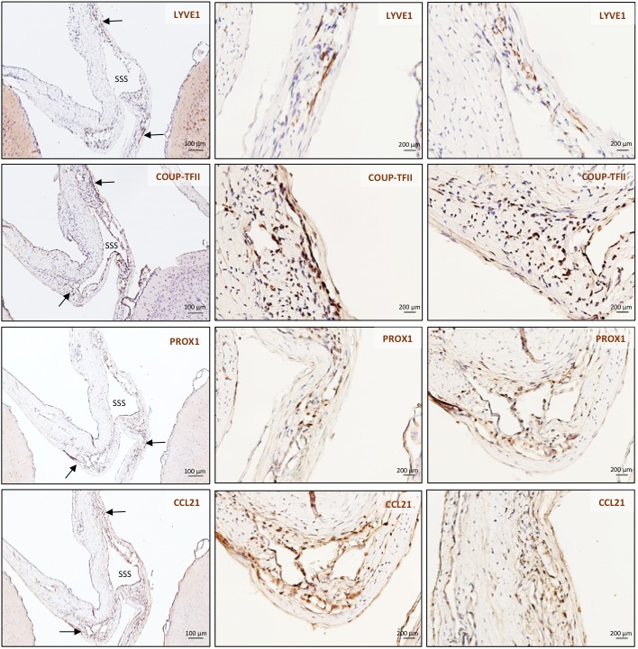
Here, we report the existence of meningeal lymphatic vessels in human and nonhuman primates (common marmoset monkeys) and the feasibility of noninvasively imaging and mapping them in vivo with high-resolution, clinical MRI. On T2-FLAIR and T1-weighted black-blood imaging, lymphatic vessels enhance with gadobutrol, a gadolinium-based contrast agent with high propensity to extravasate across a permeable capillary endothelial barrier, but not with gadofosveset, a blood-pool contrast agent. The topography of these vessels, running alongside dural venous sinuses, recapitulates the meningeal lymphatic system of rodents. In primates, meningeal lymphatics display a typical panel of lymphatic endothelial markers by immunohistochemistry. This discovery holds promise for better understanding the normal physiology of lymphatic drainage from the central nervous system and potential aberrations in neurological diseases.
Overview
The claims and statistics of this paper haven’t been checked yet.
Run the check — free, no account needed. It reads the open-access text and checks the sources it cites.
Integrity signals
Registry
Cites
Statistics
Figures
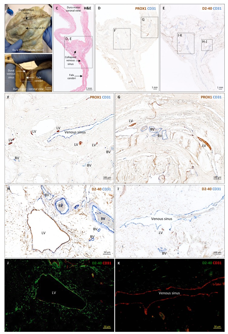
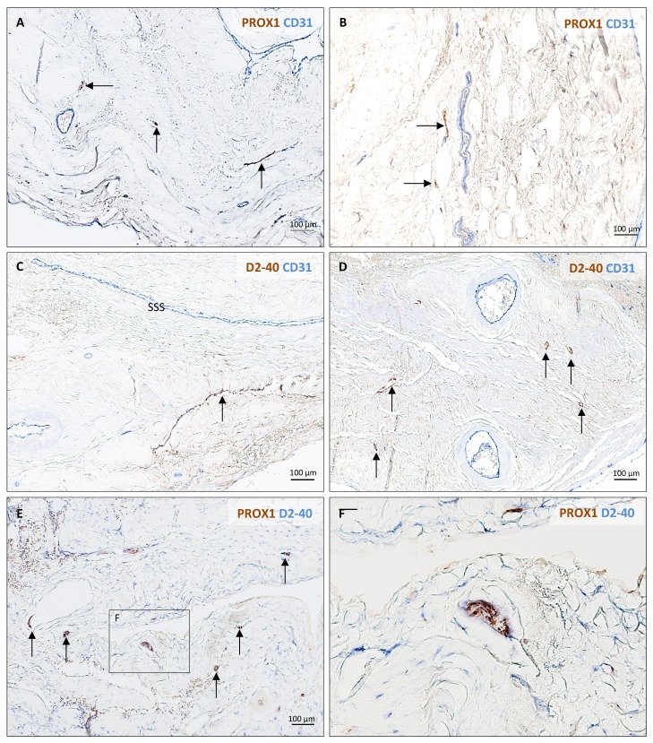
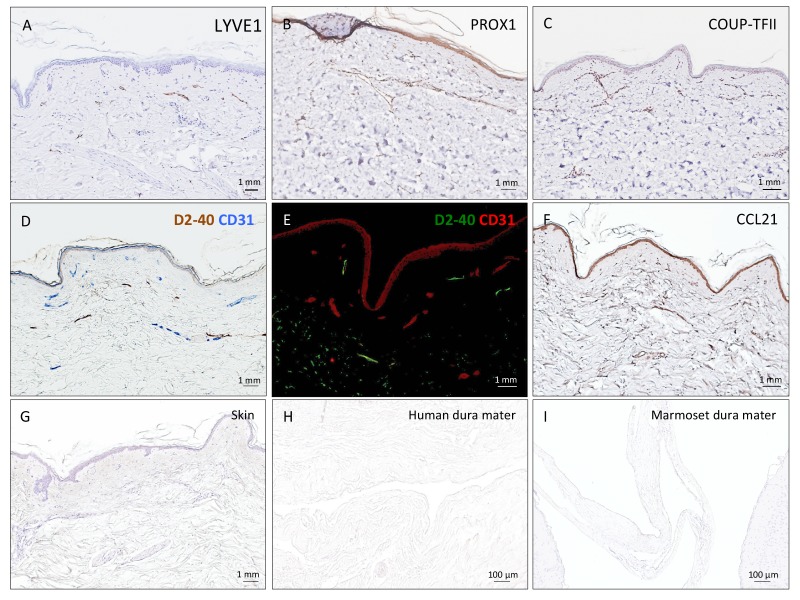
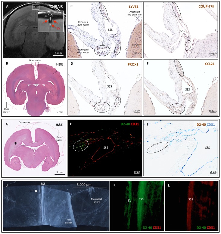
Verification
Claims the paper makes
The claims and statistics of this paper haven’t been checked yet.
Run the check — free, no account needed. It reads the open-access text and checks the sources it cites.
Claims other papers make about this one
Each is checked against this paper’s own text: does it say what the citing paper says it does?
The source backs part of the claim, not all of it — or backs it for a different population, condition or comparison.
In the citing paper
“Recent magnetic resonance imaging (MRI) studies in the living human brain and marmosets utilized a combination of gadolinium-based contrast agent (Gadovist) and blood-pool contrast agent (Vasovist) to demonstrate existence of the meningeal lymphatic system (4).”
From Blood-Brain Barrier: From Physiology to Disease and Back (2019) · cites this paper as [4]
Experimental — accuracy not yet measuredIn the citing paper
“The dural lymphatics are now recognized as a critical transport system of macromolecules, interstitial fluid, and CSF from the CNS into the cervical lymph nodes (Aspelund et al., 2015; Absinta et al., 2017; Louveau et al., 2018; Ahn et al., 2019).”
From Living on the Edge of the CNS: Meninges Cell Diversity in Health and Disease (2021) · cites this paper as Absinta et al., 2017
In the citing paper
“The extent of enhancing tissue within parasagittal dura does not resemble the previously reported appearance of true dural lymphatic vessels, which are tubular-shaped structures of sub-millimeter cross-sectional diameter near the venous sinus wall3,4,11, and also far from the classical MRI appearance of arachnoid granulations bulging into venous sinuses12.”
From Cerebrospinal fluid tracer efflux to parasagittal dura in humans (2020) · cites this paper as [11]
Self-check
Percentages, sums and reported differences re-computed from the paper’s own numbers.
The self-check needs the paper’s full text.
Citations
Cites
Iliff, J. J., Wang, M., Liao, Y., Plogg, B. A., Peng, W., Gundersen, G. A., Benveniste, H., Vates, G. E., Deane, R., Goldman, S. A., et al. (2012) · Science Translational Medicine · doi:10.1126/scitranslmed.3003748 · Cited by 5,824
Sleep Drives Metabolite Clearance from the Adult Brain
Xie, L., Kang, H., Xu, Q., Chen, M. J., Liao, Y., Thiyagarajan, M., O’Donnell, J., Christensen, D. J., Nicholson, C., Iliff, J. J., et al. (2013) · Science · doi:10.1126/science.1241224 · Cited by 5,411
Structural and functional features of central nervous system lymphatic vessels
Louveau, A., Smirnov, I., Keyes, T. J., Eccles, J. D., Rouhani, S. J., Peske, J. D., Derecki, N. C., Castle, D., Mandell, J. W., Lee, K. S., et al. (2015) · Nature · doi:10.1038/nature14432 · Cited by 4,413
A dural lymphatic vascular system that drains brain interstitial fluid and macromolecules
Aspelund, A., Antila, S., Proulx, S. T., Karlsen, T. V., Karaman, S., Detmar, M., Wiig, H., & Alitalo, K. (2015) · Journal of Experimental Medicine · doi:10.1084/jem.20142290 · Cited by 2,198
Lymphangiogenesis in development and human disease
Alitalo, K. K., Tammela, T., & Petrova, T. V. (2005) · Nature · doi:10.1038/nature04480 · Cited by 1,287
Brain-wide pathway for waste clearance captured by contrast-enhanced MRI
Iliff, J. J., Lee, H., Yu, M., Feng, T., Logan, J., Nedergaard, M., & Benveniste, H. (2013) · Journal of Clinical Investigation · doi:10.1172/jci67677 · Cited by 1,148
Lymphangiogenesis and lymphatic vessel remodelling in cancer
Stacker, S. A., Williams, S. P., Karnezis, T., Shayan, R., Fox, S. B., & Achen, M. G. (2014) · Nature reviews. Cancer · doi:10.1038/nrc3677 · Cited by 837
Mandell, D. M., Mossa‐Basha, M., Qiao, Y., Hess, C. P., Hui, F. K., Matouk, C. C., Johnson, M. H., Daemen, M. J. A. P., Vossough, A., Edjlali, M., et al. (2016) · American Journal of Neuroradiology · doi:10.3174/ajnr.a4893 · Cited by 694
Revisiting the Mechanisms of CNS Immune Privilege
Louveau, A., Harris, T. H., & Kipnis, J. (2015) · Trends in Immunology · doi:10.1016/j.it.2015.08.006 · Cited by 682
Self-citationLineage tracing demonstrates the venous origin of the mammalian lymphatic vasculaturePreprint
Srinivasan, R. S., Dillard, M. E., Lagutin, O. V., Lin, F., Tsai, S. Y., Tsai, M., Samokhvalov, I. M., & Oliver, G. C. (2007) · Genes & Development · doi:10.1101/gad.1588407 · Cited by 583
Research works printed but not linked: 1
- Mascagni, 1816, Istoria Completa Dei Vasi Linfatici, Vol. II, 195
Cited by
Sorted by citations
Items 601–643 of 643
New frontiers of lymphangiogenesis
Natale, G. (2025) · Elsevier eBooks · doi:10.1016/b978-0-443-13979-6.00006-6
Mechanisms of Dural Involvement in Cerebral Amyloid Angiopathy
Zedde, M., Piazza, F., & Pascarella, R. (2025) · Cells · doi:10.3390/cells15010026
Discovery of Abundant Nano-scale Lymphatic-like Vessels in BrainsPreprint
Gu, S., Dong, H., Chen, H., Yu, J., Liu, L., Yan, J., Wang, H., Jiang, Z., Huang, W., Wang, W., et al. (2025) · bioRxiv (Cold Spring Harbor Laboratory) · doi:10.64898/2025.12.30.697095
Glymphatic System of the Brain: Anatomy, Physiology, and Clinical Implications
Chun, M. Y., Chung, S. J., & Jeong, S. H. (2026) · Journal of the Korean Neurological Association · doi:10.17340/jkna.2025.0050
Uphoff, K., van Impel, A., Muhl, L., Stehling, M., Betsholtz, C., & Schulte‐Merker, S. (2026) · Development · doi:10.1242/dev.204988
Kraus, A., Prosper-Santiago, J. S., Potapova, A., Prevedel, J., Castranova, D., & Weinstein, B. M. (2026) · Journal of Neuroinflammation · doi:10.1186/s12974-026-03767-6 · Cited by 1
Li, H., Zhang, W., Chen, J., Lu, C., Liu, L., Zhou, Y., Jiang, H., Xue, X., Zhang, X., Zhou, D., et al. (2026) · Brain Circulation · doi:10.4103/bc.bc_79_25
Étude de la plasticité liquidienne du système cranio-spinal : de la physiologie à la pathologie
Lokossou, A. (2019) · theses.fr (ABES) · doi:10.70675/532b5045z5dd4z4268z9d4bz51e04f957bf7
Presence of innate lymphoid cells in the ischemic stroke brain
Wang, S. (2021) · theses.fr (ABES) · doi:10.70675/89448768z5d25z40b6za25az2dcf35abe6db
Holz, A., Karmann, M., Deli, S., Neumaier, V., Bonhoeffer, M., Bongratz, F., Schmitz‐Koep, B., Roßmüller, P., Zott, B., Wiestler, B., et al. (2026) · Neuroradiology · doi:10.1007/s00234-026-03912-1
Dura mater enhancement on 3T MRI is associated with cortical lesion burden in multiple sclerosis
Zivadinov, R., Semy, M., Bartnik, A., Hildesheim, F. E., Tranquille, A., Jakimovski, D., Hojnacki, D. W., Eckert, S. P., Weinstock‐Guttman, B., Dwyer, M. G., et al. (2026) · Journal of Neurology · doi:10.1007/s00415-026-13781-6
Andreo, M., Sivakolundu, D. K., Zuppichini, M. D., West, K., Spence, J. S., Gauthier, S. A., Nguyen, T., & Rypma, B. (2026) · bioRxiv (Cold Spring Harbor Laboratory) · doi:10.64898/2026.04.05.716121
Role of T-cell immunity in the pathophysiology of Alzheimer’s disease and other Tauopathies
Matta, K. (2024) · theses.fr (ABES) · doi:10.70675/cea4fe5ez67c3z4445z92f7z39af62ba059b
Age-related development of meningeal lymphatic vessels in children assessed by 3D T2 FLAIR MRI
Ying, J., Rao, Y., Chen, X., Chen, F., Zhang, T., Li, J., Wang, S., Lin, J., He, Y., Chen, J., et al. (2026) · Scientific Reports · doi:10.1038/s41598-026-49446-7
Hyperintense FLAIR signal in the anterior cranial fossa
Graf, C., Jaffray, A., Rund, A., Steinerberger, S., Li, D. K. B., & Rauscher, A. (2026) · Nature Communications · doi:10.1038/s41467-026-72196-z
Bidirectional roles of meningeal lymphatic vessels in brain metastases: friend and foe
Shao, W., Zhu, E., Chen, D., & Lu, H. (2026) · Frontiers in Oncology · doi:10.3389/fonc.2026.1793165
Ishikawa, K., Mizutani, K., & Toda, M. (2026) · Surgical Neurology International · doi:10.25259/sni_472_2026
Amin, M. R., Yu, T., Cheng, Z., Zhang, J., Chen, G., Gu, L., Jian, Z., & Xiong, X. (2026) · Experimental Neurology · doi:10.1016/j.expneurol.2026.115890 · Cited by 1
Development and prospect of treating Alzheimer's disease using the glymphatic system
Gu, J., Liu, Z., & Gu, J. (2026) · Journal of Alzheimer s Disease Reports · doi:10.1177/25424823261461528
Bridging veins govern discrepancy of CSF efflux into dura mater in mice
Yan, L., Satyanarayanan, S. K., Huang, D., Qin, Y., Wu, D., Lai, J., Qin, D., & Su, H. (2026) · The Journal of Experimental Medicine · doi:10.1084/jem.20252556
Šķuburs, G. K., Šneidere, K., Platkājis, A., Kaļva, K., Litauniece, Z. A., Ušacka, A., Freibergs, Z., Bērziņš, G. J., Gailīte, L., Isakovs, A., et al. (2026) · Diagnostics · doi:10.3390/diagnostics16152366
Tamadon, A., & Mussin, N. M. (2026) · West Kazakhstan Biomedicine Journal · doi:10.4103/wkbj.wkbj_34_26
Mănescu, M. D., Minca, T.-N., Pirici, I., Busuioc, C. J., Mateescu, V. O., & Pirici, D. N. (2026) · Frontiers in Cellular Neuroscience · doi:10.3389/fncel.2026.1903812
Ruiz-García, Á., Díaz-Benito, B., & Muñoz-García, M. I. (2026) · Journal of Alzheimer s Disease · doi:10.1177/13872877261483191
Ding, S., Yang, J., Wang, Z., Bai, R., Yin, K., Jin, L., Kueckelhaus, M., Meuth, S. G., Xiao, M., & Hu, G. (2026) · Translational Neurodegeneration · doi:10.1186/s40035-026-00579-9
La, B., Bai, Y., Qiu, Y., & Wang, L. (2026) · Frontiers in Medicine · doi:10.3389/fmed.2026.1921232
Li, W., Liang, Y., Li, L., Sun, H., & Liu, Q. (2026) · Frontiers in Immunology · doi:10.3389/fimmu.2026.1960846
Chronic Hypertension and the Brain’s Waste Drainage System
Alian, M., Le, C., Williams, A., Wardlaw, J. M., & Benveniste, H. (2026) · Hypertension · doi:10.1161/hypertensionaha.126.26767
Seyfarth, N., Elmer, N., Blottner, D., Reißhauer, A., & Liebl, M. E. (2026) · Phlebologie · doi:10.1055/a-2950-6470
Liu, J., Chen, J., Yuan, S., Chen, Y., Luo, Z., & Xie, M. (2026) · Frontiers in Immunology · doi:10.3389/fimmu.2026.1929822
Early post-contrast signal changes in CSF spaces and CSF outflow-related regions on 7 Tesla MRI
van Westen, D., Vuong, N., Elhyka, E., Zandt, R. I. ‘., Wennberg, L., Nilsson, M., & Georgiopoulos, C. (2026) · Scientific Reports · doi:10.1038/s41598-026-74235-7
Glymphatic function in humans: indicators, modulators, and measurement strategies
Nyan, T. H., Park, H., Baek, H. J., Park, E., & Cho, J. (2026) · Neurological Sciences · doi:10.1007/s10072-026-09463-w
Haq, N. U., Irshad, S. U., Bibi, Z., Rehman, S., Sohail, S., Irshad, S. R., Haider, N., Mir, S., Shareef, A., & Lucke-Wold, B. P. (2026) · Biomedicines · doi:10.3390/biomedicines14102314
Choi, B., Park, S., Lee, J.-J., & Sohn, J. (2026) · Neurobiology of Disease · doi:10.1016/j.nbd.2026.107654
Wallner, C., Königschulte, A., Pleger, B., Lehnhardt, M., & Reinkemeier, F. (2026) · Journal of Alzheimer s Disease Reports · doi:10.1177/25424823261494617
Revanth, R. B., Smitha, K. A., Jithin, S. S., Meena, P., Nair, S. S., Jayadevan, E. R., Thomas, B., & Kesavadas, C. (2026) · Journal of Magnetic Resonance Imaging · doi:10.1002/jmri.70604
Glymphatic and lymphatic systems
Kim, M., Kim, J.-H., & Park, S.-H. (2026) · Advances in magnetic resonance technology and applications · doi:10.1016/b978-0-443-21425-7.00018-x
Ringstad, G. A. (2026) · Advances in magnetic resonance technology and applications · doi:10.1016/b978-0-443-21425-7.00016-6
The characteristics of perivascular proinflammation in 4‐repeat tauopathies
Fu, M., Uruk, G., Gatto, R. G., Reichard, R. R., Whitwell, J. L., & Josephs, K. A. (2026) · Brain Pathology · doi:10.1111/bpa.70162
Neurovascular-immune-clearance remodeling in brain aging
Liu, Y., Yang, R., & Chang, H. (2026) · Frontiers in Immunology · doi:10.3389/fimmu.2026.1950144
Sha, A., Wu, Y., Peng, H., Jin, B., He, B., Zhao, Z.-K., He, F., Cao, Y., & Lu, G. (2026) · Acta Pharmacologica Sinica · doi:10.1038/s41401-026-01949-9
Metadata
- Authors
- Absinta, M., Ha, S.-K., Nair, G., Sati, P., Luciano, N. J., Palisoc, M., Louveau, A., Zaghloul, K. A., Pittaluga, S., Kipnis, J., & Reich, D. S.
- Affiliations
- National Institutes of Health (US)
- University of Virginia (US)
- Corresponding author
- Reich, Daniel S
- ORCID
- Venue
- eLife
- Publisher
- eLife Sciences Publications, Ltd
- Published
- 2017-10-03
- Volume
- 6
- ISSN
- 2050-084X (ISSN-L)
- PMC
- PMC5626482
- OpenAlex
- W2760815599
- Cited by
- 643
- References
- 32
- Citations by year
- 2026: 61 · 2025: 88 · 2024: 73 · 2023: 77 · 2022: 79 · 2021: 78 · 2020: 70 · 2019: 60 · 2018: 51 · 2017: 4
- FWCI
- 22.31
- Type
- journal article
- Language
- en
- Open access
- gold
- doi.org
- Licence
- Locations
- eLife (publishedVersion, cc-by)
- Europe PMC (PubMed Central) (publishedVersion, public-domain)
- PubMed Central (publishedVersion, public-domain)
- PubMed (publishedVersion)
- DOAJ (DOAJ: Directory of Open Access Journals) (submittedVersion, cc-by-sa)
- Funders
- National Institute of Neurological Disorders and Stroke (Intramural Research Program)
- National Multiple Sclerosis Society (Postdoctoral Fellowship)
- Conrad N. Hilton Foundation (Marilyn Hilton Award for Innovation in Multiple Sclerosis Research)
- National Cancer Institute (Intramural Research Program)
- National Institute on Aging (R01AG034113)
- Lymphatic Education and Research Network (Postdoctoral fellowship)
- Topics
- Health Sciences › Medicine › Oncology › Lymphatic System and Diseases
- Health Sciences › Medicine › Clinical Neurology › Cerebrospinal fluid and hydrocephalus
- Health Sciences › Medicine › Surgery › Lymphatic Disorders and Treatments
- Keywords
- meningeal lymphatic vessels, meningeal lymphatic system, non-human primates, Callithrix jacchus, human brain, gadobutrol, gadofosveset, contrast extravasation, lymphatic endothelial markers, dural venous sinuses, black-blood imaging
- MeSH terms
- Animals, Callithrix, Humans, Magnetic Resonance Imaging, Meninges, Lymphatic Vessels
About these checks
Registry, retraction, Cites and statistics results are computed, not judged, with one exception: a printed reference that no index lists is read once by a model before it counts as a citation error. Claims are extracted by a model and kept only when their quote appears verbatim in the text; each cited source is then checked, with a model’s help, for whether it says what the claim relies on. Those verdicts can be wrong — read the claim’s quote and the cited source before relying on one.
Statistics and arithmetic are recomputed, not judged.
Confidence bands are shown only for fields calibrated on our annotated test set. None is calibrated yet, so no band is shown.
What counts as a claim ›How we measure confidence ›How we check this ›
Are you an author, or do you see a problem with this page? Request a review
