

The DOI matches its Crossref record; 68 of the 68 references it cites were found in a registry or an index.
The inner junction complex of the cilia is an interaction hub that involves tubulin post-translational modifications
Open accessPublished version of a preprint
Abstract
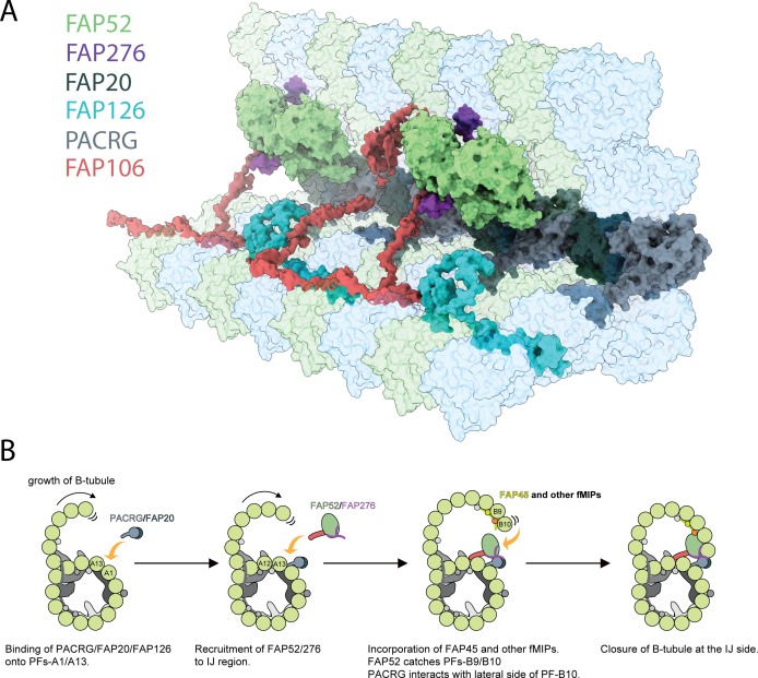
Microtubules are cytoskeletal structures involved in stability, transport and organization in the cell. The building blocks, the α- and β-tubulin heterodimers, form protofilaments that associate laterally into the hollow microtubule. Microtubule also exists as highly stable doublet microtubules in the cilia where stability is needed for ciliary beating and function. The doublet microtubule maintains its stability through interactions at its inner and outer junctions where its A- and B-tubules meet. Here, using cryo-electron microscopy, bioinformatics and mass spectrometry of the doublets of Chlamydomonas reinhardtii and Tetrahymena thermophila, we identified two new inner junction proteins, FAP276 and FAP106, and an inner junction-associated protein, FAP126, thus presenting the complete answer to the inner junction identity and localization. Our structural study of the doublets shows that the inner junction serves as an interaction hub that involves tubulin post-translational modifications. These interactions contribute to the stability of the doublet and hence, normal ciliary motility.
Overview
The claims and statistics of this paper haven’t been checked yet.
Run the check — free, no account needed. It reads the open-access text and checks the sources it cites.
Integrity signals
Registry
Cites
Statistics
Figures
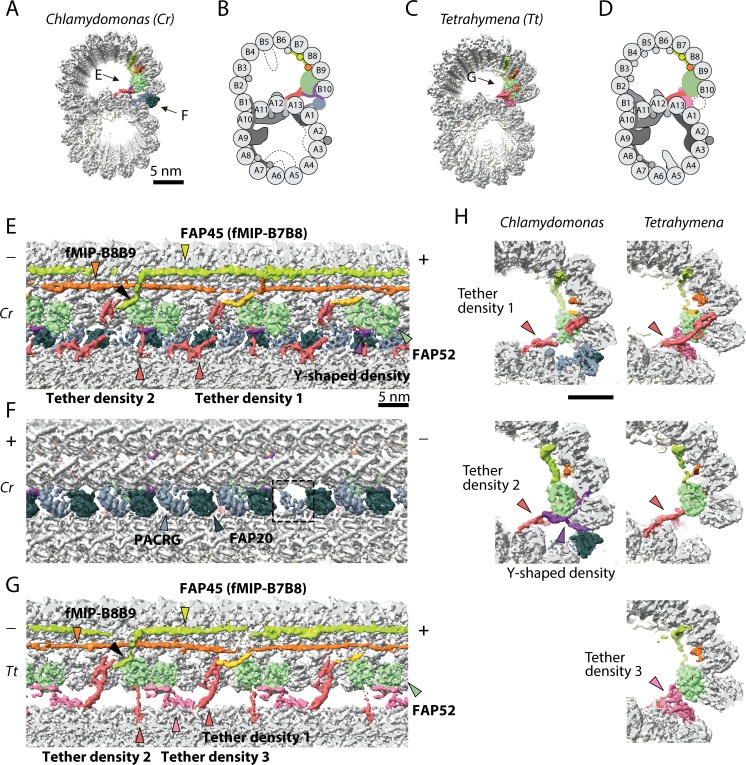
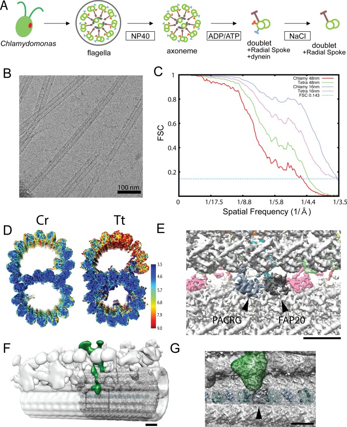
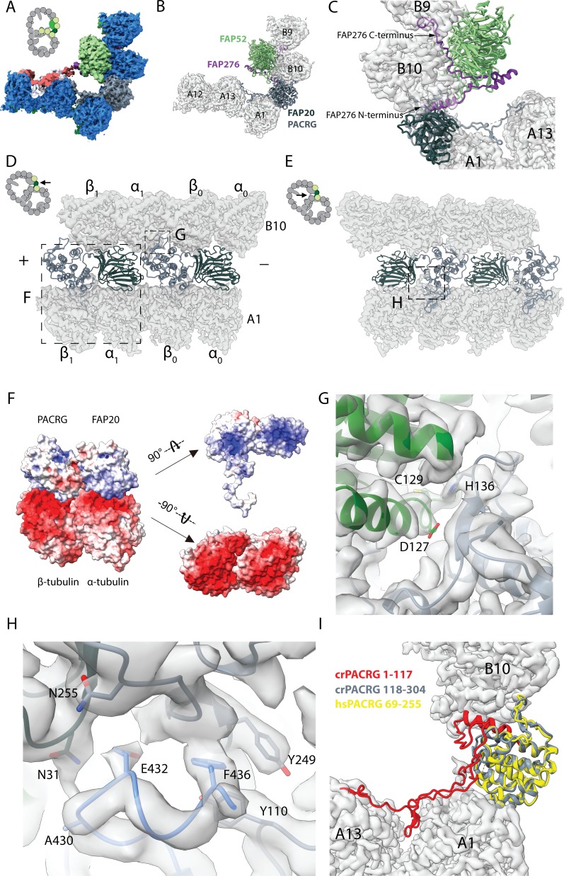
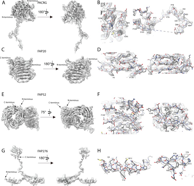
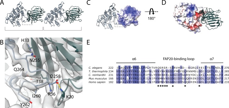
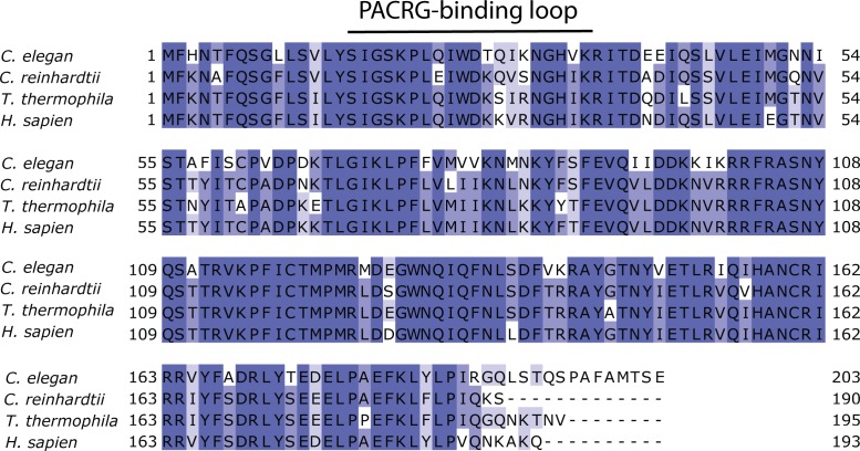
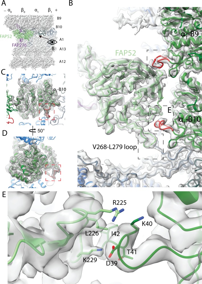
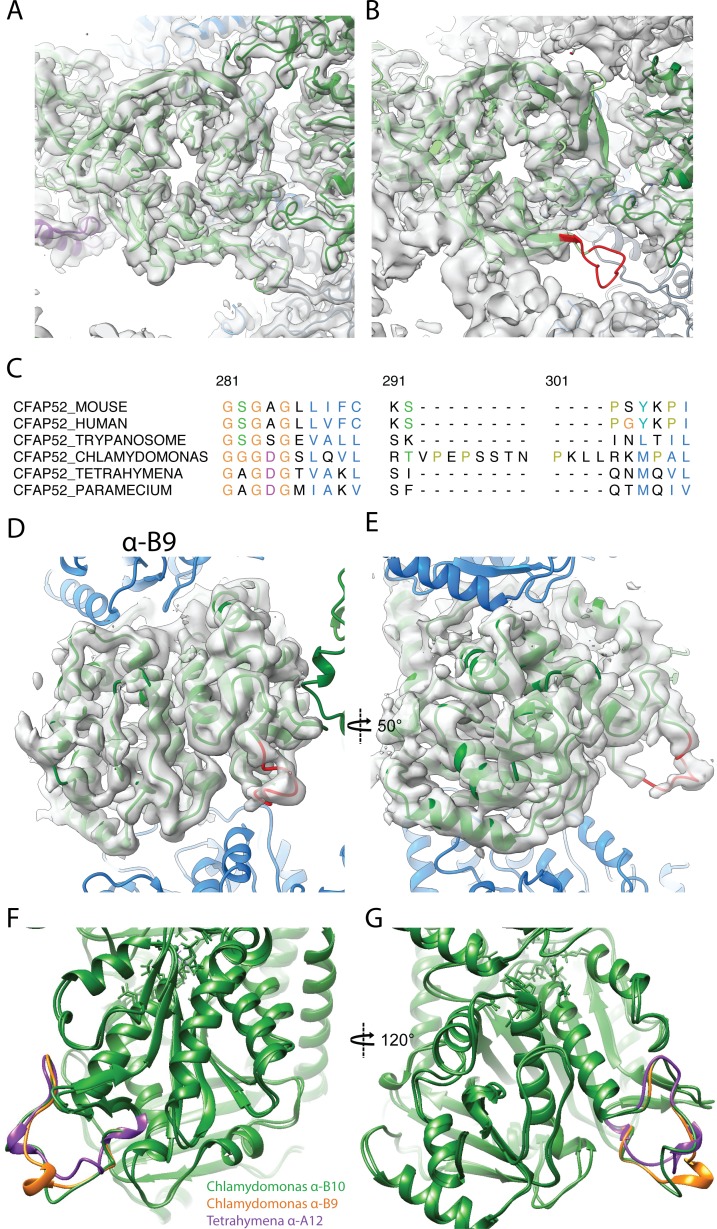
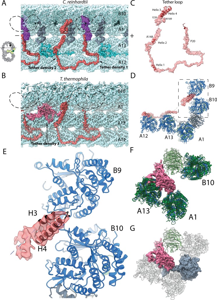
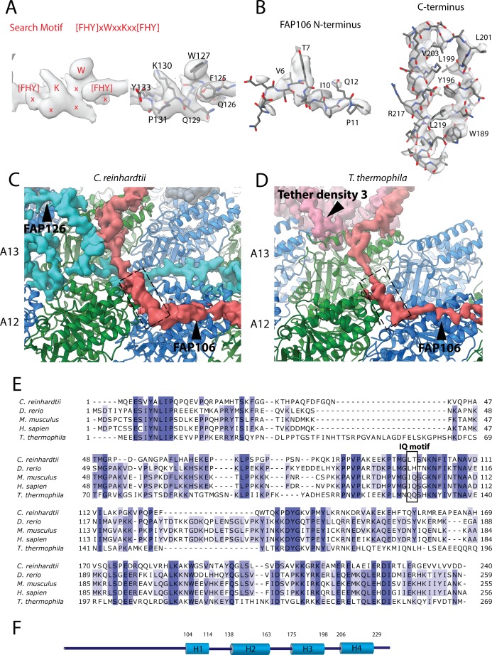
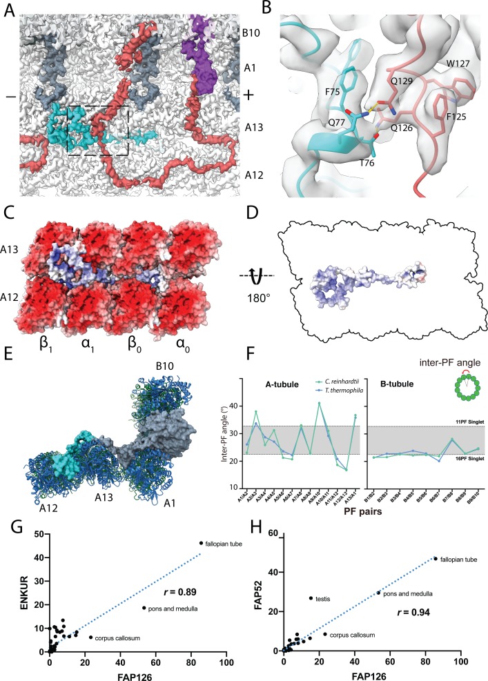
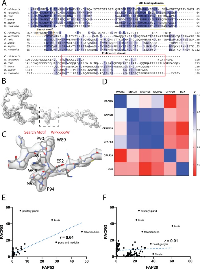
Verification
Claims the paper makes
The claims and statistics of this paper haven’t been checked yet.
Run the check — free, no account needed. It reads the open-access text and checks the sources it cites.
Claims other papers make about this one
Each is checked against this paper’s own text: does it say what the citing paper says it does?
The source backs part of the claim, not all of it — or backs it for a different population, condition or comparison.
In the citing paper
“Based on near-atomic-resolution cryo-electron microscopy reconstructions of motile cilia axonemes, the inner junction is well positioned to act as a scaffold for the radial spoke and dynein regulatory complex machinery19–22.”
From The inner junction protein CFAP20 functions in motile and non-motile cilia and is critical for vision (2022) · cites this paper as [21]
Experimental — accuracy not yet measuredThe source backs part of the claim, not all of it — or backs it for a different population, condition or comparison.
In the citing paper
“From cryo-EM structures of Chlamydomonas and Tetrahymena motile cilia, proteins at or near the inner junction, which include CFAP20, PACRG, CFAP45, CFAP52, CFAP90, CFAP126 and EFHC119–21, are all positioned to form a potential scaffold for, or functionally influence, the radial spokes and dynein regulatory complex that regulates ciliary motility (Fig. 8).”
From The inner junction protein CFAP20 functions in motile and non-motile cilia and is critical for vision (2022) · cites this paper as [21]
Experimental — accuracy not yet measuredThe cited source says what the claim attributes to it.
In the citing paper
“The multiple contacts of CFAP20 with ciliary axoneme tubulins and PACRG, as well as proximity to CFAP52 and other proteins at or near the inner junction19–21,71 (Figs. 1b and 8), could explain this degree of conservation.”
From The inner junction protein CFAP20 functions in motile and non-motile cilia and is critical for vision (2022) · cites this paper as [21]
Self-check
Percentages, sums and reported differences re-computed from the paper’s own numbers.
The self-check needs the paper’s full text.
Citations
Cites
UCSF Chimera—A visualization system for exploratory research and analysis
Pettersen, E. F., Goddard, T. D., Huang, C. C., Couch, G. S., Greenblatt, D. M., Meng, E. C., & Ferrin, T. E. (2004) · Journal of Computational Chemistry · doi:10.1002/jcc.20084 · Cited by 46,047
Features and development of Coot
Emsley, P., Lohkamp, B., Scott, W. G., & Cowtan, K. (2010) · Acta Crystallographica Section D Biological Crystallography · doi:10.1107/s0907444910007493 · Cited by 29,915
PHENIX : a comprehensive Python-based system for macromolecular structure solution
Adams, P. D., Afonine, P. V., Bunkóczi, G., Chen, V. B., Davis, I. W., Echols, N., Headd, J. J., Hung, L.-W., Kapral, G. J., Grosse-Kunstleve, R. W., et al. (2010) · Acta Crystallographica Section D Biological Crystallography · doi:10.1107/s0907444909052925 · Cited by 23,716
MotionCor2: anisotropic correction of beam-induced motion for improved cryo-electron microscopy
Zheng, S. Q., Palovcak, E. J., Armache, J., Verba, K. A., Cheng, Y., & Agard, D. A. (2017) · Nature Methods · doi:10.1038/nmeth.4193 · Cited by 7,496
PHENIX: a comprehensive Python-based system for macromolecular structure solution
Adams, P. D., Afonine, P. V., Bunkòczi, G., Chen, V. B., Davis, I., Echols, N., Headd, J. J., Hung, L., Kapral, G. J., Grosse‐Kunstleve, R. W., et al. (2012) · International Tables for Crystallography · doi:10.1107/97809553602060000865 · Cited by 7,334
The I-TASSER Suite: protein structure and function prediction
Yang, J., Yan, R., Roy, A., Xu, D., Poisson, J., & Zhang, Y. (2015) · Nature Methods · doi:10.1038/nmeth.3213 · Cited by 5,596
UCSF ChimeraX: Meeting modern challenges in visualization and analysis
Goddard, T. D., Huang, C. C., Meng, E. C., Pettersen, E. F., Couch, G. S., Morris, J. H., & Ferrin, T. E. (2018) · Protein Science · doi:10.1002/pro.3235 · Cited by 5,267
Mutations in the parkin gene cause autosomal recessive juvenile parkinsonism
Kitada, T., Asakawa, S., Hattori, N., Matsumine, H., Yamamura, Y., Minoshima, S., Yokochi, M., Mizuno, Y., & Shimizu, N. (1998) · Nature · doi:10.1038/33416 · Cited by 5,227
New tools for automated high-resolution cryo-EM structure determination in RELION-3
Zivanov, J., Nakane, T., Forsberg, B., Kimanius, D., Hagen, W., Lindahl, E., & Scheres, S. (2018) · eLife · doi:10.7554/elife.42166 · Cited by 4,377
Gctf: Real-time CTF determination and correction
Zhang, K. (2015) · Journal of Structural Biology · doi:10.1016/j.jsb.2015.11.003 · Cited by 3,417
Real-space refinement in PHENIX for cryo-EM and crystallography
Afonine, P. V., Poon, B. K., Read, R. J., Sobolev, O. V., Terwilliger, T. C., Urzhumtsev, A., & Adams, P. D. (2018) · Acta Crystallographica Section D Structural Biology · doi:10.1107/s2059798318006551 · Cited by 3,234
EMAN2: An extensible image processing suite for electron microscopy
Tang, G., Peng, L., Baldwin, P. R., Mann, D. S., Jiang, W., Rees, I., & Ludtke, S. J. (2006) · Journal of Structural Biology · doi:10.1016/j.jsb.2006.05.009 · Cited by 2,914
Protein Structure Modeling with MODELLER
Eswar, N., Eramian, D., Webb, B., Shen, M., & Šali, A. (2008) · Methods in molecular biology · doi:10.1007/978-1-60327-058-8_8 · Cited by 1,781
JPred4: a protein secondary structure prediction server
Drozdetskiy, A., Cole, C., Procter, J. B., & Barton, G. J. (2015) · Nucleic Acids Research · doi:10.1093/nar/gkv332 · Cited by 1,714
Overview of Structure and Function of Mammalian Cilia
Satir, P., & Christensen, S. T. (2007) · Annual Review of Physiology · doi:10.1146/annurev.physiol.69.040705.141236 · Cited by 1,142
Proteomic analysis of a eukaryotic cilium
Pazour, G. J., Agrin, N. S., Leszyk, J., & Witman, G. B. (2005) · The Journal of Cell Biology · doi:10.1083/jcb.200504008 · Cited by 1,028
Protein Structure Modeling with MODELLER
Webb, B., & Šali, A. (2014) · Methods in molecular biology · doi:10.1007/978-1-4939-0366-5_1 · Cited by 600
XMAP215 Is a Processive Microtubule Polymerase
Brouhard, G. J., Stear, J. H., Noetzel, T. L., Al‐Bassam, J., Kinoshita, K., Harrison, S. C., Howard, J., & Hyman, A. A. (2008) · Cell · doi:10.1016/j.cell.2007.11.043 · Cited by 570
Microtubules acquire resistance from mechanical breakage through intralumenal acetylation
Xu, Z., Schaedel, L., Portran, D., Aguilar, A., Gaillard, J., Marinkovich, M. P., Théry, M., & Nachury, M. V. (2017) · Science · doi:10.1126/science.aai8764 · Cited by 505
MICROTUBULES: EVIDENCE FOR 13 PROTOFILAMENTS
Tilney, L. G., Bryan, J., Bush, D. J., Fujiwara, K., Mooseker, M. S., Murphy, D. B., & Snyder, D. H. (1973) · The Journal of Cell Biology · doi:10.1083/jcb.59.2.267 · Cited by 496
Tubulin acetylation protects long-lived microtubules against mechanical ageing
Portran, D., Schaedel, L., Xu, Z., Théry, M., & Nachury, M. V. (2017) · Nature Cell Biology · doi:10.1038/ncb3481 · Cited by 493
Near-atomic model of microtubule-tau interactions
Kellogg, E. H., Hejab, N. M. A., Poepsel, S., Downing, K. H., DiMaio, F., & Nogales, E. (2018) · Science · doi:10.1126/science.aat1780 · Cited by 416
Effects of α-tubulin acetylation on microtubule structure and stability
Eshun-Wilson, L., Zhang, R., Portran, D., Nachury, M. V., Toso, D. B., Löhr, T., Vendruscolo, M., Bonomi, M., Fraser, J. S., & Nogales, E. (2019) · Proceedings of the National Academy of Sciences · doi:10.1073/pnas.1900441116 · Cited by 367
The dynein regulatory complex is the nexin link and a major regulatory node in cilia and flagella
Heuser, T., Raytchev, M., Krell, J., Porter, M. E., & Nicastro, D. (2009) · The Journal of Cell Biology · doi:10.1083/jcb.200908067 · Cited by 352
[28] Isolation of Chlamydomonas flagella and flagellar axonemes
Witman, G. B. (1986) · Methods in enzymology on CD-ROM/Methods in enzymology · doi:10.1016/0076-6879(86)34096-5 · Cited by 321
Identification of an acetylation site of Chlamydomonas alpha-tubulin.
Ledizet, M., & Piperno, G. (1987) · Proceedings of the National Academy of Sciences · doi:10.1073/pnas.84.16.5720 · Cited by 319
Hervé, C., Rogowski, A., Blake, A. W., Marcus, S. E., Gilbert, H. J., & Knox, J. P. (2010) · Proceedings of the National Academy of Sciences · doi:10.1073/pnas.1005732107 · Cited by 305
αTAT1 is the major α-tubulin acetyltransferase in mice
Kalebic, N., Sorrentino, S., Perlas, E., Bolasco, G., Martinez, C., & Heppenstall, P. A. (2013) · Nature Communications · doi:10.1038/ncomms2962 · Cited by 241
MonoRes: Automatic and Accurate Estimation of Local Resolution for Electron Microscopy Maps
Vilas, J. L., Gómez-Blanco, J., Conesa, P., Melero, R., de la Rosa-Trevin, J. M., Otón, J., Cuenca-Alba, J., Marabini, R., Carazo, J. M., Vargas, J., et al. (2018) · Structure · doi:10.1016/j.str.2017.12.018 · Cited by 217
Self-citationThe ZebrafishfleerGene Encodes an Essential Regulator of Cilia Tubulin Polyglutamylation
Pathak, N. H., Obara, T., Mangos, S., Liu, Y., & Drummond, I. A. S. (2007) · Molecular Biology of the Cell · doi:10.1091/mbc.e07-06-0537 · Cited by 207
Polarity of midbody and phragmoplast microtubules.
Euteneuer, U., & McIntosh, J. R. (1980) · The Journal of Cell Biology · doi:10.1083/jcb.87.2.509 · Cited by 186
Effects of tubulin acetylation and tubulin acetyltransferase binding on microtubule structure
Howes, S. C., Alushin, G. M., Shida, T., Nachury, M. V., & Nogales, E. (2014) · Molecular Biology of the Cell · doi:10.1091/mbc.e13-07-0387 · Cited by 183
Posttranslational Modifications of Tubulin and Cilia
Wloga, D., Joachimiak, E., Louka, P., & Gaertig, J. (2017) · Cold Spring Harbor Perspectives in Biology · doi:10.1101/cshperspect.a028159 · Cited by 183
Bui, K. H., Yagi, T., Yamamoto, R., Kamiya, R., & Ishikawa, T. (2012) · The Journal of Cell Biology · doi:10.1083/jcb.201201120 · Cited by 181
Self-citationA molecular ruler determines the repeat length in eukaryotic cilia and flagella
Oda, T., Yanagisawa, H., Kamiya, R., & Kikkawa, M. (2014) · Science · doi:10.1126/science.1260214 · Cited by 179
Molecular architecture of inner dynein arms in situ in Chlamydomonas reinhardtii flagella
Bui, K. H., Sakakibara, H., Movassagh, T., Oiwa, K., & Ishikawa, T. (2008) · The Journal of Cell Biology · doi:10.1083/jcb.200808050 · Cited by 167
Self-citationCryo-electron tomography reveals conserved features of doublet microtubules in flagella
Nicastro, D., Fu, X., Heuser, T., Tso, A., Porter, M. E., & Linck, R. W. (2011) · Proceedings of the National Academy of Sciences · doi:10.1073/pnas.1106178108 · Cited by 147
Doublecortin Recognizes the 13-Protofilament Microtubule Cooperatively and Tracks Microtubule Ends
Bechstedt, S., & Brouhard, G. J. (2012) · Developmental Cell · doi:10.1016/j.devcel.2012.05.006 · Cited by 146
Self-citationThree-dimensional architecture of epithelial primary cilia
Sun, S., Fisher, R. L., Bowser, S. S., Pentecost, B. T., & Sui, H. (2019) · Proceedings of the National Academy of Sciences · doi:10.1073/pnas.1821064116 · Cited by 142
Inner lumen proteins stabilize doublet microtubules in cilia and flagella
Owa, M., Uchihashi, T., Yanagisawa, H.-A., Yamano, T., Iguchi, H., Fukuzawa, H., Wakabayashi, K.-I., Ando, T., & Kikkawa, M. (2019) · Nature Communications · doi:10.1038/s41467-019-09051-x · Cited by 140
Asymmetry of inner dynein arms and inter-doublet links in Chlamydomonas flagella
Bui, K. H., Sakakibara, H., Movassagh, T., Oiwa, K., & Ishikawa, T. (2009) · The Journal of Cell Biology · doi:10.1083/jcb.200903082 · Cited by 131
Self-citationEvolutionary Proteomics Uncovers Ancient Associations of Cilia with Signaling Pathways
Sigg, M. A., Menchen, T., Lee, C., Johnson, J., Jungnickel, M. K., Choksi, S. P., Garcia, G., Busengdal, H., Dougherty, G. W., Pennekamp, P., et al. (2017) · Developmental Cell · doi:10.1016/j.devcel.2017.11.014 · Cited by 130
Enkurin is a novel calmodulin and TRPC channel binding protein in sperm
Sutton, K. A., Jungnickel, M. K., Wang, Y., Cullen, K. J., Lambert, S., & Florman, H. M. (2004) · Developmental Biology · doi:10.1016/j.ydbio.2004.07.031 · Cited by 125
Identification of a Novel Gene Linked to Parkin via a Bi-directional Promoter
West, A. B., Lockhart, P. J., O’Farrell, C., & Farrer, M. J. (2003) · Journal of Molecular Biology · doi:10.1016/s0022-2836(02)01376-1 · Cited by 119
Ichikawa, M., Liu, D., Kastritis, P. L., Basu, K., Hsu, T. C., Yang, S.-K., & Bui, K. H. (2017) · Nature Communications · doi:10.1038/ncomms15035 · Cited by 115
Self-citationMechanisms of Nephronophthisis and Related Ciliopathies
Hurd, T. W., & Hildebrandt, F. (2010) · Nephron Experimental Nephrology · doi:10.1159/000320888 · Cited by 99
Deletion of the Parkin coregulated gene causes male sterility in the quaking viable mouse mutant
Lorenzetti, D., Bishop, C. E., & Justice, M. J. (2004) · Proceedings of the National Academy of Sciences · doi:10.1073/pnas.0401832101 · Cited by 93
Friedmann, D. R., Aguilar, A., Fan, J., Nachury, M. V., & Marmorstein, R. (2012) · Proceedings of the National Academy of Sciences · doi:10.1073/pnas.1209357109 · Cited by 90
Yanagisawa, H.-A., Mathis, G., Oda, T., Hirono, M., Richey, E. A., Ishikawa, H., Marshall, W. F., Kikkawa, M., & Qin, H. (2014) · Molecular Biology of the Cell · doi:10.1091/mbc.e13-08-0464 · Cited by 89
Tubulin lattice in cilia is in a stressed form regulated by microtubule inner proteins
Ichikawa, M., Khalifa, A. A. Z., Kubo, S., Dai, D., Basu, K., Maghrebi, M. A. F., Vargas, J., & Bui, K. H. (2019) · Proceedings of the National Academy of Sciences · doi:10.1073/pnas.1911119116 · Cited by 83
Self-citationThe Motility of Axonemal Dynein Is Regulated by the Tubulin Code
Alper, J., Decker, F., Agana, B. A., & Howard, J. (2014) · Biophysical Journal · doi:10.1016/j.bpj.2014.10.061 · Cited by 80
A human laterality disorder associated with a homozygous WDR16 deletion
Ta-Shma, A., Perles, Z., Yaacov, B., Werner, M., Frumkin, A., Rein, A. J., & Elpeleg, O. (2015) · European Journal of Human Genetics · doi:10.1038/ejhg.2014.265 · Cited by 70
Wilson, G. R. A., Wang, H. X., Egan, G. F., Robinson, P. J., Delatycki, M. B., O’Bryan, M. K., & Lockhart, P. J. (2010) · Human Molecular Genetics · doi:10.1093/hmg/ddq031 · Cited by 65
Dymek, E. E., Lin, J., Fu, G., Porter, M. E., Nicastro, D., & Smith, E. F. (2019) · Molecular Biology of the Cell · doi:10.1091/mbc.e19-01-0063 · Cited by 61
Luminal material in microtubules of frog olfactory axons: structure and distribution.
Burton, P. R. (1984) · The Journal of Cell Biology · doi:10.1083/jcb.99.2.520 · Cited by 59
Flattop regulates basal body docking and positioning in mono- and multiciliated cells
Gegg, M., Böttcher, A., Burtscher, I., Hasenöder, S., Campenhout, C. V., Aichler, M., Walch, A. K., Grant, S. G. N., & Lickert, H. (2014) · eLife · doi:10.7554/elife.03842 · Cited by 56
Cernohorska, M. S., Zhernov, I., Steib, E., Guennec, M. L., Achek, R., Borgers, S., Demurtas, D., Mouawad, L., Lánský, Z., Hamel, V., et al. (2019) · Science · doi:10.1126/science.aav2567 · Cited by 54
Functional protofilament numbering of ciliary, flagellar, and centriolar microtubules
Linck, R. W., & Stephens, R. E. (2007) · Cell Motility and the Cytoskeleton · doi:10.1002/cm.20202 · Cited by 44
Identification and mapping of central pair proteins by proteomic analysis
Dai, D., Ichikawa, M., Peri, K., Rebinsky, R., & Bui, K. H. (2020) · Biophysics and Physicobiology · doi:10.2142/biophysico.bsj-2019048 · Cited by 42
Self-citationStauber, M., Weidemann, M., Dittrich‐Breiholz, O., Lobschat, K., Alten, L., Mai, M., Beckers, A., Kracht, M., & Gossler, A. (2016) · Developmental Biology · doi:10.1016/j.ydbio.2016.11.019 · Cited by 41
Laligné, C., Klotz, C., Garreau de Loubresse, N., Lemullois, M., Hori, M., Laurent, F. X., Papon, J. F., Louis, B., Cohen, J., & Koll, F. (2010) · Eukaryotic Cell · doi:10.1128/ec.00368-09 · Cited by 35
Jungnickel, M. K., Sutton, K. A., Baker, M. A., Cohen, M. G., Sanderson, M. J., & Florman, H. M. (2018) · Biology of Reproduction · doi:10.1093/biolre/ioy105 · Cited by 27
Khan, N., Pelletier, D., McAlear, T. S., Croteau, N., Veyron, S., Bayne, A. N., Black, C., Ichikawa, M., Khalifa, A. A. Z., Chaaban, S., et al. (2021) · Structure · doi:10.1016/j.str.2021.01.001 · Cited by 24
Self-citationStructures of TOG1 and TOG2 from the human microtubule dynamics regulator CLASP1
Leano, J. B., & Slep, K. C. (2019) · PLoS ONE · doi:10.1371/journal.pone.0219823 · Cited by 23
Identification of a Novel Gene Linked to Parkin via a Bidirectional Promoter
Lockhart, P. J., West, A. B., O’Farrell, C., & Farrer, M. J. (2003) · Annals of the New York Academy of Sciences · doi:10.1111/j.1749-6632.2003.tb07493.x · Cited by 20
Ciliary and non-ciliary expression and function of PACRGduring vertebrate development
Thumberger, T., Hagenlocher, C., Tisler, M., Beyer, T., Tietze, N., Schweickert, A., Feistel, K., & Blum, M. (2012) · Cilia · doi:10.1186/2046-2530-1-13 · Cited by 18
Crystal structure of human PACRG in complex with MEIG1
(2019) · doi:10.1101/783373 · Cited by 3
Duong (2000) · doi:10.1002/1097-0169(200011)47:3
Cited by
Sorted by citations
A pan-cancer blueprint of the heterogeneous tumor microenvironment revealed by single-cell profiling
Qian, J., Olbrecht, S., Boeckx, B., Vos, H., Laoui, D., Etlioglu, E., Wauters, E., Pomella, V., Verbandt, S., Busschaert, P., et al. (2020) · Cell Research · doi:10.1038/s41422-020-0355-0 · Cited by 845
Sampling alternative conformational states of transporters and receptors with AlphaFold2
del Alamo, D., Sala, D., Mchaourab, H. S., & Meiler, J. (2022) · eLife · doi:10.7554/elife.75751 · Cited by 511
Higgins-Chen, A. T., Thrush, K. L., Wang, Y., Minteer, C. J., Kuo, P.-L., Wang, M., Niimi, P., Sturm, G., Lin, J., Moore, A. Z., et al. (2022) · Nature Aging · doi:10.1038/s43587-022-00248-2 · Cited by 502
The connectome of an insect brain
Winding, M., Pedigo, B. D., Barnes, C. L., Patsolic, H. G., Park, Y., Kazimiers, T., Fushiki, A., Andrade, I. V., Khandelwal, A., Valdes-Aleman, J., et al. (2023) · Science · doi:10.1126/science.add9330 · Cited by 435
Quantifying the local adaptive landscape of a nascent bacterial community
Ascensao, J. A., Wetmore, K. M., Good, B. H., Arkin, A. P., & Hallatschek, O. (2023) · Nature Communications · doi:10.1038/s41467-022-35677-5 · Cited by 447
Metadata
- Authors
- Khalifa, A. A. Z., Ichikawa, M., Dai, D., Kubo, S., Black, C. S., Peri, K., McAlear, T. S., Veyron, S., Yang, S. K., Vargas, J., Bechstedt, S., Trempe, J.-F., & Bui, K. H.
- Affiliations
- McGill University (CA)
- Kyoto University (JP)
- Corresponding author
- Bui, Khanh Huy
- ORCID
- Venue
- eLife
- Publisher
- eLife Sciences Publications, Ltd
- Published
- 2020-01-17
- Volume
- 9
- ISSN
- 2050-084X (ISSN-L)
- PMC
- PMC6994238
- OpenAlex
- W3000377151
- Cited by
- 1,676
- References
- 67
- Citations by year
- 2026: 159 · 2025: 264 · 2024: 275 · 2023: 301 · 2022: 336 · 2021: 273 · 2020: 64
- FWCI
- 142.78
- Type
- journal article
- Language
- en
- Open access
- gold
- doi.org
- Licence
- Locations
- eLife (publishedVersion, cc-by)
- PubMed Central (publishedVersion, cc-by)
- PubMed (publishedVersion)
- DOAJ (DOAJ: Directory of Open Access Journals) (submittedVersion, cc-by-sa)
- Funders
- Canadian Institutes of Health Research (PJT-156354)
- Natural Sciences and Engineering Research Council of Canada (RGPIN-2016-04954, RGPIN-2018-04813, RGPIN-2017-04649)
- Canadian Institute for Advanced Research (Arzieli Global Scholar Program)
- Japan Society for the Promotion of Science
- Abdulla Al Ghurair Foundation for Education (Al Ghurair STEM program scholarship)
- Canada Research Chairs (950-229792)
- Topics
- Life Sciences › Biochemistry, Genetics and Molecular Biology › Genetics › Genetic and Kidney Cyst Diseases
- Life Sciences › Biochemistry, Genetics and Molecular Biology › Cell Biology › Microtubule and mitosis dynamics
- Health Sciences › Medicine › Pulmonary and Respiratory Medicine › Cystic Fibrosis Research Advances
- Keywords
- tubulin post-translational modifications, Chlamydomonas reinhardtii, Tetrahymena thermophila, ciliary motility, microtubule stability, cryo-electron microscopy
- MeSH terms
- Cilia, Microtubules, Plant Proteins, Protein Processing, Post-Translational, Mass Spectrometry, Protozoan Proteins, Tetrahymena thermophila, Chlamydomonas reinhardtii, Computational Biology, Cryoelectron Microscopy
About these checks
Registry, retraction, Cites and statistics results are computed, not judged, with one exception: a printed reference that no index lists is read once by a model before it counts as a citation error. Claims are extracted by a model and kept only when their quote appears verbatim in the text; each cited source is then checked, with a model’s help, for whether it says what the claim relies on. Those verdicts can be wrong — read the claim’s quote and the cited source before relying on one.
Statistics and arithmetic are recomputed, not judged.
Confidence bands are shown only for fields calibrated on our annotated test set. None is calibrated yet, so no band is shown.
What counts as a claim ›How we measure confidence ›How we check this ›
Are you an author, or do you see a problem with this page? Request a review
