Cited by 101

The DOI matches its Crossref record; 5 of the 5 references it cites were found in a registry or an index.

Reduction of elevated proton leak rejuvenates mitochondria in the aged cardiomyocyte

Zhang, Huiliang, Alder, Nathan N, Wang, Wang, Szeto, Hazel, Marcinek, David J, Rabinovitch, Peter S·2020·eLife

Abstract

Aging-associated diseases, including cardiac dysfunction, are increasingly common in the population. However, the mechanisms of physiologic aging in general, and cardiac aging in particular, remain poorly understood. Age-related heart impairment is lacking a clinically effective treatment. Using the model of naturally aging mice and rats, we show direct evidence of increased proton leak in the aged heart mitochondria. Moreover, our data suggested ANT1 as the most likely site of mediating increased mitochondrial proton permeability in old cardiomyocytes. Most importantly, the tetra-peptide SS-31 prevents age-related excess proton entry, decreases the mitochondrial flash activity and mitochondrial permeability transition pore opening, rejuvenates mitochondrial function by direct association with ANT1 and the mitochondrial ATP synthasome, and leads to substantial reversal of diastolic dysfunction. Our results uncover the excessive proton leak as a novel mechanism of age-related cardiac dysfunction and elucidate how SS-31 can reverse this clinically important complication of cardiac aging.

Overview

The claims and statistics of this paper haven’t been checked yet.

Run the check — free, no account needed. It reads the open-access text and checks the sources it cites.

A few free checks a day · papers already verified open instantly · results are public

Integrity signals

Registry

Crossref · 10.7554/elife.60827

Cites

5 of 5 references verified0 citation errors · 0 not checkable

Statistics

Figures

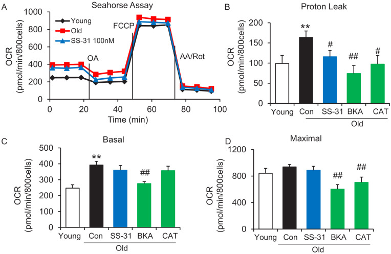
Figure 1. SS-31 alleviates the excessive mitochondrial proton leak of cardiomyocytes from 24 mo old mice. (A) Representative Seahorse assay traces of cardiomyocytes isolated from untreated young and old mice, then exposed or not to 100 nM SS-31 for 2 hr in vitro. Aging-increased basal respiration (C), which was attributable to the augmentation of proton leak (B), but did not affect maximal respiration (D). ANT1 inhibitors Bongkrekic acid (BKA, 10 μM) and carboxyatractyloside (CAT, 20 μM) 2 hr treatment decreased the proton leak (B) and basal respiration (C) but also decreased maximal respiration (D) in old cardiomyocytes. N = 5–14 mice in each group; one-way ANOVA followed by Fisher’s LSD test. *p<0.05, **p<0.01 vs young; #p<0.05, ##p<0.01 vs old controls.
Figure 1. SS-31 alleviates the excessive mitochondrial proton leak of cardiomyocytes from 24 mo old mice. (A) Representative Seahorse assay traces of cardiomyocytes isolated from untreated young and old mice, then exposed or not to 100 nM SS-31 for 2 hr in vitro. Aging-increased basal respiration (C), which was attributable to the augmentation of proton leak (B), but did not affect maximal respiration (D). ANT1 inhibitors Bongkrekic acid (BKA, 10 μM) and carboxyatractyloside (CAT, 20 μM) 2 hr treatment decreased the proton leak (B) and basal respiration (C) but also decreased maximal respiration (D) in old cardiomyocytes. N = 5–14 mice in each group; one-way ANOVA followed by Fisher’s LSD test. *p<0.05, **p<0.01 vs young; #p<0.05, ##p<0.01 vs old controls.CC BY · PubMed Central
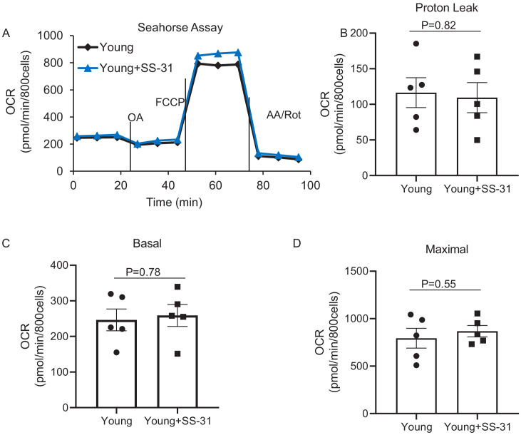
Figure 1—figure supplement 1. SS-31 has a minor effect on mitochondrial respiration in the young cardiomyocytes. None of the differences are significant. N = 5 mice in each group. Student’s t-test was applied to determine the statistical significance.
Figure 1—figure supplement 1. SS-31 has a minor effect on mitochondrial respiration in the young cardiomyocytes. None of the differences are significant. N = 5 mice in each group. Student’s t-test was applied to determine the statistical significance.CC BY · PubMed Central
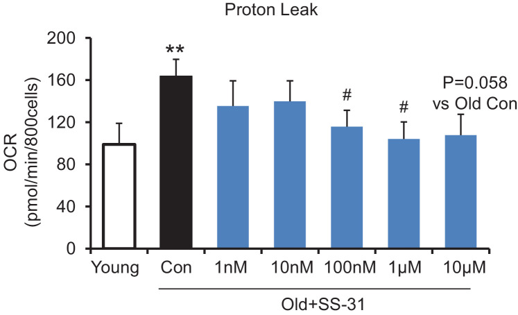
Figure 1—figure supplement 2. SS-31 reaches its inhibitory effect on proton leak suppression at low concentrations. SS-31 decreased the mitochondrial proton leak. N = 6–14 mice in each group. **p<0.01 vs young, #p<0.05 vs old controls. One-way ANOVA with Fisher’s LSD test was applied.
Figure 1—figure supplement 2. SS-31 reaches its inhibitory effect on proton leak suppression at low concentrations. SS-31 decreased the mitochondrial proton leak. N = 6–14 mice in each group. **p<0.01 vs young, #p<0.05 vs old controls. One-way ANOVA with Fisher’s LSD test was applied.CC BY · PubMed Central
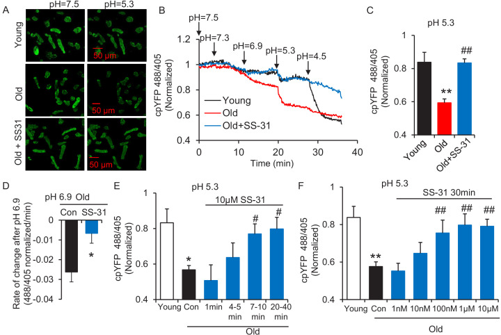
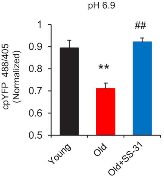
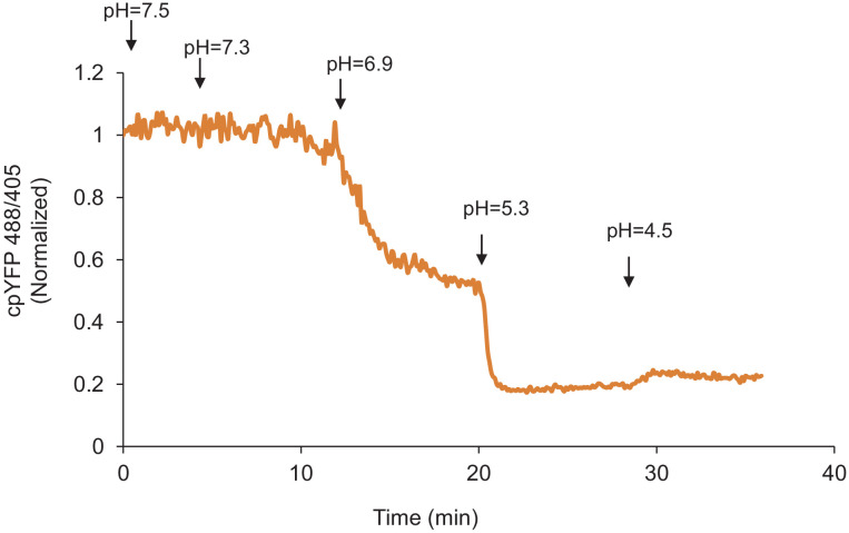
Figure 2. SS-31 restores the resistance of cardiomyocytes from old rat to proton entry into the mitochondrial matrix during external pH gradient stress. (A) Typical image of the effects of pH gradient stress on permeabilized rat cardiomyocyte mt-cpYFP fluorescence. Upper panel, young, middle panel, old, lower panel, old+SS-31 (10 μM, 3 days) visualized after exposure of the cells to pH 7.5 and, later, to pH 5.3. The excitation is 488 nm and collection is at 505–730 nm. (B) Saponin (50 μg/ml) permeabilized cardiomyocytes expressing mt-cpYFP were exposed to progressively lower external pH. Proton permeability of old mitochondria was greater than that of young mitochondria, but preincubation of old cells with 10 μM SS-31 for 3 days enhanced the mitochondrial inner membrane resistance to the pH stress. The traces were averaged from 4 to 19 experiments. The arrows indicate the changes of pH. (C) Quantitation of the SS-31 treatment effect on the mitochondrial matrix cpYFP ratio at pH 5.3. The data are from 7 to 8 min after the pH was adjusted to 5.3. The mean value of the normalized 488/405 ratio in old group is 0.59 with a SD ±0.10 and the range is from 0.46 to 0.77. N = 5–19 rats in each group, analyzed by one-way ANOVA with Dunnett’s test. (D) SS-31 decreased the rate of cpYFP 488/405 ratio drop at pH 6.9. N = 4–10 rats in each group. The rate is calculated as indicated in Figure 2—figure supplement 3. Student’s t-test was applied to determine the statistical significance. The time dependence (N = 3–4 rats in each group) (E) and dose dependence (N = 3–14 rats in each group) (F) of SS-31 protection of mitochondrial resistance to pH gradient stress are shown and analyzed by one-way ANOVA with Fisher’s LSD test. After cardiomyocyte permeabilization, 10 μM SS-31 was added for the times shown in (E) or at the doses shown in (F) for 30 min, followed by pH stress. *p<0.05, **p<0.01 vs Young; #p<0.05, ##p<0.01 vs Old.
Figure 2. SS-31 restores the resistance of cardiomyocytes from old rat to proton entry into the mitochondrial matrix during external pH gradient stress. (A) Typical image of the effects of pH gradient stress on permeabilized rat cardiomyocyte mt-cpYFP fluorescence. Upper panel, young, middle panel, old, lower panel, old+SS-31 (10 μM, 3 days) visualized after exposure of the cells to pH 7.5 and, later, to pH 5.3. The excitation is 488 nm and collection is at 505–730 nm. (B) Saponin (50 μg/ml) permeabilized cardiomyocytes expressing mt-cpYFP were exposed to progressively lower external pH. Proton permeability of old mitochondria was greater than that of young mitochondria, but preincubation of old cells with 10 μM SS-31 for 3 days enhanced the mitochondrial inner membrane resistance to the pH stress. The traces were averaged from 4 to 19 experiments. The arrows indicate the changes of pH. (C) Quantitation of the SS-31 treatment effect on the mitochondrial matrix cpYFP ratio at pH 5.3. The data are from 7 to 8 min after the pH was adjusted to 5.3. The mean value of the normalized 488/405 ratio in old group is 0.59 with a SD ±0.10 and the range is from 0.46 to 0.77. N = 5–19 rats in each group, analyzed by one-way ANOVA with Dunnett’s test. (D) SS-31 decreased the rate of cpYFP 488/405 ratio drop at pH 6.9. N = 4–10 rats in each group. The rate is calculated as indicated in Figure 2—figure supplement 3. Student’s t-test was applied to determine the statistical significance. The time dependence (N = 3–4 rats in each group) (E) and dose dependence (N = 3–14 rats in each group) (F) of SS-31 protection of mitochondrial resistance to pH gradient stress are shown and analyzed by one-way ANOVA with Fisher’s LSD test. After cardiomyocyte permeabilization, 10 μM SS-31 was added for the times shown in (E) or at the doses shown in (F) for 30 min, followed by pH stress. *p<0.05, **p<0.01 vs Young; #p<0.05, ##p<0.01 vs Old.CC BY · PubMed Central
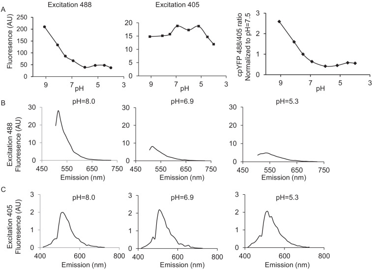
Figure 2—figure supplement 1. pH calibration of mt-cpYFP in adult cardiac myocytes with Nigericin. 10 μM Nigericin was added to the mitochondrial permeabilization buffer. (A) shows the pH calibration of 488 nm and 405 nm, the ratio of 488/405 (normalized to that of the value at pH = 7.5) as the pH is gradually lowered. The ratio of 488/405 reaches the lowest point at pH 6.0, followed by a small increase starting at pH 5.5, consistent with the known pH response of this dye (Wei-LaPierre et al., 2013). (B) shows the emission spectra of mt-cpYFP pH 8.0, 6.9, and 5.3 when excited at 488 nm (C) shows the emission spectra of mt-cpYFP pH 8.0, 6.9, and 5.3 when excited at 405 nm. The 488 nm excitation is sensitive to the pH change but the 405 nm excitation is much less pH dependent.
Figure 2—figure supplement 1. pH calibration of mt-cpYFP in adult cardiac myocytes with Nigericin. 10 μM Nigericin was added to the mitochondrial permeabilization buffer. (A) shows the pH calibration of 488 nm and 405 nm, the ratio of 488/405 (normalized to that of the value at pH = 7.5) as the pH is gradually lowered. The ratio of 488/405 reaches the lowest point at pH 6.0, followed by a small increase starting at pH 5.5, consistent with the known pH response of this dye (Wei-LaPierre et al., 2013). (B) shows the emission spectra of mt-cpYFP pH 8.0, 6.9, and 5.3 when excited at 488 nm (C) shows the emission spectra of mt-cpYFP pH 8.0, 6.9, and 5.3 when excited at 405 nm. The 488 nm excitation is sensitive to the pH change but the 405 nm excitation is much less pH dependent.CC BY · PubMed Central
Figure 2—figure supplement 2. SS31 restores the resistance to external pH gradient stress in the old cardiomyocytes at pH 6.9. N = 5–18 in each group. One-way ANOVA with Dunnett’s test was applied. **p<0.01 vs young, #p<0.05 vs old controls.
Figure 2—figure supplement 2. SS31 restores the resistance to external pH gradient stress in the old cardiomyocytes at pH 6.9. N = 5–18 in each group. One-way ANOVA with Dunnett’s test was applied. **p<0.01 vs young, #p<0.05 vs old controls.CC BY · PubMed Central
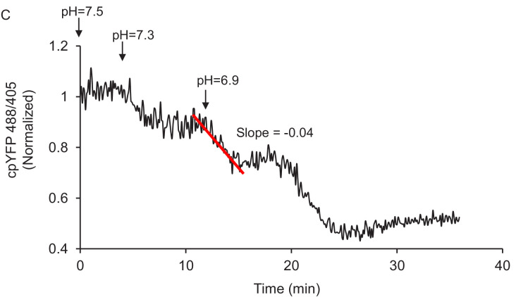
Figure 2—figure supplement 3. Method of quantitation of the slope of mt-cpYFP fluorescence ratio change after permeabilized cells are exposed to external pH 6.9. The trace is from old rat cardiomyocytes.
Figure 2—figure supplement 3. Method of quantitation of the slope of mt-cpYFP fluorescence ratio change after permeabilized cells are exposed to external pH 6.9. The trace is from old rat cardiomyocytes.CC BY · PubMed Central
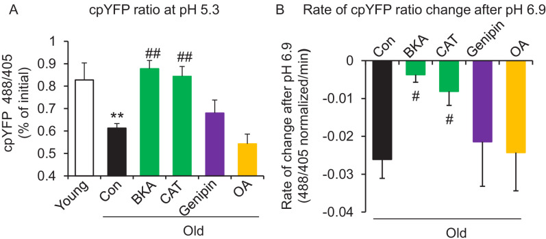
Figure 3. ANT1 inhibitors restore resistance to proton leak of mitochondria in old rat cardiomyocytes. ANT1 inhibitors 10 μM Bongkrekic Acid (BKA) and 20 μM carboxyatractyloside (CAT), but not 50 μM Genipin (UCP2 inhibitor) or 1 μM oligomycin A (OA, ATPase inhibitor), protected the mitochondrial matrix from decreased pH after exposure to external pH 5.3 (N = 4–19 rats in each group) (A) and reduced the rate of 488/405 decline after exposure to pH 6.9 (N = 4–10 rats in each group) (B). BKA, CAT, Genipin, or OA were added immediately after the mitochondria permeabilization. One-way ANOVA with Fisher’s LSD test was applied. *p<0.05, **p<0.01 vs Young, #p<0.05, ##p<0.01 vs Old.
Figure 3. ANT1 inhibitors restore resistance to proton leak of mitochondria in old rat cardiomyocytes. ANT1 inhibitors 10 μM Bongkrekic Acid (BKA) and 20 μM carboxyatractyloside (CAT), but not 50 μM Genipin (UCP2 inhibitor) or 1 μM oligomycin A (OA, ATPase inhibitor), protected the mitochondrial matrix from decreased pH after exposure to external pH 5.3 (N = 4–19 rats in each group) (A) and reduced the rate of 488/405 decline after exposure to pH 6.9 (N = 4–10 rats in each group) (B). BKA, CAT, Genipin, or OA were added immediately after the mitochondria permeabilization. One-way ANOVA with Fisher’s LSD test was applied. *p<0.05, **p<0.01 vs Young, #p<0.05, ##p<0.01 vs Old.CC BY · PubMed Central
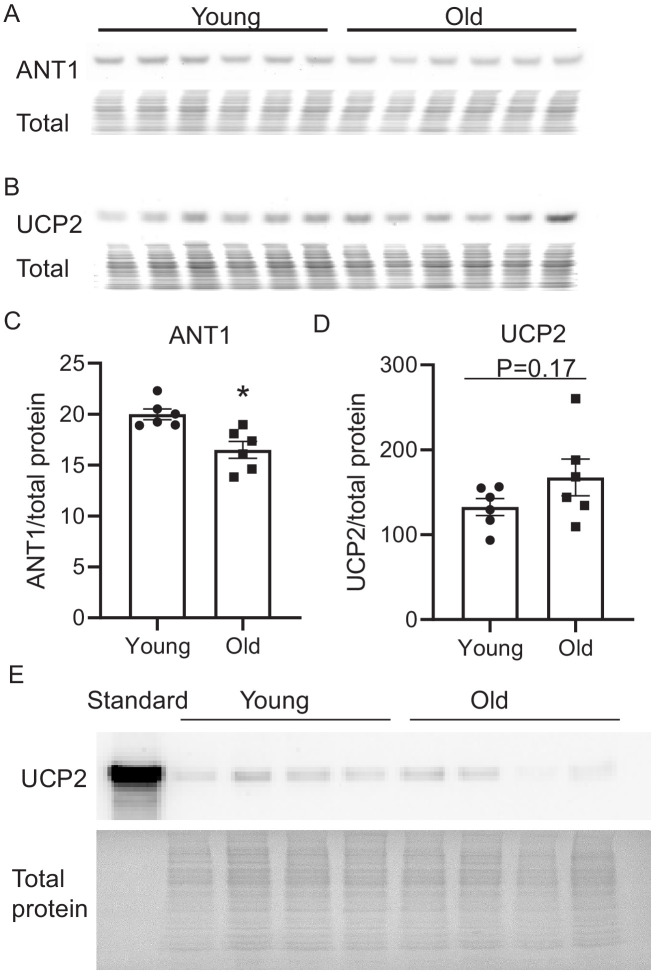
Figure 3—figure supplement 1. Aging effect on ANT1 and UCP2 cardiac protein abundance. Aging slightly, but significantly decreases ANT1 levels in the mouse heart. Western blot of ANT1 (A, C) and UCP2 (B, D). N = 6 mice in each group. Student’s t-test was applied to determine the statistical significance. *p<0.05 vs young. The total protein loading control was stained with MemCode Reversible Protein Stain Kit for Polyvinylidene difluoride Membranes (Pierce, Rockford, IL). The identity of the band as UCP2 in B was confirmed using a recombinant UCP2 protein standard (Origene, CAT#: TP761109), as shown in E.
Figure 3—figure supplement 1. Aging effect on ANT1 and UCP2 cardiac protein abundance. Aging slightly, but significantly decreases ANT1 levels in the mouse heart. Western blot of ANT1 (A, C) and UCP2 (B, D). N = 6 mice in each group. Student’s t-test was applied to determine the statistical significance. *p<0.05 vs young. The total protein loading control was stained with MemCode Reversible Protein Stain Kit for Polyvinylidene difluoride Membranes (Pierce, Rockford, IL). The identity of the band as UCP2 in B was confirmed using a recombinant UCP2 protein standard (Origene, CAT#: TP761109), as shown in E.CC BY · PubMed Central
Figure 3—figure supplement 2. Mitochondrial proton entry in buffer without phosphate. Mitochondrial pH stress on permeabilized rat cardiomyocytes in a buffer that did not contain phosphate.
Figure 3—figure supplement 2. Mitochondrial proton entry in buffer without phosphate. Mitochondrial pH stress on permeabilized rat cardiomyocytes in a buffer that did not contain phosphate.CC BY · PubMed Central
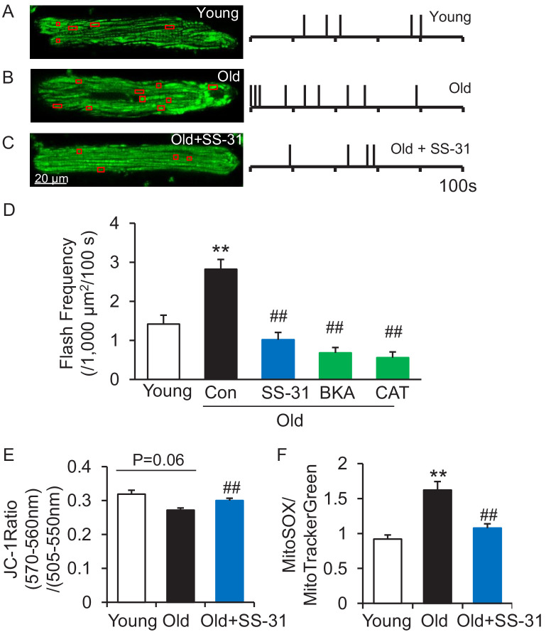
Figure 4. SS-31 attenuates excessive mitoflash activity in aged rat cardiomyocytes. (A–C) Mitoflash events within the regions shown in the red boxes took place at the times shown by vertical bars during the 100 s scanning time in the representative cardiomyocytes from young (A), old (B) and old+SS-31 (C) rat hearts. (D) The rate of mitoflash activity was increased in old rat cardiomyocytes compared to young, but 1 hr SS-31, Bongkrekic Acid (BKA) (10 μM) and Carboxyatractyloside (CAT) (20 μM) treatments decreased the mitoflash frequency in old cells to or below that of young cells. N = 26–87 cells from 3 to 14 rats. **p<0.05 vs young. ##p<0.05 vs old. (E) JC-1 red to green fluorescence ratio, indicative of mitochondrial membrane potential, in cells from young and old mice and old mouse cardiomyocytes treated with SS-31 (10 μM for 12 hr). N = 84–218 cells from 3 to 4 mice. p=0.06 vs young. ##p<0.01 vs old. (F) Mitochondrial ROS production in mouse cardiomyocytes measured by the fluorescence ratio of MitoSOX (5 μM, excitation 540 nm, emission >560 nm) to Mitotracker green (200 nM, excitation 488 nm, emission 505–530 nm). One-way ANOVA with Dunnett’s test was applied. N = 40–84 cells from 3 to 5 mice. **p<0.05 vs young. ##p<0.05 vs old.
Figure 4. SS-31 attenuates excessive mitoflash activity in aged rat cardiomyocytes. (A–C) Mitoflash events within the regions shown in the red boxes took place at the times shown by vertical bars during the 100 s scanning time in the representative cardiomyocytes from young (A), old (B) and old+SS-31 (C) rat hearts. (D) The rate of mitoflash activity was increased in old rat cardiomyocytes compared to young, but 1 hr SS-31, Bongkrekic Acid (BKA) (10 μM) and Carboxyatractyloside (CAT) (20 μM) treatments decreased the mitoflash frequency in old cells to or below that of young cells. N = 26–87 cells from 3 to 14 rats. **p<0.05 vs young. ##p<0.05 vs old. (E) JC-1 red to green fluorescence ratio, indicative of mitochondrial membrane potential, in cells from young and old mice and old mouse cardiomyocytes treated with SS-31 (10 μM for 12 hr). N = 84–218 cells from 3 to 4 mice. p=0.06 vs young. ##p<0.01 vs old. (F) Mitochondrial ROS production in mouse cardiomyocytes measured by the fluorescence ratio of MitoSOX (5 μM, excitation 540 nm, emission >560 nm) to Mitotracker green (200 nM, excitation 488 nm, emission 505–530 nm). One-way ANOVA with Dunnett’s test was applied. N = 40–84 cells from 3 to 5 mice. **p<0.05 vs young. ##p<0.05 vs old.CC BY · PubMed Central
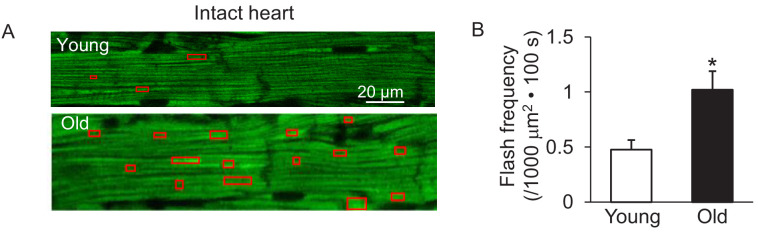
Figure 4—figure supplement 1. Increased mitochondrial flash activity in the intact perfused mouse aged heart. (A) Typical images from the young (upper panel) and old (lower panel) mouse hearts. (B) Statistical analysis of mitoflash frequency in the analyzed regions indicated by the red boxes in panel A. Young: N = 18 regions from three mice. Old: N = 16 regions from four mice. Student’s t-test was applied to determine the statistical significance. *p<0.05 vs Young.
Figure 4—figure supplement 1. Increased mitochondrial flash activity in the intact perfused mouse aged heart. (A) Typical images from the young (upper panel) and old (lower panel) mouse hearts. (B) Statistical analysis of mitoflash frequency in the analyzed regions indicated by the red boxes in panel A. Young: N = 18 regions from three mice. Old: N = 16 regions from four mice. Student’s t-test was applied to determine the statistical significance. *p<0.05 vs Young.CC BY · PubMed Central
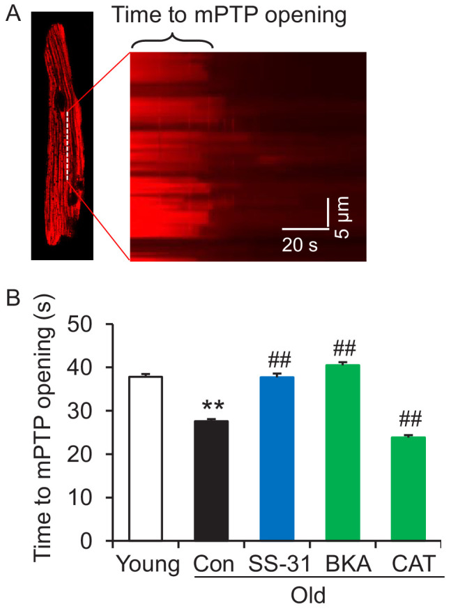
Figure 5. SS-31 reverses the increased speed of mitochondrial permeability transition pore (mPTP) opening in aged mouse cardiomyocytes. (A) A typical image shows 1 Hz line-scanning photo-excitation induced mPTP opening in a cardiomyocyte loaded with the mitochondrial membrane potential (Δψm) dye Tetramethylrhodamine methyl ester (TMRM). The sudden decline of TMRM fluorescence with time (rightward) indicates mPTP opening and Δψm loss. (B) 1 hr SS-31 and Bongkrekic Acid (BKA), but not carboxyatractyloside (CAT) treatments protect the photo-excitation induced mPTP opening. Quantification of time to mPTP opening from 418 to 658 mitochondria from 19 to 32 cells isolated from 3 to 4 mice in each group. One-way ANOVA with Dunnett’s test was applied. **p<0.01 vs young, ##p<0.01 vs old.
Figure 5. SS-31 reverses the increased speed of mitochondrial permeability transition pore (mPTP) opening in aged mouse cardiomyocytes. (A) A typical image shows 1 Hz line-scanning photo-excitation induced mPTP opening in a cardiomyocyte loaded with the mitochondrial membrane potential (Δψm) dye Tetramethylrhodamine methyl ester (TMRM). The sudden decline of TMRM fluorescence with time (rightward) indicates mPTP opening and Δψm loss. (B) 1 hr SS-31 and Bongkrekic Acid (BKA), but not carboxyatractyloside (CAT) treatments protect the photo-excitation induced mPTP opening. Quantification of time to mPTP opening from 418 to 658 mitochondria from 19 to 32 cells isolated from 3 to 4 mice in each group. One-way ANOVA with Dunnett’s test was applied. **p<0.01 vs young, ##p<0.01 vs old.CC BY · PubMed Central
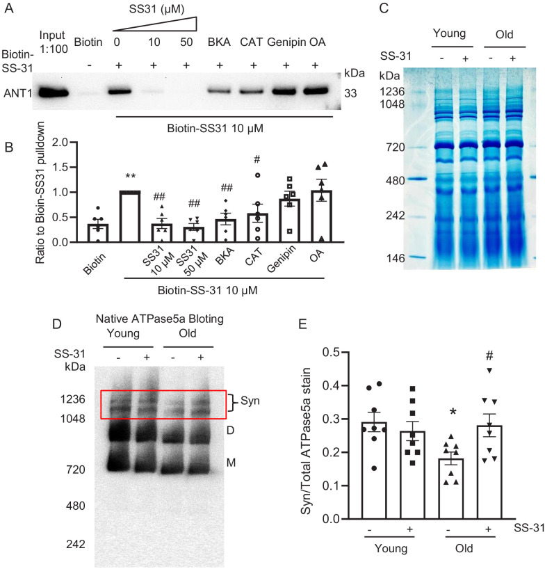
Figure 6. SS-31 interacts with ANT1 and stabilizes the ATP synthasome in the old mouse heart mitochondria. (A, B) Biotin-SS-31 pulldown shows the association of biotin-SS-31 to ANT1. Free SS-31 competes with this interaction, while Bongkrekic Acid (BKA) and carboxyatractyloside (CAT) inhibit the interaction of biotin-SS-31 with ANT1. Panel A shows a representative Western blot. N = 6 mice in each group. One-way ANOVA with Fisher’s LSD test was applied. **p<0.01 vs Biotin control, #p<0.05, ##p<0.01 vs Biotin-SS31 pulldown. (C) Coomassie blue staining of isolated mitochondria in a native gel. (D) Left panel is the total protein loading control for the Native gel blot. (D, E) Native gel blotting shows that 10 μM SS-31 stabilizes the mitochondrial synthasome (Syn) in isolated mitochondria. The Syn is highlighted in the red box. The Syn and ATPase Dimer (D) and Monomer (M) were labeled using anti-ATP5A. N = 8 mice in each group. One-way ANOVA with Fisher’s LSD test was applied. *p<0.05 vs young, #p<0.05 vs old.
Figure 6. SS-31 interacts with ANT1 and stabilizes the ATP synthasome in the old mouse heart mitochondria. (A, B) Biotin-SS-31 pulldown shows the association of biotin-SS-31 to ANT1. Free SS-31 competes with this interaction, while Bongkrekic Acid (BKA) and carboxyatractyloside (CAT) inhibit the interaction of biotin-SS-31 with ANT1. Panel A shows a representative Western blot. N = 6 mice in each group. One-way ANOVA with Fisher’s LSD test was applied. **p<0.01 vs Biotin control, #p<0.05, ##p<0.01 vs Biotin-SS31 pulldown. (C) Coomassie blue staining of isolated mitochondria in a native gel. (D) Left panel is the total protein loading control for the Native gel blot. (D, E) Native gel blotting shows that 10 μM SS-31 stabilizes the mitochondrial synthasome (Syn) in isolated mitochondria. The Syn is highlighted in the red box. The Syn and ATPase Dimer (D) and Monomer (M) were labeled using anti-ATP5A. N = 8 mice in each group. One-way ANOVA with Fisher’s LSD test was applied. *p<0.05 vs young, #p<0.05 vs old.CC BY · PubMed Central
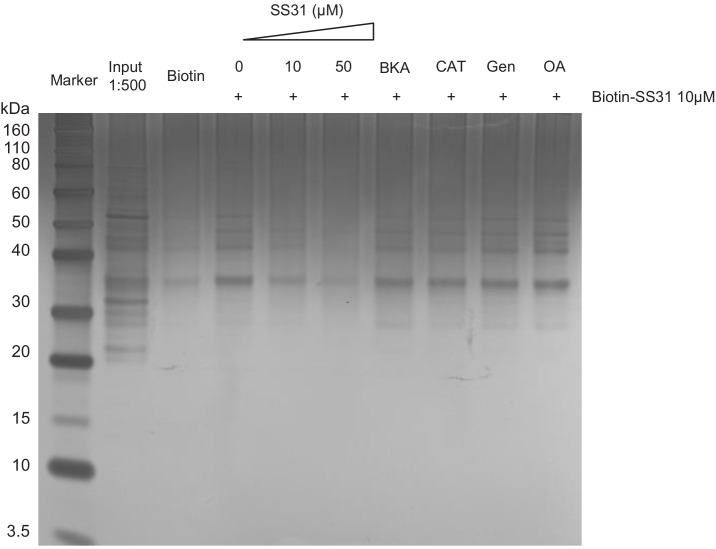
Figure 6—figure supplement 1. Silver staining of the Biotin-SS-31 pulldown of Figure 6A from the old mouse heart mitochondria. The great majority of bands present in the input are not present in the biotin-SS-31 pulldown; those that are show suppression by SS-31 competition.
Figure 6—figure supplement 1. Silver staining of the Biotin-SS-31 pulldown of Figure 6A from the old mouse heart mitochondria. The great majority of bands present in the input are not present in the biotin-SS-31 pulldown; those that are show suppression by SS-31 competition.CC BY · PubMed Central
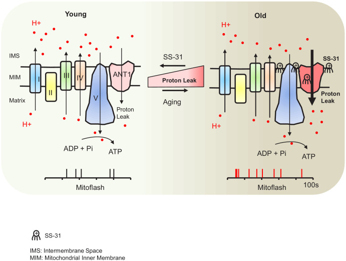
Figure 7. Schematic of the mechanism of SS-31 protection of proton leak and rejuvenation of mitochondrial function. Due to increased mitochondrial proton leak, the mitochondria work harder to maintain ATP production, and thus the work load is increased in the aged heart.
Figure 7. Schematic of the mechanism of SS-31 protection of proton leak and rejuvenation of mitochondrial function. Due to increased mitochondrial proton leak, the mitochondria work harder to maintain ATP production, and thus the work load is increased in the aged heart.CC BY · PubMed Central

Verification

Claims the paper makes

The claims and statistics of this paper haven’t been checked yet.

Run the check — free, no account needed. It reads the open-access text and checks the sources it cites.

A few free checks a day · papers already verified open instantly · results are public

Claims other papers make about this one

Each is checked against this paper’s own text: does it say what the citing paper says it does?

Self-check

Percentages, sums and reported differences re-computed from the paper’s own numbers.

The self-check needs the paper’s full text.

Citations

Cites

Research works printed but not linked: 52

  1. Adams, 2000, Cardiomyocyte apoptosis induced by Galphaq signaling is mediated by permeability transition pore formation and activation of the mitochondrial death pathway, Circulation Research, 87, 1180, 10.1161/01.RES.87.12.1180 · DOI
  2. Bertholet, 2019, H+ transport is an integral function of the mitochondrial ADP/ATP carrier, Nature, 571, 515, 10.1038/s41586-019-1400-3 · DOI
  3. Birk, 2013, The mitochondrial-targeted compound SS-31 re-energizes ischemic mitochondria by interacting with cardiolipin, Journal of the American Society of Nephrology, 24, 1250, 10.1681/ASN.2012121216 · DOI · Paper page
  4. Brand, 2000, Uncoupling to survive? the role of mitochondrial inefficiency in ageing, Experimental Gerontology, 35, 811, 10.1016/S0531-5565(00)00135-2 · DOI · Paper page
  5. Chavez, 2020, Mitochondrial protein interaction landscape of SS-31, PNAS, 31, 10.1101/739128 · DOI
  6. Chiao, 2016, Rapamycin transiently induces mitochondrial remodeling to reprogram energy metabolism in old hearts, Aging, 8, 314, 10.18632/aging.100881 · DOI · Paper page
  7. Chiao, 2020, Late-life restoration of mitochondrial function reverses cardiac dysfunction in old mice, 10.1101/2020.01.02.893008 · DOI
  8. Dai, 2013, Global proteomics and pathway analysis of pressure-overload-induced heart failure and its attenuation by mitochondrial-targeted peptides, Circulation: Heart Failure, 6, 1067, 10.1161/CIRCHEARTFAILURE.113.000406 · DOI
  9. Dai, 2014, Mitochondrial oxidative stress in aging and healthspan, Longevity & Healthspan, 3, 10.1186/2046-2395-3-6 · DOI
  10. Demaurex, 2016, Mitochondrial flashes: dump superoxide and dance with protons now, Antioxidants & Redox Signaling, 25, 550, 10.1089/ars.2016.6819 · DOI
  11. Divakaruni, 2011, The regulation and physiology of mitochondrial proton leak, Physiology, 26, 192, 10.1152/physiol.00046.2010 · DOI
  12. Feng, 2017, Handbook of Experimental Pharmacology, 403, 10.1007/164_2016_129 · DOI
  13. Hafner, 2010, Regulation of the mPTP by SIRT3-mediated deacetylation of CypD at lysine 166 suppresses age-related cardiac hypertrophy, Aging, 2, 914, 10.18632/aging.100252 · DOI
  14. Halestrap, 1997, Oxidative stress, thiol reagents, and membrane potential modulate the mitochondrial permeability transition by affecting nucleotide binding to the Adenine nucleotide translocase, Journal of Biological Chemistry, 272, 3346, 10.1074/jbc.272.6.3346 · DOI
  15. Hao, 2017, SS31 ameliorates age-related activation of NF-κB signaling in senile mice model, SAMP8, Oncotarget, 8, 1983, 10.18632/oncotarget.14077 · DOI
  16. Harper, 1998, Age-related increase in mitochondrial proton leak and decrease in ATP turnover reactions in mouse hepatocytes, American Journal of Physiology-Endocrinology and Metabolism, 275, E197, 10.1152/ajpendo.1998.275.2.E197 · DOI
  17. Hou, 2014, Mitochondrial flashes: new insights into mitochondrial ROS signalling and beyond, The Journal of Physiology, 592, 3703, 10.1113/jphysiol.2014.275735 · DOI
  18. Johnson, 2013, mTOR inhibition alleviates mitochondrial disease in a mouse model of leigh syndrome, Science, 342, 1524, 10.1126/science.1244360 · DOI · Paper page
  19. Karaa, 2018, Randomized dose-escalation trial of elamipretide in adults with primary mitochondrial myopathy, Neurology, 90, e1212, 10.1212/WNL.0000000000005255 · DOI
  20. Ko, 2003, Mitochondrial ATP synthasome. Cristae-enriched membranes and a multiwell detergent screening assay yield dispersed single complexes containing the ATP synthase and carriers for pi and ADP/ATP, The Journal of Biological Chemistry, 278, 12305, 10.1074/jbc.C200703200 · DOI
  21. Lesnefsky, 2016, Mitochondrial metabolism in aging heart, Circulation Research, 118, 1593, 10.1161/CIRCRESAHA.116.307505 · DOI
  22. Liu, 2013, Adenine nucleotide translocase, mitochondrial stress, and degenerative cell death, Oxidative Medicine and Cellular Longevity, 2013, 1, 10.1155/2013/146860 · DOI
  23. Lukyanenko, 1999, Ca2+ sparks and Ca2+ waves in saponin-permeabilized rat ventricular myocytes, The Journal of Physiology, 521 Pt 3, 575, 10.1111/j.1469-7793.1999.00575.x · DOI
  24. Marcu, 2012, Multi-parameter measurement of the permeability transition pore opening in isolated mouse heart mitochondria, Journal of Visualized Experiments, 1, 10.3791/4131 · DOI
  25. Mitchell, 2019, Molecular mechanism of action of mitochondrial therapeutic SS-31 (Elamipretide): Membrane interactions and effects on surface electrostatics, 10.1101/735001 · DOI
  26. Olgar, 2019, Mitochondria-Targeting antioxidant provides cardioprotection through regulation of cytosolic and mitochondrial Zn2+levels with Re-Distribution of Zn2+-Transporters in aged rat cardiomyocytes, International Journal of Molecular Sciences, 20, 10.3390/ijms20153783 · DOI
  27. Pebay-Peyroula, 2003, Structure of mitochondrial ADP/ATP carrier in complex with carboxyatractyloside, Nature, 426, 39, 10.1038/nature02056 · DOI
  28. Pérez, 2009, Is the oxidative stress theory of aging dead?, Biochimica et Biophysica Acta (BBA) - General Subjects, 1790, 1005, 10.1016/j.bbagen.2009.06.003 · DOI · Paper page
  29. Rolfe, 1996, Contribution of mitochondrial proton leak to skeletal muscle respiration and to standard metabolic rate, American Journal of Physiology-Cell Physiology, 271, C1380, 10.1152/ajpcell.1996.271.4.C1380 · DOI
  30. Ruprecht, 2019, The molecular mechanism of transport by the mitochondrial ADP/ATP carrier, Cell, 176, 435, 10.1016/j.cell.2018.11.025 · DOI
  31. Schwarzländer, 2012, Mitochondrial ‘flashes’: a radical concept repHined, Trends in Cell Biology, 22, 503, 10.1016/j.tcb.2012.07.007 · DOI
  32. Serviddio, 2007, Bioenergetics in aging: mitochondrial proton leak in aging rat liver, kidney and heart, Redox Report, 12, 91, 10.1179/135100007X162112 · DOI
  33. Shen, 2014, Mitoflash frequency in early adulthood predicts lifespan in Caenorhabditis elegans, Nature, 508, 128, 10.1038/nature13012 · DOI
  34. Siegel, 2013, Mitochondrial-targeted peptide rapidly improves mitochondrial energetics and skeletal muscle performance in aged mice, Aging Cell, 12, 763, 10.1111/acel.12102 · DOI
  35. Sweetwyne, 2017, The mitochondrial-targeted peptide, SS-31, improves glomerular architecture in mice of advanced age, Kidney International, 91, 1126, 10.1016/j.kint.2016.10.036 · DOI
  36. Szeto, 2008, Mitochondria-Targeted Cytoprotective Peptides for Ischemia–Reperfusion Injury, Antioxidants & Redox Signaling, 10, 601, 10.1089/ars.2007.1892 · DOI
  37. Szeto, 2014, First-in-class cardiolipin-protective compound as a therapeutic agent to restore mitochondrial bioenergetics, British Journal of Pharmacology, 171, 2029, 10.1111/bph.12461 · DOI
  38. Szeto, 2014, Serendipity and the Discovery of Novel Compounds That Restore Mitochondrial Plasticity, Clinical Pharmacology & Therapeutics, 96, 672, 10.1038/clpt.2014.174 · DOI
  39. Vignais, 1976, Binding of radioactively labeled carboxyatractyloside, atractyloside and bongkrekic acid to the ADP translocator of potato mitochondria, Biochimica et Biophysica Acta (BBA) - Bioenergetics, 440, 688, 10.1016/0005-2728(76)90051-7 · DOI
  40. Wang, 2008, Superoxide Flashes in Single Mitochondria, Cell, 134, 279, 10.1016/j.cell.2008.06.017 · DOI · Paper page
  41. Wang, 2016, Mitochondrial Flash: Integrative Reactive Oxygen Species and pH Signals in Cell and Organelle Biology, Antioxidants & Redox Signaling, 25, 534, 10.1089/ars.2016.6739 · DOI
  42. Wang, 2016, Mitochondrial flashes: From indicator characterization to in vivo imaging, Methods, 109, 12, 10.1016/j.ymeth.2016.06.004 · DOI
  43. Wang, 2016, Protons Trigger Mitochondrial Flashes, Biophysical Journal, 111, 386, 10.1016/j.bpj.2016.05.052 · DOI
  44. Wang, 2017, Mitochondrial flashes regulate ATP homeostasis in the heart, eLife, 6, 10.7554/eLife.23908 · DOI
  45. Wei-LaPierre, 2013, Respective Contribution of Mitochondrial Superoxide and pH to Mitochondria-targeted Circularly Permuted Yellow Fluorescent Protein (mt-cpYFP) Flash Activity, Journal of Biological Chemistry, 288, 10567, 10.1074/jbc.M113.455709 · DOI
  46. Winter, 2016, Adenine nucleotide translocase 1 overexpression protects cardiomyocytes against hypoxia via increased ERK1/2 and AKT activation, Cellular Signalling, 28, 152, 10.1016/j.cellsig.2015.11.002 · DOI
  47. Yang, 2009, Mitochondria targeted peptides protect against 1-methyl-4-phenyl-1,2,3,6-tetrahydropyridine neurotoxicity, Antioxidants & Redox Signaling, 11, 2095, 10.1089/ars.2009.2445 · DOI
  48. Zhang, 2013, Β-adrenergic-stimulated L-type channel Ca²+ entry mediates hypoxic Ca²+ overload in intact heart, Journal of Molecular and Cellular Cardiology, 65, 51, 10.1016/j.yjmcc.2013.09.002 · DOI
  49. Zhang, 2015, Remodeling of mitochondrial flashes in muscular development and dystrophy in zebrafish, PLOS ONE, 10, 10.1371/journal.pone.0132567 · DOI
  50. Zhang, 2017, A novel fission-independent role of dynamin-related protein 1 in cardiac mitochondrial respiration, Cardiovascular Research, 113, 160, 10.1093/cvr/cvw212 · DOI
  51. Zhang, 2018, Heart specific knockout of Ndufs4 ameliorates ischemia reperfusion injury, Journal of Molecular and Cellular Cardiology, 123, 38, 10.1016/j.yjmcc.2018.08.022 · DOI
  52. Zorov, 2000, Reactive oxygen species (ROS)-induced ROS release: a new phenomenon accompanying induction of the mitochondrial permeability transition in cardiac myocytes, The Journal of Experimental Medicine, 192, 1001, 10.1084/jem.192.7.1001 · DOI · Paper page

Cited by

Sorted by citations

See all 101

Metadata

Authors
Zhang, H., Alder, N. N., Wang, W., Szeto, H., Marcinek, D. J., & Rabinovitch, P. S.
Affiliations
  • Department of Laboratory Medicine and Pathology, University of Washington, Seattle, United States
  • Department of Molecular and Cell Biology, University of Connecticut, Storrs, United States
  • Social Profit Network Research Lab, Alexandria LaunchLabs, New York, United States
  • Department of Radiology, University of Washington, Seattle, United States
Venue
eLife
Published
2020-12-15
Volume
9
ISSN
2050-084X
Cited by
101
References
5
Language
en
Funders
  • NIA (P01AG001751, R56AG055114)
  • NHLBI (HL114760, HL137266)
  • AHA (18EIA33900041, 19CDA34660311)
  • Glenn/AFAR Foundation (Medical Research Postdoctoral Fellowship)

About these checks

Registry, retraction, Cites and statistics results are computed, not judged, with one exception: a printed reference that no index lists is read once by a model before it counts as a citation error. Claims are extracted by a model and kept only when their quote appears verbatim in the text; each cited source is then checked, with a model’s help, for whether it says what the claim relies on. Those verdicts can be wrong — read the claim’s quote and the cited source before relying on one.

Statistics and arithmetic are recomputed, not judged.

Confidence bands are shown only for fields calibrated on our annotated test set. None is calibrated yet, so no band is shown.

What counts as a claim ›How we measure confidence ›How we check this ›

Are you an author, or do you see a problem with this page? Request a review