Cited by 219

The DOI matches its Crossref record; 11 of the 11 references it cites were found in a registry or an index.

Apoptotic vesicles restore liver macrophage homeostasis to counteract type 2 diabetes

Zheng, Chenxi, Sui, Bingdong, Zhang, Xiao, Hu, Jiachen, Chen, Ji, Liu, Jin, Wu, Di, Ye, Qingyuan, Xiang, Lei, Qiu, Xinyu, Liu, Siying, Deng, Zhihong, Zhou, Jun, Liu, Shiyu, Shi, Songtao, Jin, Yan·2021·Journal of Extracellular Vesicles

Abstract

Apoptosis is a naturally occurring process generating plenty of apoptotic vesicles (apoVs), but the feature, fate and function of apoVs remain largely unknown. Notably, as an appealing source for cell therapy, mesenchymal stem cells (MSCs) undergo necessary apoptosis and release apoVs during therapeutic application. In this study, we characterized and used MSC‐derived apoVs to treat type 2 diabetes (T2D) mice, and we found that apoVs were efferocytosed by macrophages and functionally modulated liver macrophage homeostasis to counteract T2D. We showed that apoVs can induce macrophage reprogramming at the transcription level in an efferocytosis‐dependent manner, leading to inhibition of macrophage accumulation and transformation of macrophages towards an anti‐inflammation phenotype in T2D liver. At the molecular level, we discovered that calreticulin (CRT) was exposed on the surface of apoVs to act as a critical ‘eat‐me’ signal mediating apoV efferocytosis and macrophage regulatory effects. Importantly, we demonstrated that CRT‐mediated efferocytosis of MSC‐derived apoVs contributes to T2D therapy with alleviation of T2D phenotypes including glucose intolerance and insulin resistance. These findings uncover that functional efferocytosis of apoVs restores liver macrophage homeostasis and ameliorates T2D.

Overview

The claims and statistics of this paper haven’t been checked yet.

Run the check — free, no account needed. It reads the open-access text and checks the sources it cites.

A few free checks a day · papers already verified open instantly · results are public

Integrity signals

Registry

Crossref · 10.1002/jev2.12109

Cites

11 of 11 references verified0 citation errors · 0 not checkable

Statistics

Figures

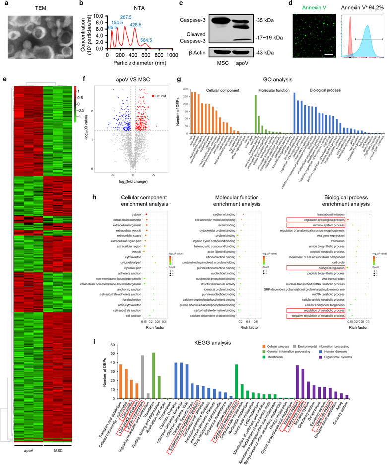
FIGURE 1 Characterization and proteomic analysis of mesenchymal stem cell (MSC)‐derived apoptotic vesicles (apoVs). (a) Representative transmission electron microscope (TEM) image showing the morphology of apoVs. Scale bar, 125 nm. (b) Nanoparticle tracking analysis (NTA) showing the size distribution of apoVs. (c) Western blotting analysis showing the presence of Caspase‐3/Cleaved Caspase‐3 in MSCs and apoVs. (d) Representative confocal microscopy images and flow cytometric analysis of Annexin V (green) staining in apoVs. Scale bars, 10 μm. (e) Hierarchical clustering of differentially expressed proteins (DEPs) (Fold change > 1.5 and Q value < 0.05) between MSCs and apoVs, with protein abundance being Z‐score normalized. Rows represent proteins and columns represent individual replicates. (f) Volcano plot showing significantly upregulated (red dots) and downregulated (blue dots) proteins in apoVs, compared to MSCs. (g) Gene ontology (GO) analysis of significantly upregulated proteins in apoVs, categorized into ‘Cellular component’, ‘Molecular function’ and ‘Biological process’. (h) GO enrichment analysis of significantly upregulated proteins in apoVs. The top twenty enriched terms of the three categories in (g) were respectively presented as bubble charts. The Y‐axis represents GO terms and the X‐axis represents rich factor. The colour of the bubble represents enrichment significance and the size of the bubble represents number of upregulated proteins. (i) Kyoto Encyclopedia of Genes and Genomes (KEGG) pathway analysis of significantly upregulated proteins in apoVs
FIGURE 1 Characterization and proteomic analysis of mesenchymal stem cell (MSC)‐derived apoptotic vesicles (apoVs). (a) Representative transmission electron microscope (TEM) image showing the morphology of apoVs. Scale bar, 125 nm. (b) Nanoparticle tracking analysis (NTA) showing the size distribution of apoVs. (c) Western blotting analysis showing the presence of Caspase‐3/Cleaved Caspase‐3 in MSCs and apoVs. (d) Representative confocal microscopy images and flow cytometric analysis of Annexin V (green) staining in apoVs. Scale bars, 10 μm. (e) Hierarchical clustering of differentially expressed proteins (DEPs) (Fold change > 1.5 and Q value < 0.05) between MSCs and apoVs, with protein abundance being Z‐score normalized. Rows represent proteins and columns represent individual replicates. (f) Volcano plot showing significantly upregulated (red dots) and downregulated (blue dots) proteins in apoVs, compared to MSCs. (g) Gene ontology (GO) analysis of significantly upregulated proteins in apoVs, categorized into ‘Cellular component’, ‘Molecular function’ and ‘Biological process’. (h) GO enrichment analysis of significantly upregulated proteins in apoVs. The top twenty enriched terms of the three categories in (g) were respectively presented as bubble charts. The Y‐axis represents GO terms and the X‐axis represents rich factor. The colour of the bubble represents enrichment significance and the size of the bubble represents number of upregulated proteins. (i) Kyoto Encyclopedia of Genes and Genomes (KEGG) pathway analysis of significantly upregulated proteins in apoVsCC BY · PubMed Central
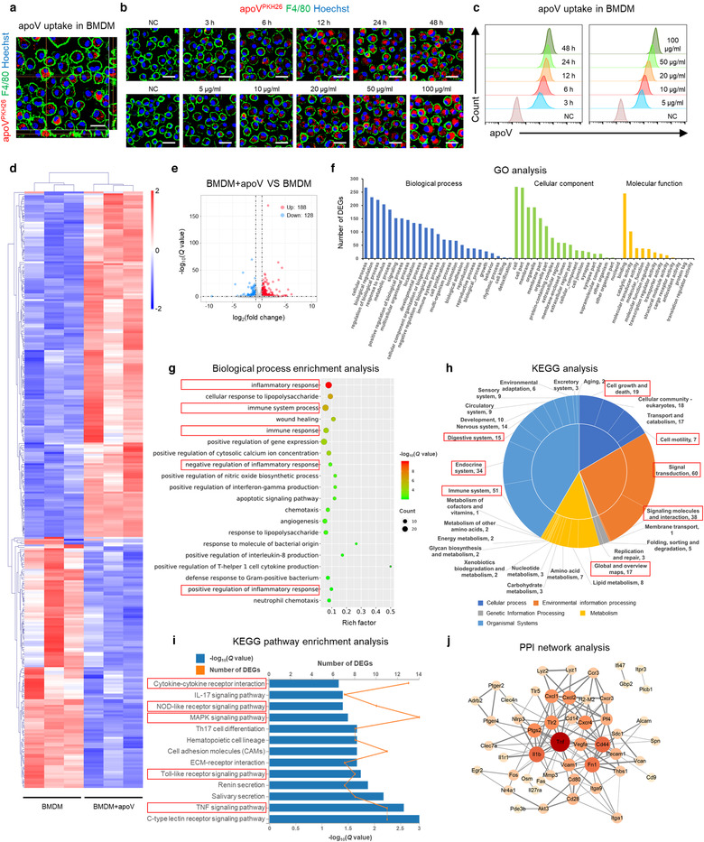
FIGURE 2 Efferocytosis of MSC‐derived apoVs induces transcriptional reprogramming in bone marrow‐derived macrophages (BMDMs). (a) Representative confocal orthogonal view showing uptake of PKH26‐labeded apoVs (red) by BMDMs (green), counterstained by Hoechst (blue). Scale bar, 20 μm. (b) Representative confocal microscopy images showing time‐dependent and concentration‐dependent uptake of PKH26‐labeded apoVs (red) by BMDMs (green), counterstained by Hoechst (blue). After removal of unbound PKH, the stained apoVs were resuspended in PBS and underwent centrifugation, after which the supernatant was used as the negative control (NC) and added to BMDMs. Scale bars, 25 μm. (c) Flow cytometric analysis of time‐dependent and concentration‐dependent uptake of PKH67‐labeded apoVs by macrophages. NC was prepared as stated above. (d) Hierarchical clustering of differentially expressed genes (DEGs) (Fold change > 1.5 and Q value < 0.05) between BMDMs and apoV‐treated BMDMs (BMDM+apoV), with gene abundance being Z‐score normalized. Rows represent genes and columns represent individual replicates. (e) Volcano plot showing DEGs in BMDM+apoV compared to BMDMs. The blue and red dots indicate downregulated and upregulated genes, respectively. (f) Gene ontology (GO) analysis of the DEGs in (e), categorized into ‘Cellular component’, ‘Molecular function’ and ‘Biological process’. (g) ‘Biological process’ enrichment analysis of the DEGs in (e). The top twenty enriched terms were presented as a bubble chart. The Y‐axis represents GO terms and the X‐axis represents rich factor. The colour of the bubble represents enrichment significance and the size of the bubble represents the number of DEGs. (h) Kyoto Encyclopedia of Genes and Genomes (KEGG) pathway analysis of the DEGs in (e). The number of genes annotated into different categories is shown. (i) KEGG pathway enrichment analysis of the DEGs in (e). The enriched KEGG pathways were presented as a bar chart. The Y‐axis represents KEGG pathways and the X‐axes represents number of DEGs (top) and enrichment significance (low), respectively. (j) Protein‐protein interaction (PPI) network analysis of the DEGs belonging to the enriched KEGG pathways in (i)
FIGURE 2 Efferocytosis of MSC‐derived apoVs induces transcriptional reprogramming in bone marrow‐derived macrophages (BMDMs). (a) Representative confocal orthogonal view showing uptake of PKH26‐labeded apoVs (red) by BMDMs (green), counterstained by Hoechst (blue). Scale bar, 20 μm. (b) Representative confocal microscopy images showing time‐dependent and concentration‐dependent uptake of PKH26‐labeded apoVs (red) by BMDMs (green), counterstained by Hoechst (blue). After removal of unbound PKH, the stained apoVs were resuspended in PBS and underwent centrifugation, after which the supernatant was used as the negative control (NC) and added to BMDMs. Scale bars, 25 μm. (c) Flow cytometric analysis of time‐dependent and concentration‐dependent uptake of PKH67‐labeded apoVs by macrophages. NC was prepared as stated above. (d) Hierarchical clustering of differentially expressed genes (DEGs) (Fold change > 1.5 and Q value < 0.05) between BMDMs and apoV‐treated BMDMs (BMDM+apoV), with gene abundance being Z‐score normalized. Rows represent genes and columns represent individual replicates. (e) Volcano plot showing DEGs in BMDM+apoV compared to BMDMs. The blue and red dots indicate downregulated and upregulated genes, respectively. (f) Gene ontology (GO) analysis of the DEGs in (e), categorized into ‘Cellular component’, ‘Molecular function’ and ‘Biological process’. (g) ‘Biological process’ enrichment analysis of the DEGs in (e). The top twenty enriched terms were presented as a bubble chart. The Y‐axis represents GO terms and the X‐axis represents rich factor. The colour of the bubble represents enrichment significance and the size of the bubble represents the number of DEGs. (h) Kyoto Encyclopedia of Genes and Genomes (KEGG) pathway analysis of the DEGs in (e). The number of genes annotated into different categories is shown. (i) KEGG pathway enrichment analysis of the DEGs in (e). The enriched KEGG pathways were presented as a bar chart. The Y‐axis represents KEGG pathways and the X‐axes represents number of DEGs (top) and enrichment significance (low), respectively. (j) Protein‐protein interaction (PPI) network analysis of the DEGs belonging to the enriched KEGG pathways in (i)CC BY · PubMed Central
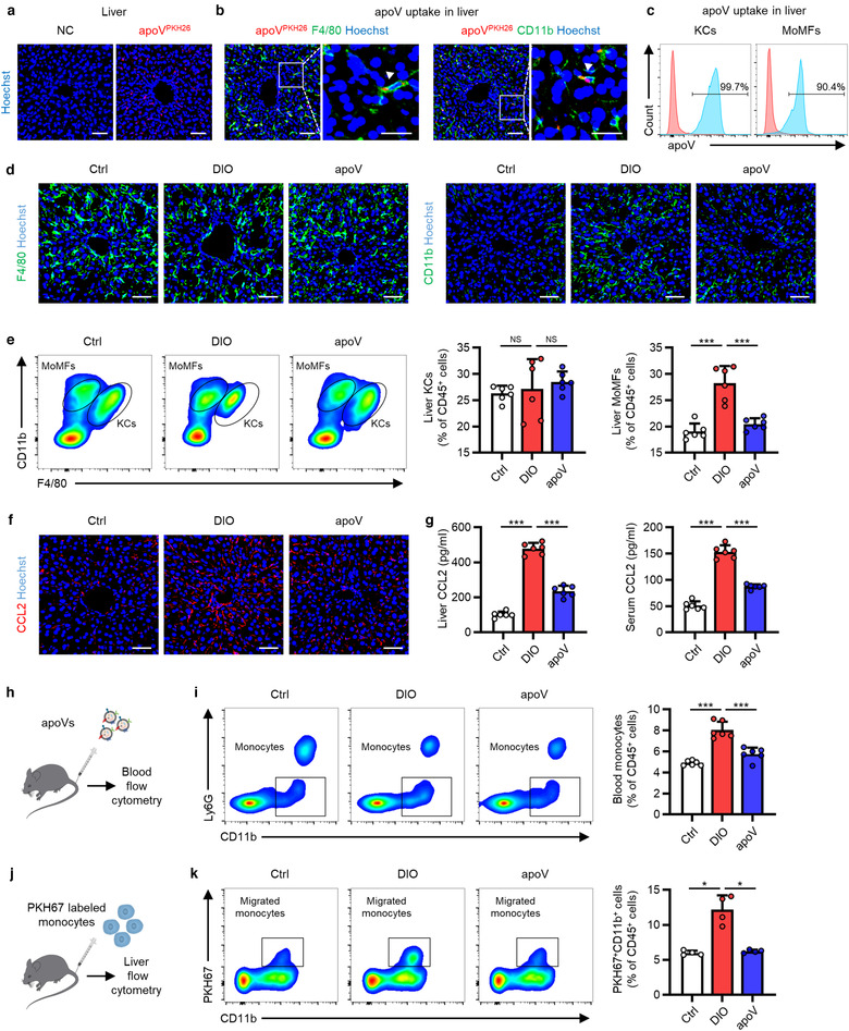
FIGURE 3 Efferocytosis of MSC‐derived apoVs by liver macrophages alleviates macrophage infiltration in the type 2 diabetes (T2D) liver. (a) Representative confocal microscopy images showing distribution of PKH26‐labeled apoVs (red) in the liver, counterstained by Hoechst (blue). After removal of unbound PKH, the stained apoVs were resuspended in PBS and underwent centrifugation, after which the supernatant was used as the negative control (NC) and injected. Scale bars, 50 μm. (b) Representative confocal microscopy images showing uptake of apoVs (red) by macrophages (green) in the liver, counterstained by Hoechst (blue). Scale bars, 50 μm in low magnification images and 25 μm in high magnification images. (c) Flow cytometric analysis showing the uptake of apoVs by macrophages in the liver. KCs, Kupffer cells; MoMFs, monocyte‐derived macrophages. (d) Representative immunofluorescent (IF) staining images of F4/80 (green) and CD11b (green) in the liver, counterstained by Hoechst (blue). Ctrl, control mice; DIO, mice with diet‐induced obesity; apoV, DIO mice treated by apoVs. Scale bars, 50 μm. (e) Flow cytometric analysis and the corresponding quantification of the percentages of KCs and MoMFs in hepatic CD45+ cells. N = 6 per group. (f) Representative IF staining images of chemokine (C‐C motif) ligand 2 (CCL2) (red) in the liver, counterstained by Hoechst (blue). Scale bars, 50 μm. (g) Enzyme‐linked immunosorbent assay (ELISA) analysis of CCL2 in liver lysate and serum. N = 6 per group. (h) Schematic diagram showing systemic injection of apoVs into mice which undergo blood flow cytometric analysis. (i) Flow cytometric analysis and the corresponding quantification of the percentages of monocytes in the peripheral blood CD45+ cells. N = 6 per group. (j) Schematic diagram showing injection and in vivo tracking of PKH67‐labeled bone marrow monocytes. (k) Flow cytometric analysis and the corresponding quantification of the percentages of PKH67‐labeled monocytes migrating to the liver. N = 4 per group. Data are presented as mean ± standard deviation (SD). Statistical analyses are performed by One‐way ANOVA with Tukey's post hoc test or Welch's ANOVA with Tamhane's T2 post hoc test. *, P < 0.05; ***, P < 0.001; NS, P > 0.05
FIGURE 3 Efferocytosis of MSC‐derived apoVs by liver macrophages alleviates macrophage infiltration in the type 2 diabetes (T2D) liver. (a) Representative confocal microscopy images showing distribution of PKH26‐labeled apoVs (red) in the liver, counterstained by Hoechst (blue). After removal of unbound PKH, the stained apoVs were resuspended in PBS and underwent centrifugation, after which the supernatant was used as the negative control (NC) and injected. Scale bars, 50 μm. (b) Representative confocal microscopy images showing uptake of apoVs (red) by macrophages (green) in the liver, counterstained by Hoechst (blue). Scale bars, 50 μm in low magnification images and 25 μm in high magnification images. (c) Flow cytometric analysis showing the uptake of apoVs by macrophages in the liver. KCs, Kupffer cells; MoMFs, monocyte‐derived macrophages. (d) Representative immunofluorescent (IF) staining images of F4/80 (green) and CD11b (green) in the liver, counterstained by Hoechst (blue). Ctrl, control mice; DIO, mice with diet‐induced obesity; apoV, DIO mice treated by apoVs. Scale bars, 50 μm. (e) Flow cytometric analysis and the corresponding quantification of the percentages of KCs and MoMFs in hepatic CD45+ cells. N = 6 per group. (f) Representative IF staining images of chemokine (C‐C motif) ligand 2 (CCL2) (red) in the liver, counterstained by Hoechst (blue). Scale bars, 50 μm. (g) Enzyme‐linked immunosorbent assay (ELISA) analysis of CCL2 in liver lysate and serum. N = 6 per group. (h) Schematic diagram showing systemic injection of apoVs into mice which undergo blood flow cytometric analysis. (i) Flow cytometric analysis and the corresponding quantification of the percentages of monocytes in the peripheral blood CD45+ cells. N = 6 per group. (j) Schematic diagram showing injection and in vivo tracking of PKH67‐labeled bone marrow monocytes. (k) Flow cytometric analysis and the corresponding quantification of the percentages of PKH67‐labeled monocytes migrating to the liver. N = 4 per group. Data are presented as mean ± standard deviation (SD). Statistical analyses are performed by One‐way ANOVA with Tukey's post hoc test or Welch's ANOVA with Tamhane's T2 post hoc test. *, P < 0.05; ***, P < 0.001; NS, P > 0.05CC BY · PubMed Central
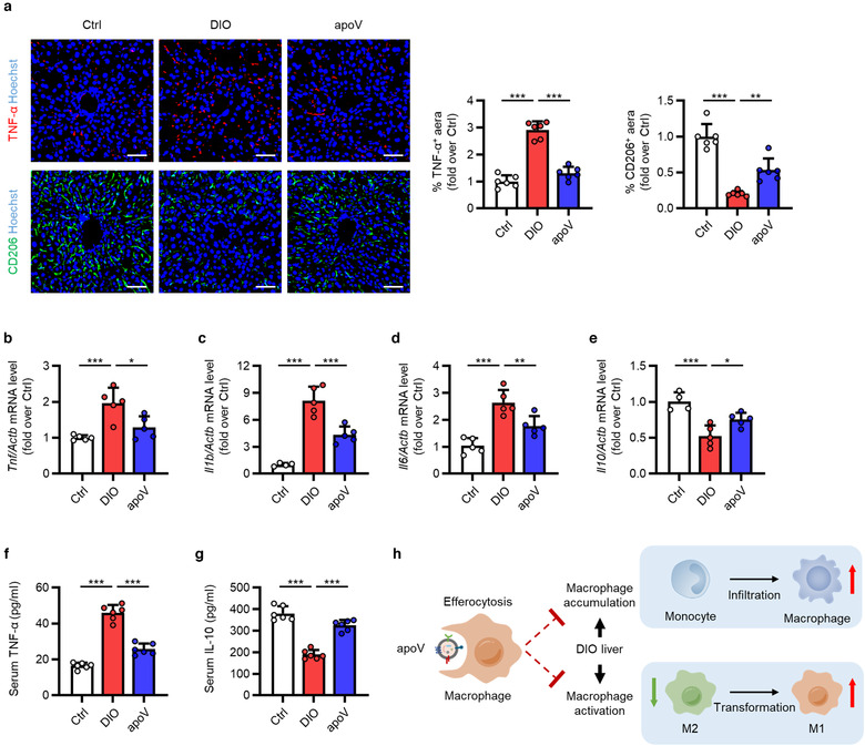
FIGURE 4 Efferocytosis of MSC‐derived apoVs by liver macrophages alleviates macrophage activation in the T2D liver. (a) Representative immunofluorescent (IF) staining images of tumor necrosis factor‐alpha (TNF‐α) (red) and CD206 (green) in the liver, counterstained by Hoechst (blue), and the corresponding quantification of fold changes over the Ctrl group. Ctrl, control mice; DIO, mice with diet‐induced obesity; apoV, DIO mice treated by apoVs. Scale bars, 50 μm. N = 6 per group. (b‐e) Quantitative real time polymerase chain reaction (qRT‐PCR) analysis of the mRNA expression levels of Tnf (b), interleukin 1 beta (Il1b) (c), interleukin 6 (Il6) (d) and interleukin 10 (Il10) (e) in the liver, normalized to β‐actin (Actb), and quantification of fold changes over the Ctrl group. N = 4–5 per group. (f, g) Enzyme‐linked immunosorbent assay (ELISA) analysis of TNF‐α (f) and IL‐10 (g) in serum. N = 6 per group. (h) Schematic diagram showing that efferocytosis of apoVs inhibits macrophage accumulation and activation in the liver of DIO mice. M1, pro‐inflammatory macrophages; M2, anti‐inflammatory macrophages. Data are presented as mean ± standard deviation (SD). Statistical analyses are performed by One‐way ANOVA with Tukey's post hoc test. *, P < 0.05; **, P < 0.01; ***, P < 0.001
FIGURE 4 Efferocytosis of MSC‐derived apoVs by liver macrophages alleviates macrophage activation in the T2D liver. (a) Representative immunofluorescent (IF) staining images of tumor necrosis factor‐alpha (TNF‐α) (red) and CD206 (green) in the liver, counterstained by Hoechst (blue), and the corresponding quantification of fold changes over the Ctrl group. Ctrl, control mice; DIO, mice with diet‐induced obesity; apoV, DIO mice treated by apoVs. Scale bars, 50 μm. N = 6 per group. (b‐e) Quantitative real time polymerase chain reaction (qRT‐PCR) analysis of the mRNA expression levels of Tnf (b), interleukin 1 beta (Il1b) (c), interleukin 6 (Il6) (d) and interleukin 10 (Il10) (e) in the liver, normalized to β‐actin (Actb), and quantification of fold changes over the Ctrl group. N = 4–5 per group. (f, g) Enzyme‐linked immunosorbent assay (ELISA) analysis of TNF‐α (f) and IL‐10 (g) in serum. N = 6 per group. (h) Schematic diagram showing that efferocytosis of apoVs inhibits macrophage accumulation and activation in the liver of DIO mice. M1, pro‐inflammatory macrophages; M2, anti‐inflammatory macrophages. Data are presented as mean ± standard deviation (SD). Statistical analyses are performed by One‐way ANOVA with Tukey's post hoc test. *, P < 0.05; **, P < 0.01; ***, P < 0.001CC BY · PubMed Central
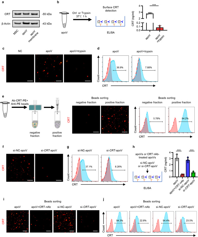
FIGURE 5 Surface expression of calreticulin (CRT) on apoVs. (a) Western blotting analysis showing the presence of CRT in membrane proteins of apoVs as well as in total proteins of MSCs and apoVs. (b) Schematic diagram showing detection of surface CRT via enzyme‐linked immunosorbent assay (ELISA) analysis and quantification of the concentration of CRT. apoV+trypsin, apoVs pre‐treated with trypsin at 37℃ for 1 h. N = 3–4 per group. (c) Representative immunofluorescent (IF) staining images of CRT (red) on apoVs. ApoVs incubated with only secondary antibody was used as the negative control (NC). Scale bars, 30 μm. (d) Flow cytometric analysis of CRT on the surface of apoVs. (e) Enrichment of CRT‐positive apoV subpopulations via magnetic beads sorting. Left: schematic diagram; Medium: fluorescent images of CRT (red); Right: flow cytometric analysis of CRT. Scale bars, 30 μm. (f) Representative IF staining images of CRT (red). si‐NC‐apoV, apoVs derived from MSCs treated by siRNA‐NC; si‐CRT‐apoV, apoVs derived from MSCs treated by siRNA‐CRT. Scale bars, 30 μm. (g) Flow cytometric analysis of CRT on the surface of apoVs. (h) ELISA analysis of CRT on the surface of apoVs. apoV+CRT‐nAb, apoVs pre‐treated with CRT neutralizing antibody at 37℃ for 1 h. N = 3–4 per group. (i) Representative fluorescent images of CRT (red) after magnetic beads sorting of CRT‐positive apoV subpopulations. Scale bars, 30 μm. (j) Flow cytometric analysis of CRT on the surface of apoVs after magnetic beads sorting of CRT‐positive apoV subpopulations. Data are presented as mean ± standard deviation (SD). Statistical analyses are performed by Student's t test (two‐tailed). ***, P < 0.001
FIGURE 5 Surface expression of calreticulin (CRT) on apoVs. (a) Western blotting analysis showing the presence of CRT in membrane proteins of apoVs as well as in total proteins of MSCs and apoVs. (b) Schematic diagram showing detection of surface CRT via enzyme‐linked immunosorbent assay (ELISA) analysis and quantification of the concentration of CRT. apoV+trypsin, apoVs pre‐treated with trypsin at 37℃ for 1 h. N = 3–4 per group. (c) Representative immunofluorescent (IF) staining images of CRT (red) on apoVs. ApoVs incubated with only secondary antibody was used as the negative control (NC). Scale bars, 30 μm. (d) Flow cytometric analysis of CRT on the surface of apoVs. (e) Enrichment of CRT‐positive apoV subpopulations via magnetic beads sorting. Left: schematic diagram; Medium: fluorescent images of CRT (red); Right: flow cytometric analysis of CRT. Scale bars, 30 μm. (f) Representative IF staining images of CRT (red). si‐NC‐apoV, apoVs derived from MSCs treated by siRNA‐NC; si‐CRT‐apoV, apoVs derived from MSCs treated by siRNA‐CRT. Scale bars, 30 μm. (g) Flow cytometric analysis of CRT on the surface of apoVs. (h) ELISA analysis of CRT on the surface of apoVs. apoV+CRT‐nAb, apoVs pre‐treated with CRT neutralizing antibody at 37℃ for 1 h. N = 3–4 per group. (i) Representative fluorescent images of CRT (red) after magnetic beads sorting of CRT‐positive apoV subpopulations. Scale bars, 30 μm. (j) Flow cytometric analysis of CRT on the surface of apoVs after magnetic beads sorting of CRT‐positive apoV subpopulations. Data are presented as mean ± standard deviation (SD). Statistical analyses are performed by Student's t test (two‐tailed). ***, P < 0.001CC BY · PubMed Central
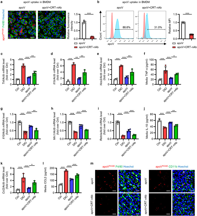
FIGURE 6 CRT blockade inhibits functional efferocytosis of MSC‐derived apoVs by T2D macrophages in vitro. (a) Representative confocal microscopy images showing uptake of apoVs (red) by bone marrow‐derived macrophages (BMDMs) (green) in vitro, counterstained by Hoechst (blue), and the corresponding quantification of fold changes of relative fluorescence intensity. apoV+CRT‐nAb, apoVs pre‐treated with CRT neutralizing antibody at 37℃ for 1 h. Scale bars, 25 μm. N = 5–6 per group. (b) Flow cytometric analysis showing uptake of apoVs by BMDMs in vitro, and the corresponding quantification of fold changes of mean fluorescence intensity (MFI). N = 6 per group. (C‐E) Quantitative real time polymerase chain reaction (qRT‐PCR) analysis of mRNA expression levels of tumor necrosis factor (Tnf) (c), interleukin 1 beta (Il1b) (d) and nitric oxide synthase 2, inducible (Nos2) (e) in cultured BMDMs, normalized to β‐actin (Actb), and quantification of fold changes over the Ctrl group. Ctrl, BMDMs derived from control mice; DIO, BMDMs derived from mice with diet‐induced obesity; apoV, BMDMs derived from DIO mice and treated with apoVs; apoV+CRT‐nAb, BMDMs derived from DIO mice and treated with apoVs which were pre‐treated with CRT‐nAb. N = 3–4 per group. (f) Enzyme‐linked immunosorbent assay (ELISA) analysis of TNF‐α in media from cultured BMDMs. N = 4 per group. (g‐i) qRT‐PCR analysis of mRNA expression levels of interleukin 10 (Il10) (g), mannose receptor, C type 1 (Mrc1) (h) and resistin like alpha (Retnla) (i) in cultured BMDMs, normalized to Actb, and quantification of fold changes over the Ctrl group. N = 3–4 per group. (j) ELISA analysis of IL‐10 in media from cultured BMDMs. N = 4 per group. (k) qRT‐PCR analysis of mRNA expression levels of chemokine (C‐C motif) ligand 2 (Ccl2) in cultured BMDMs, normalized to Actb, and quantification of fold changes over the Ctrl group. N = 3–4 per group. (l) ELISA analysis of CCL2 in media from cultured BMDMs. N = 4 per group. (m) Representative confocal microscopy images showing uptake of apoVs (red) by macrophages (green) in the liver, counterstained by Hoechst (blue). Scale bars, 25 μm. Data are presented as mean ± standard deviation (SD). Statistical analyses are performed by Student's t test with Welch correction (two‐tailed) for two group comparisons and One‐way ANOVA with Tukey's post hoc test for multiple group comparisons. *, P < 0.05; **, P < 0.01; ***, P < 0.001
FIGURE 6 CRT blockade inhibits functional efferocytosis of MSC‐derived apoVs by T2D macrophages in vitro. (a) Representative confocal microscopy images showing uptake of apoVs (red) by bone marrow‐derived macrophages (BMDMs) (green) in vitro, counterstained by Hoechst (blue), and the corresponding quantification of fold changes of relative fluorescence intensity. apoV+CRT‐nAb, apoVs pre‐treated with CRT neutralizing antibody at 37℃ for 1 h. Scale bars, 25 μm. N = 5–6 per group. (b) Flow cytometric analysis showing uptake of apoVs by BMDMs in vitro, and the corresponding quantification of fold changes of mean fluorescence intensity (MFI). N = 6 per group. (C‐E) Quantitative real time polymerase chain reaction (qRT‐PCR) analysis of mRNA expression levels of tumor necrosis factor (Tnf) (c), interleukin 1 beta (Il1b) (d) and nitric oxide synthase 2, inducible (Nos2) (e) in cultured BMDMs, normalized to β‐actin (Actb), and quantification of fold changes over the Ctrl group. Ctrl, BMDMs derived from control mice; DIO, BMDMs derived from mice with diet‐induced obesity; apoV, BMDMs derived from DIO mice and treated with apoVs; apoV+CRT‐nAb, BMDMs derived from DIO mice and treated with apoVs which were pre‐treated with CRT‐nAb. N = 3–4 per group. (f) Enzyme‐linked immunosorbent assay (ELISA) analysis of TNF‐α in media from cultured BMDMs. N = 4 per group. (g‐i) qRT‐PCR analysis of mRNA expression levels of interleukin 10 (Il10) (g), mannose receptor, C type 1 (Mrc1) (h) and resistin like alpha (Retnla) (i) in cultured BMDMs, normalized to Actb, and quantification of fold changes over the Ctrl group. N = 3–4 per group. (j) ELISA analysis of IL‐10 in media from cultured BMDMs. N = 4 per group. (k) qRT‐PCR analysis of mRNA expression levels of chemokine (C‐C motif) ligand 2 (Ccl2) in cultured BMDMs, normalized to Actb, and quantification of fold changes over the Ctrl group. N = 3–4 per group. (l) ELISA analysis of CCL2 in media from cultured BMDMs. N = 4 per group. (m) Representative confocal microscopy images showing uptake of apoVs (red) by macrophages (green) in the liver, counterstained by Hoechst (blue). Scale bars, 25 μm. Data are presented as mean ± standard deviation (SD). Statistical analyses are performed by Student's t test with Welch correction (two‐tailed) for two group comparisons and One‐way ANOVA with Tukey's post hoc test for multiple group comparisons. *, P < 0.05; **, P < 0.01; ***, P < 0.001CC BY · PubMed Central
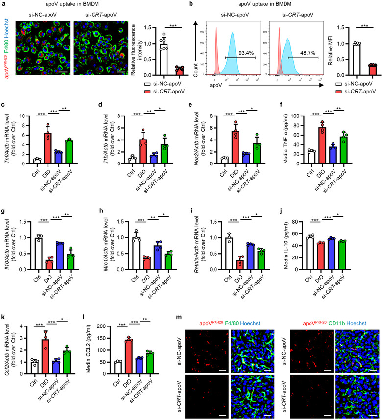
FIGURE 7 CRT downregulation inhibits functional efferocytosis of MSC‐derived apoVs by T2D macrophages in vitro. (a) Representative confocal microscopy images showing uptake of apoVs (red) by bone marrow‐derived macrophages (BMDMs) (green) in vitro, counterstained by Hoechst (blue), and the corresponding quantification of fold changes of relative fluorescence intensity. si‐NC‐apoV, apoVs derived from MSCs treated by siRNA‐negative control; si‐CRT‐apoV, apoVs derived from MSCs treated by siRNA‐CRT. Scale bars, 25 μm. N = 5–6 per group. (b) Flow cytometric analysis showing uptake of apoVs by BMDMs in vitro, and the corresponding quantification of fold changes of mean fluorescence intensity (MFI). N = 6 per group. (C‐E) Quantitative real time polymerase chain reaction (qRT‐PCR) analysis of the mRNA expression levels of tumor necrosis factor (Tnf) (c), interleukin 1 beta (Il1b) (d) and nitric oxide synthase 2, inducible (Nos2) (e) in cultured BMDMs, normalized to β‐actin (Actb), and quantification of fold changes over the Ctrl group. Ctrl, BMDMs derived from control mice; DIO, BMDMs derived from mice with diet‐induced obesity; si‐NC‐apoV, BMDMs derived from DIO mice and treated with si‐NC‐apoVs; si‐CRT‐apoV, BMDMs derived from DIO mice and treated with si‐CRT‐apoVs. N = 3–4 per group. (f) Enzyme‐linked immunosorbent assay (ELISA) analysis of TNF‐α in media from cultured BMDMs. N = 4 per group. (G‐I) qRT‐PCR analysis of mRNA expression levels of interleukin 10 (Il10) (g), mannose receptor, C type 1 (Mrc1) (h) and resistin like alpha (Retnla) (i) in cultured BMDMs, normalized to Actb, and quantification of fold changes over the Ctrl group. N = 3–4 per group. (j) ELISA analysis of IL‐10 in media from cultured BMDMs. N = 4 per group. (k) qRT‐PCR analysis of mRNA expression levels of chemokine (C‐C motif) ligand 2 (Ccl2) in cultured BMDMs, normalized to Actb, and quantification of fold changes over the Ctrl group. N = 3–4 per group. (l) ELISA analysis of CCL2 in media from cultured BMDMs. N = 4 per group. (m) Representative confocal microscopy images showing uptake of apoVs (red) by macrophages (green) in the liver, counterstained by Hoechst (blue). Scale bars, 25 μm. Data are presented as mean ± standard deviation (SD). Statistical analyses are performed by Student's t test (two‐tailed) or Student's t test with Welch correction (two‐tailed) for two group comparisons and One‐way ANOVA with Tukey's post hoc test for multiple group comparisons. *, P < 0.05; **, P < 0.01; ***, P < 0.001
FIGURE 7 CRT downregulation inhibits functional efferocytosis of MSC‐derived apoVs by T2D macrophages in vitro. (a) Representative confocal microscopy images showing uptake of apoVs (red) by bone marrow‐derived macrophages (BMDMs) (green) in vitro, counterstained by Hoechst (blue), and the corresponding quantification of fold changes of relative fluorescence intensity. si‐NC‐apoV, apoVs derived from MSCs treated by siRNA‐negative control; si‐CRT‐apoV, apoVs derived from MSCs treated by siRNA‐CRT. Scale bars, 25 μm. N = 5–6 per group. (b) Flow cytometric analysis showing uptake of apoVs by BMDMs in vitro, and the corresponding quantification of fold changes of mean fluorescence intensity (MFI). N = 6 per group. (C‐E) Quantitative real time polymerase chain reaction (qRT‐PCR) analysis of the mRNA expression levels of tumor necrosis factor (Tnf) (c), interleukin 1 beta (Il1b) (d) and nitric oxide synthase 2, inducible (Nos2) (e) in cultured BMDMs, normalized to β‐actin (Actb), and quantification of fold changes over the Ctrl group. Ctrl, BMDMs derived from control mice; DIO, BMDMs derived from mice with diet‐induced obesity; si‐NC‐apoV, BMDMs derived from DIO mice and treated with si‐NC‐apoVs; si‐CRT‐apoV, BMDMs derived from DIO mice and treated with si‐CRT‐apoVs. N = 3–4 per group. (f) Enzyme‐linked immunosorbent assay (ELISA) analysis of TNF‐α in media from cultured BMDMs. N = 4 per group. (G‐I) qRT‐PCR analysis of mRNA expression levels of interleukin 10 (Il10) (g), mannose receptor, C type 1 (Mrc1) (h) and resistin like alpha (Retnla) (i) in cultured BMDMs, normalized to Actb, and quantification of fold changes over the Ctrl group. N = 3–4 per group. (j) ELISA analysis of IL‐10 in media from cultured BMDMs. N = 4 per group. (k) qRT‐PCR analysis of mRNA expression levels of chemokine (C‐C motif) ligand 2 (Ccl2) in cultured BMDMs, normalized to Actb, and quantification of fold changes over the Ctrl group. N = 3–4 per group. (l) ELISA analysis of CCL2 in media from cultured BMDMs. N = 4 per group. (m) Representative confocal microscopy images showing uptake of apoVs (red) by macrophages (green) in the liver, counterstained by Hoechst (blue). Scale bars, 25 μm. Data are presented as mean ± standard deviation (SD). Statistical analyses are performed by Student's t test (two‐tailed) or Student's t test with Welch correction (two‐tailed) for two group comparisons and One‐way ANOVA with Tukey's post hoc test for multiple group comparisons. *, P < 0.05; **, P < 0.01; ***, P < 0.001CC BY · PubMed Central
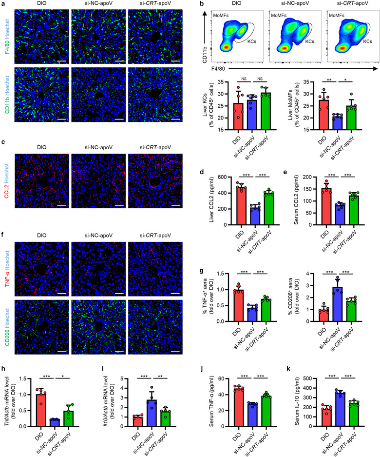
FIGURE 8 CRT mediates efferocytosis of MSC‐derived apoVs to modulate T2D liver macrophages in vivo. (a) Representative immunofluorescent (IF) staining images of F4/80 (green) and CD11b (green) in the liver, counterstained by Hoechst (blue). DIO, mice with diet‐induced obesity; si‐NC‐apoV, DIO mice treated by apoVs derived from MSCs transfected by siRNA‐negative control; si‐CRT‐apoV, DIO mice treated by apoVs derived from MSCs transfected by siRNA‐CRT. Scale bars, 50 μm. (b) Flow cytometric analysis and the corresponding quantification of the percentages of KCs and MoMFs in hepatic CD45+ cells. KCs, Kupffer cells; MoMFs, monocyte‐derived macrophages. N = 5–6 per group. (c) Representative IF staining images of chemokine (C‐C motif) ligand 2 (CCL2) (red) in the liver, counterstained by Hoechst (blue). Scale bars, 50 μm. (d and e) ELISA analysis of CCL2 in liver lysate (d) and serum (e). N = 6 per group. (f and g) Representative IF staining images of tumor necrosis factor‐alpha (TNF‐α) (red) and CD206 (green) in the liver, counterstained by Hoechst (blue), and the corresponding quantification of fold changes over the DIO group. Scale bars, 50 μm. N = 5–6 per group. (h and i) Quantitative real time polymerase chain reaction (qRT‐PCR) analysis of mRNA expression levels of Tnf (h) and interleukin 10 (Il10) (i) in the liver, normalized to β‐actin (Actb), and quantification of fold changes over the DIO group. N = 5 per group. (j and k) Enzyme‐linked immunosorbent assay (ELISA) analysis of TNF‐α (j) and IL‐10 (k) in serum. N = 6 per group. Data are presented as mean ± standard deviation (SD). Statistical analyses are performed by One‐way ANOVA with Tukey's post hoc test or Kruskal‐Wallis H test. *, P < 0.05; **, P < 0.01; ***, P < 0.001; NS, P > 0.05
FIGURE 8 CRT mediates efferocytosis of MSC‐derived apoVs to modulate T2D liver macrophages in vivo. (a) Representative immunofluorescent (IF) staining images of F4/80 (green) and CD11b (green) in the liver, counterstained by Hoechst (blue). DIO, mice with diet‐induced obesity; si‐NC‐apoV, DIO mice treated by apoVs derived from MSCs transfected by siRNA‐negative control; si‐CRT‐apoV, DIO mice treated by apoVs derived from MSCs transfected by siRNA‐CRT. Scale bars, 50 μm. (b) Flow cytometric analysis and the corresponding quantification of the percentages of KCs and MoMFs in hepatic CD45+ cells. KCs, Kupffer cells; MoMFs, monocyte‐derived macrophages. N = 5–6 per group. (c) Representative IF staining images of chemokine (C‐C motif) ligand 2 (CCL2) (red) in the liver, counterstained by Hoechst (blue). Scale bars, 50 μm. (d and e) ELISA analysis of CCL2 in liver lysate (d) and serum (e). N = 6 per group. (f and g) Representative IF staining images of tumor necrosis factor‐alpha (TNF‐α) (red) and CD206 (green) in the liver, counterstained by Hoechst (blue), and the corresponding quantification of fold changes over the DIO group. Scale bars, 50 μm. N = 5–6 per group. (h and i) Quantitative real time polymerase chain reaction (qRT‐PCR) analysis of mRNA expression levels of Tnf (h) and interleukin 10 (Il10) (i) in the liver, normalized to β‐actin (Actb), and quantification of fold changes over the DIO group. N = 5 per group. (j and k) Enzyme‐linked immunosorbent assay (ELISA) analysis of TNF‐α (j) and IL‐10 (k) in serum. N = 6 per group. Data are presented as mean ± standard deviation (SD). Statistical analyses are performed by One‐way ANOVA with Tukey's post hoc test or Kruskal‐Wallis H test. *, P < 0.05; **, P < 0.01; ***, P < 0.001; NS, P > 0.05CC BY · PubMed Central
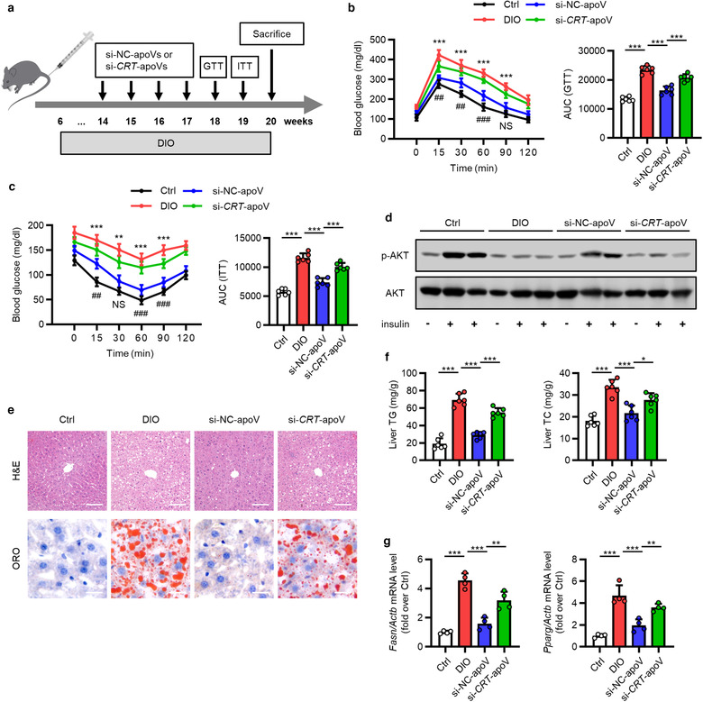
FIGURE 9 CRT‐mediated efferocytosis of MSC‐derived apoVs contributes to T2D therapy. (a) Schematic diagram indicating the study design of apoV efferocytosis‐mediated treatment for T2D in C57 mice. DIO, diet‐induced obesity; si‐NC‐apoVs, apoVs derived from MSCs treated by siRNA‐negative control; si‐CRT‐apoVs, apoVs derived from MSCs treated by siRNA‐CRT; GTT, glucose tolerance test; ITT, insulin tolerance test. (b) Blood glucose levels during GTT and quantification of area under the curve (AUC). Ctrl, control mice; DIO, mice with DIO; si‐NC‐apoV, DIO mice treated by si‐NC‐apoVs; si‐CRT‐apoV, DIO mice treated by si‐CRT‐apoVs. *, comparison between DIO and si‐NC‐apoV; #, comparison between si‐NC‐apoV and si‐CRT‐apoV. N = 6 per group. (c) Blood glucose levels during ITT and quantification of AUC. *, comparison between DIO and si‐NC‐apoV; #, comparison between si‐NC‐apoV and si‐CRT‐apoV. N = 6 per group. (d) Western blotting analysis of the expression levels of phosphorylated AKT (p‐AKT) and AKT in liver tissues. (e) Representative haematoxylin and eosin (H&E) and oil red O (ORO) staining images of liver tissues. Scale bars, 100 μm (top) and 25 μm (bottom). (f) Quantification of liver triglyceride (TG) and total cholesterol (TC) levels. N = 6 per group. (g) Quantitative real time polymerase chain reaction (qRT‐PCR) analysis of mRNA expression levels of fatty acid synthase (Fasn) and peroxisome proliferator‐activated receptor gamma (Pparg) in the liver, normalized to β‐actin (Actb), and quantification of fold changes over the Ctrl group. N = 4 per group. Data are presented as mean ± standard deviation (SD). Statistical analyses are performed by One‐way ANOVA with Tukey's post hoc test or Kruskal‐Wallis H test. *, P < 0.05; **, P < 0.01; ***, P < 0.001; ##, P < 0.01; ###, P < 0.001; NS, P > 0.05
FIGURE 9 CRT‐mediated efferocytosis of MSC‐derived apoVs contributes to T2D therapy. (a) Schematic diagram indicating the study design of apoV efferocytosis‐mediated treatment for T2D in C57 mice. DIO, diet‐induced obesity; si‐NC‐apoVs, apoVs derived from MSCs treated by siRNA‐negative control; si‐CRT‐apoVs, apoVs derived from MSCs treated by siRNA‐CRT; GTT, glucose tolerance test; ITT, insulin tolerance test. (b) Blood glucose levels during GTT and quantification of area under the curve (AUC). Ctrl, control mice; DIO, mice with DIO; si‐NC‐apoV, DIO mice treated by si‐NC‐apoVs; si‐CRT‐apoV, DIO mice treated by si‐CRT‐apoVs. *, comparison between DIO and si‐NC‐apoV; #, comparison between si‐NC‐apoV and si‐CRT‐apoV. N = 6 per group. (c) Blood glucose levels during ITT and quantification of AUC. *, comparison between DIO and si‐NC‐apoV; #, comparison between si‐NC‐apoV and si‐CRT‐apoV. N = 6 per group. (d) Western blotting analysis of the expression levels of phosphorylated AKT (p‐AKT) and AKT in liver tissues. (e) Representative haematoxylin and eosin (H&E) and oil red O (ORO) staining images of liver tissues. Scale bars, 100 μm (top) and 25 μm (bottom). (f) Quantification of liver triglyceride (TG) and total cholesterol (TC) levels. N = 6 per group. (g) Quantitative real time polymerase chain reaction (qRT‐PCR) analysis of mRNA expression levels of fatty acid synthase (Fasn) and peroxisome proliferator‐activated receptor gamma (Pparg) in the liver, normalized to β‐actin (Actb), and quantification of fold changes over the Ctrl group. N = 4 per group. Data are presented as mean ± standard deviation (SD). Statistical analyses are performed by One‐way ANOVA with Tukey's post hoc test or Kruskal‐Wallis H test. *, P < 0.05; **, P < 0.01; ***, P < 0.001; ##, P < 0.01; ###, P < 0.001; NS, P > 0.05CC BY · PubMed Central
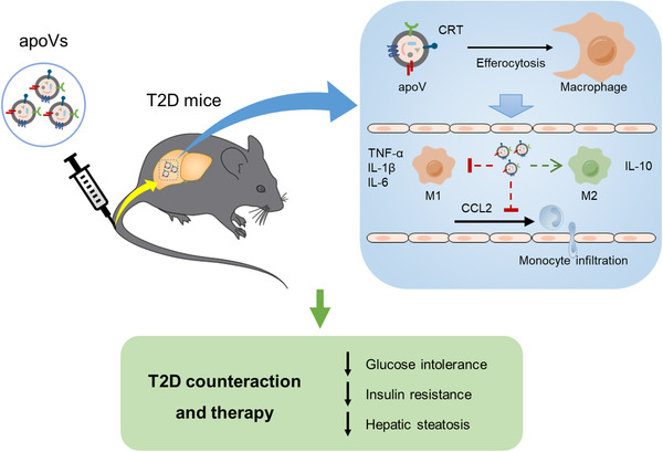
FIGURE 10 Schematic diagram showing the synopsis of the findings. Systemic infusion of MSC‐derived apoVs modulates liver macrophage function via calreticulin (CRT)‐mediated efferocytosis, thus providing a promising therapy for T2D. M1, pro‐inflammatory macrophages; M2, anti‐inflammatory macrophages; TNF‐α, tumor necrosis factor‐alpha; IL‐1β, interleukin‐1 beta; IL‐6, interleukin‐6; IL‐10, interleukin‐10; CCL2, chemokine (C‐C motif) ligand 2
FIGURE 10 Schematic diagram showing the synopsis of the findings. Systemic infusion of MSC‐derived apoVs modulates liver macrophage function via calreticulin (CRT)‐mediated efferocytosis, thus providing a promising therapy for T2D. M1, pro‐inflammatory macrophages; M2, anti‐inflammatory macrophages; TNF‐α, tumor necrosis factor‐alpha; IL‐1β, interleukin‐1 beta; IL‐6, interleukin‐6; IL‐10, interleukin‐10; CCL2, chemokine (C‐C motif) ligand 2CC BY · PubMed Central

Verification

Claims the paper makes

The claims and statistics of this paper haven’t been checked yet.

Run the check — free, no account needed. It reads the open-access text and checks the sources it cites.

A few free checks a day · papers already verified open instantly · results are public

Claims other papers make about this one

Each is checked against this paper’s own text: does it say what the citing paper says it does?

Self-check

Percentages, sums and reported differences re-computed from the paper’s own numbers.

The self-check needs the paper’s full text.

Citations

Cites

Show all 11 references

Research works printed but not linked: 90

  1. 10.1136/gutjnl-2011-300304 · DOI · Paper page
  2. 10.1002/eji.200424898 · DOI
  3. 10.1038/nm.3028 · DOI
  4. 10.1038/s41580-020-0232-1 · DOI
  5. 10.1038/s41467-019-09010-6 · DOI
  6. 10.1053/jhep.2003.50472 · DOI
  7. 10.3389/fimmu.2018.01486 · DOI · Paper page
  8. 10.1016/j.pharmthera.2020.107521 · DOI
  9. 10.1038/s41586-018-0392-8 · DOI · Paper page
  10. 10.1038/s41598-019-42063-7 · DOI
  11. 10.1016/j.stemcr.2020.06.007 · DOI
  12. 10.1093/gigascience/gix120 · DOI
  13. 10.1002/jcb.22838 · DOI
  14. 10.1080/20013078.2020.1757209 · DOI
  15. 10.1038/nbt.1511 · DOI · Paper page
  16. 10.1002/1878-0261.12644 · DOI
  17. 10.1038/nrdp.2015.19 · DOI · Paper page
  18. 10.1126/scitranslmed.aac9816 · DOI
  19. 10.1038/s41577-019-0240-6 · DOI · Paper page
  20. 10.1126/sciadv.aba2987 · DOI
  21. 10.1517/14728222.2016.1164695 · DOI
  22. 10.1016/j.stem.2019.01.013 · DOI
  23. 10.1161/ATVBAHA.119.313506 · DOI
  24. 10.1007/978-3-319-39406-0_2 · DOI
  25. 10.1016/j.stem.2018.05.004 · DOI · Paper page
  26. 10.1126/scitranslmed.aam7828 · DOI
  27. 10.1016/j.cell.2005.08.032 · DOI · Paper page
  28. 10.1042/BST20180080 · DOI
  29. 10.1016/j.jhep.2019.07.019 · DOI
  30. 10.1016/j.cell.2020.07.009 · DOI
  31. 10.1038/s12276-019-0362-8 · DOI
  32. 10.1038/s41575-018-0082-x · DOI · Paper page
  33. 10.1084/jem.20160459 · DOI
  34. 10.1126/science.278.5336.294 · DOI
  35. 10.1038/nri.2017.11 · DOI · Paper page
  36. 10.1083/jcb.201702048 · DOI
  37. 10.2337/db14-1668 · DOI
  38. 10.1016/j.cell.2017.12.025 · DOI
  39. 10.1186/1471-2105-12-323 · DOI · Paper page
  40. 10.1016/j.cell.2016.10.025 · DOI
  41. 10.1038/nm.3800 · DOI
  42. 10.7150/thno.16911 · DOI
  43. 10.1038/s41422-018-0070-2 · DOI · Paper page
  44. 10.1080/15548627.2020.1717128 · DOI
  45. 10.1007/978-1-4939-6700-1_21 · DOI
  46. 10.1038/jid.2014.169 · DOI
  47. Liu W., 2018, Alkaline phosphatase controls lineage switching of mesenchymal stem cells by regulating the LRP6/GSK3beta complex in hypophosphatasia, Theranostics, 8, 5575, 10.7150/thno.27372 · DOI
  48. 10.1186/s13059-014-0550-8 · DOI · Paper page
  49. Ma Q., 2020, Osteoclast‐derived apoptotic bodies show extended biological effects of parental cell in promoting bone defect healing, Theranostics, 10, 6825, 10.7150/thno.45170 · DOI
  50. 10.1016/j.immuni.2014.05.010 · DOI
  51. 10.1038/nprot.2015.017 · DOI
  52. 10.1038/nprot.2013.055 · DOI
  53. 10.1016/j.immuni.2019.04.018 · DOI · Paper page
  54. 10.1146/annurev-physiol-022516-034339 · DOI · Paper page
  55. 10.1146/annurev-immunol-042617-053010 · DOI
  56. 10.1038/nm1523 · DOI · Paper page
  57. 10.2337/db09-1403 · DOI
  58. 10.1016/j.cmet.2008.04.003 · DOI
  59. Okin D., 2016, The effect of sustained inflammation on hepatic mevalonate pathway results in hyperglycemia, Cell, 165, 343, 10.1016/j.cell.2016.02.023 · DOI
  60. 10.1038/nature05935 · DOI
  61. 10.1016/j.jmb.2011.02.029 · DOI
  62. 10.1073/pnas.0801394105 · DOI
  63. 10.1016/j.ccell.2018.05.012 · DOI
  64. 10.1038/s41556-019-0431-1 · DOI
  65. 10.1080/20013078.2019.1608786 · DOI
  66. 10.1021/acsnano.9b01383 · DOI
  67. 10.1016/j.immuni.2018.07.014 · DOI
  68. 10.1038/s41586-019-1797-8 · DOI · Paper page
  69. 10.1016/j.diabres.2019.107843 · DOI · Paper page
  70. 10.1016/j.tcb.2015.08.003 · DOI
  71. 10.1016/j.addr.2017.04.012 · DOI
  72. 10.1038/11387 · DOI · Paper page
  73. 10.1016/j.immuni.2009.01.001 · DOI · Paper page
  74. 10.7150/thno.18181 · DOI
  75. 10.1093/nar/gky1131 · DOI · Paper page
  76. 10.1016/j.jhep.2017.02.026 · DOI · Paper page
  77. 10.2337/db17-0371 · DOI
  78. 10.1038/nmat4718 · DOI
  79. 10.1038/cddis.2011.94 · DOI
  80. 10.1016/j.jhep.2010.04.010 · DOI
  81. 10.1038/s41589-019-0306-6 · DOI
  82. 10.1002/hep.26607 · DOI · Paper page
  83. 10.1038/ni.3002 · DOI · Paper page
  84. 10.3389/fimmu.2019.01228 · DOI
  85. 10.1038/cdd.2013.139 · DOI
  86. 10.1038/s41551-018-0325-8 · DOI
  87. 10.1186/s13098-017-0233-1 · DOI
  88. 10.1126/scisignal.2000610 · DOI
  89. 10.1161/STROKEAHA.115.009792 · DOI
  90. 10.1038/s41598-018-23098-8 · DOI

Cited by

Sorted by citations

Metadata

Authors
Zheng, C., Sui, B., Zhang, X., Hu, J., Chen, J., Liu, J., Wu, D., Ye, Q., Xiang, L., Qiu, X., Liu, S., Deng, Z., Zhou, J., Liu, S., Shi, S., & Jin, Y.
Venue
Journal of Extracellular Vesicles
Publisher
Wiley
Published
2021-05
Volume
10
Issue
7
ISSN
2001-3078
Cited by
219
References
11
Language
en
Funders
  • National Basic Research Program of China (2016YFC1101400)
  • National Natural Science Foundation of China (32000974, 31800817, 81670915, 31870970)

About these checks

Registry, retraction, Cites and statistics results are computed, not judged, with one exception: a printed reference that no index lists is read once by a model before it counts as a citation error. Claims are extracted by a model and kept only when their quote appears verbatim in the text; each cited source is then checked, with a model’s help, for whether it says what the claim relies on. Those verdicts can be wrong — read the claim’s quote and the cited source before relying on one.

Statistics and arithmetic are recomputed, not judged.

Confidence bands are shown only for fields calibrated on our annotated test set. None is calibrated yet, so no band is shown.

What counts as a claim ›How we measure confidence ›How we check this ›

Are you an author, or do you see a problem with this page? Request a review