

The DOI matches its Crossref record; 29 of the 29 references it cites were found in a registry or an index.
Vascular basement membranes as pathways for the passage of fluid into and out of the brain
Open accessAbstract
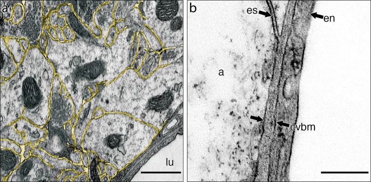
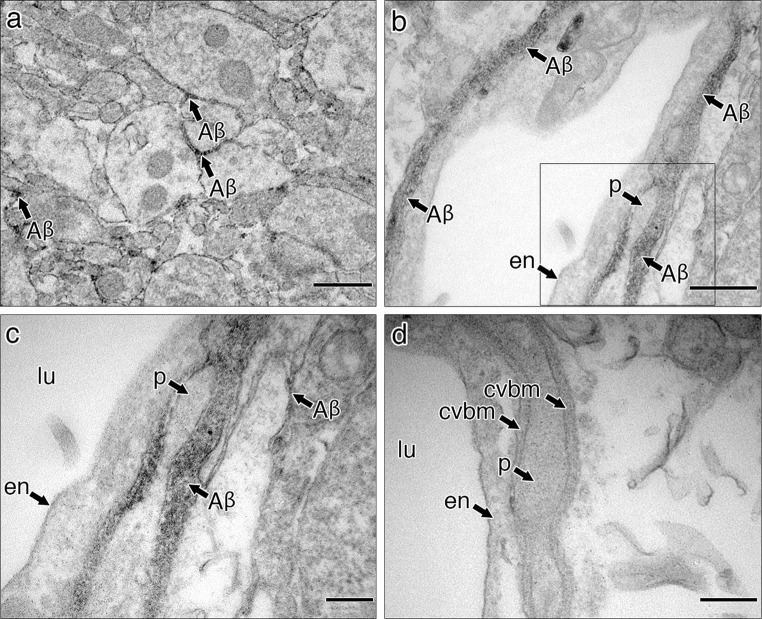
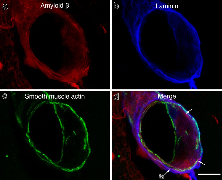
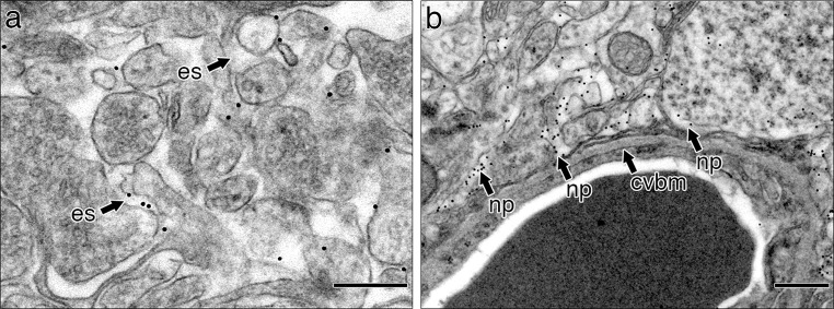
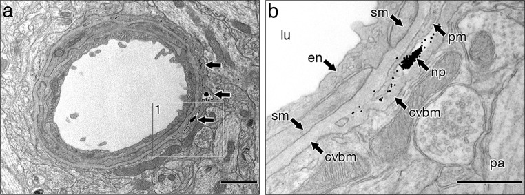
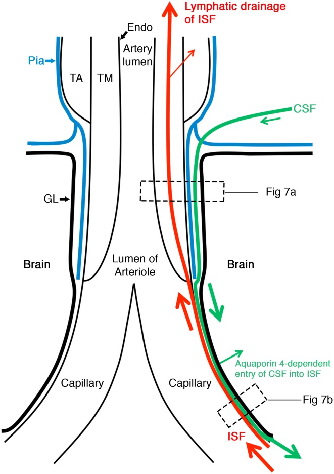
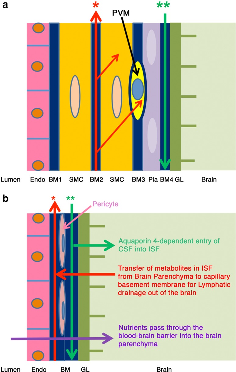
In the absence of conventional lymphatics, drainage of interstitial fluid and solutes from the brain parenchyma to cervical lymph nodes is along basement membranes in the walls of cerebral capillaries and tunica media of arteries. Perivascular pathways are also involved in the entry of CSF into the brain by the convective influx/glymphatic system. The objective of this study is to differentiate the cerebral vascular basement membrane pathways by which fluid passes out of the brain from the pathway by which CSF enters the brain. Experiment 1: 0.5 µl of soluble biotinylated or fluorescent Aβ, or 1 µl 15 nm gold nanoparticles was injected into the mouse hippocampus and their distributions determined at 5 min by transmission electron microscopy. Aβ was distributed within the extracellular spaces of the hippocampus and within basement membranes of capillaries and tunica media of arteries. Nanoparticles did not enter capillary basement membranes from the extracellular spaces. Experiment 2: 2 µl of 15 nm nanoparticles were injected into mouse CSF. Within 5 min, groups of nanoparticles were present in the pial-glial basement membrane on the outer aspect of cortical arteries between the investing layer of pia mater and the glia limitans. The results of this study and previous research suggest that cerebral vascular basement membranes form the pathways by which fluid passes into and out of the brain but that different basement membrane layers are involved. The significance of these findings for neuroimmunology, Alzheimer's disease, drug delivery to the brain and the concept of the Virchow-Robin space are discussed.
Overview
The claims and statistics of this paper haven’t been checked yet.
Run the check — free, no account needed. It reads the open-access text and checks the sources it cites.
Integrity signals
Registry
Cites
Statistics
Figures
Verification
Claims the paper makes
The claims and statistics of this paper haven’t been checked yet.
Run the check — free, no account needed. It reads the open-access text and checks the sources it cites.
Claims other papers make about this one
Each is checked against this paper’s own text: does it say what the citing paper says it does?
The source backs part of the claim, not all of it — or backs it for a different population, condition or comparison.
In the citing paper
“Moreover, animal studies report diverging findings regarding the sites of perivascular brain influx and efflux and direction of perivascular flow (7, 15, 16).”
From Brain-wide glymphatic enhancement and clearance in humans assessed with MRI. (2018) · cites this paper as [16]
Experimental — accuracy not yet measuredIn the citing paper
“For example, in the cortex, a packed barrier of endothelial cells, basement membrane, and glia limitans surrounds penetrating arteries, leaving little to no fluid-filled space, whereas the lenticulostriate branches in the basal ganglia are surrounded by a larger perivascular space (13).”
From The anatomy and immunology of vasculature in the central nervous system (2019) · cites this paper as [13]
In the citing paper
“Movement of ISF from the brain parenchyma occurs primarily between basement membranes in the perivascular spaces of capillaries and arteries. This continues along basement membranes in the tunica media of cerebral and leptomeningeal arteries (13).”
From The anatomy and immunology of vasculature in the central nervous system (2019) · cites this paper as [13]
Self-check
Percentages, sums and reported differences re-computed from the paper’s own numbers.
The self-check needs the paper’s full text.
Citations
Cites
Iliff, J. J., Wang, M., Liao, Y., Plogg, B. A., Peng, W., Gundersen, G. A., Benveniste, H., Vates, G. E., Deane, R., Goldman, S. A., et al. (2012) · Science Translational Medicine · doi:10.1126/scitranslmed.3003748 · Cited by 5,824
Turkevich Method for Gold Nanoparticle Synthesis Revisited
Kimling, J., Maier, M., Okenve, B., Kotaidis, V., Ballot, H., & Plech, A. (2006) · The Journal of Physical Chemistry B · doi:10.1021/jp061667w · Cited by 2,283
Direct brain infusion of glial cell line–derived neurotrophic factor in Parkinson disease
Gill, S. S., Patel, N. K., Hotton, G. R., O'Sullivan, K., McCarter, R., Bunnage, M., Brooks, D. J., Svendsen, C. N., & Heywood, P. (2003) · Nature Medicine · doi:10.1038/nm850 · Cited by 1,290
A new look at cerebrospinal fluid circulation
Brinker, T., Stopa, E., Morrison, J., & Klinge, P. (2014) · Fluids and Barriers of the CNS · doi:10.1186/2045-8118-11-10 · Cited by 819
Evidence for bulk flow of brain interstitial fluid: significance for physiology and pathology
Abbott, N. J. (2004) · Neurochemistry International · doi:10.1016/j.neuint.2003.11.006 · Cited by 786
Rennels, M. L., Gregory, T. F., Blaumanis, O. R., Fujimoto, K., & Grady, P. A. (1985) · Brain Research · doi:10.1016/0006-8993(85)91383-6 · Cited by 752
Carare, R. O., Bernardes‐Silva, M., Newman, T. A., Page, A. M., Nicoll, J. A. R., Perry, V. H., & Weller, R. O. (2008) · Neuropathology and Applied Neurobiology · doi:10.1111/j.1365-2990.2007.00926.x · Cited by 665
Self-citationKida, S., Pantazis, A., & Weller, R. O. (1993) · Neuropathology and Applied Neurobiology · doi:10.1111/j.1365-2990.1993.tb00476.x · Cited by 520
Self-citationDynamics of CNS Barriers: Evolution, Differentiation, and Modulation
Abbott, N. J. (2005) · Cellular and Molecular Neurobiology · doi:10.1007/s10571-004-1374-y · Cited by 471
(1990) · Europe PMC (PubMed Central) · Cited by 463
Self-citation
Cited by
Sorted by citations
Items 201–289 of 289
Brain Fluid Channels for Metabolite Removal
Maloveská, M., Humeník, F., Vikartovská, Z., Hudáková, N., Almášiová, V., Krešáková, L., & Čı́žková, D. (2022) · Physiological Research · doi:10.33549/physiolres.934802 · Cited by 8
Tsuji, M., Ohshima, M., Yamamoto, Y., Saitô, S., Hattori, Y., Tanaka, E., Taguchi, A., Ihara, M., & Ogawa, Y. (2020) · Frontiers in Aging Neuroscience · doi:10.3389/fnagi.2020.00106 · Cited by 8
Chronic neuroinflammation impairs waste clearance in the rat brain
Suresh, S., Larson, J., & Jenrow, K. A. (2022) · Frontiers in Neuroanatomy · doi:10.3389/fnana.2022.1013808 · Cited by 7
Sharp, M. M., Cassidy, J., Thornton, T., Lyles, J., Keable, A., Gatherer, M., Yasui, M., Abe, Y., Shibata, S., Weller, R. O., et al. (2021) · Acta Neuropathologica Communications · doi:10.1186/s40478-021-01274-8 · Cited by 7
Self-citationYim, Y., & Moon, W. (2022) · Journal of the Korean Society of Radiology · doi:10.3348/jksr.2022.0049 · Cited by 6
Pathogenesis of cerebral amyloid angiopathy caused by chaotic glymphatics—Mini-review
Lui, F., Alcaide, J., Knowlton, S., Ysit, M., & Zhong, N. (2023) · Frontiers in Neuroscience · doi:10.3389/fnins.2023.1180237 · Cited by 8
Quantitative Assessment of Cerebral Basement Membranes Using Electron Microscopy
Sharp, M. M., Page, A. M., Morris, A. W. J., Weller, R. O., & Carare, R. O. (2017) · Methods in molecular biology · doi:10.1007/978-1-4939-6786-5_25 · Cited by 7
Self-citationCerebrovascular pathology: the dark side of neurodegeneration
Carare, R. O., & Kalaria, R. N. (2016) · Acta Neuropathologica · doi:10.1007/s00401-016-1573-x · Cited by 7
Self-citationLadrón-de-Guevara, A., Shang, J., Nedergaard, M., & Kelley, D. H. (2020) · bioRxiv · doi:10.1101/2020.07.02.183608 · Cited by 8
TUBE Project: Transport-Derived Ultrafines and the Brain Effects
Martikainen, M., Aakko-Saksa, P. T., van den Broek, L., Cassee, F. R., Carare, R. O., Chew, S., Dinnyés, A., Giugno, R., Kanninen, K. M., Malm, T., et al. (2021) · International Journal of Environmental Research and Public Health · doi:10.3390/ijerph19010311 · Cited by 6
Self-citationThe glycosaminoglycan chains of perlecan regulate the perivascular fluid transport
Singh, A., Kaakinen, M., Elamaa, H., Kiviniemi, V. J., & Eklund, L. (2025) · Fluids and Barriers of the CNS · doi:10.1186/s12987-025-00648-7 · Cited by 7
Rossi, L., Preziosa, P., Filippi, M., & Rocca, M. A. (2025) · Multiple Sclerosis and Related Disorders · doi:10.1016/j.msard.2025.106596 · Cited by 6
Brain lymphatic drainage system – visualization opportunities and current state of the art
Yankova, G., & Bogomyakova, O. B. (2020) · Complex Issues of Cardiovascular Diseases · doi:10.17802/2306-1278-2020-9-3-81-89 · Cited by 5
Research Progress on Intracranial Lymphatic Circulation and Its Involvement in Disorders
Chen, F., Xie, X., & Wang, L. (2022) · Frontiers in Neurology · doi:10.3389/fneur.2022.865714 · Cited by 6
Dudchenko, N. G., Chimagomedova, A. S., Васенина, Е. Е., & Levin, O. S. (2022) · S S Korsakov Journal of Neurology and Psychiatry · doi:10.17116/jnevro202212207120 · Cited by 6
Längrich, T., Bork, K., Horstkorte, R., Weber, V., Hofmann, B., Fuszard, M., & Olzscha, H. (2022) · Proteomes · doi:10.3390/proteomes10030028 · Cited by 6
Is cerebral small vessel disease a central nervous system interstitial fluidopathy?
Hamid, H. A., Hambali, A., Okon, U. E., Nassir, C. M. N. C. M., Mehat, M. Z., Norazit, A., & Mustapha, M. (2023) · IBRO Neuroscience Reports · doi:10.1016/j.ibneur.2023.12.004 · Cited by 6
Michalski, D., Hofmann, S., Pitsch, R., Grosche, J., & Härtig, W. (2017) · Journal of Alzheimer s Disease · doi:10.3233/jad-170185 · Cited by 6
Nanoscience: Convergence with Biomedical and Biological Applications
Dalal, V., & Biswas, S. (2020) · Nanotechnology in the life sciences · doi:10.1007/978-3-030-41464-1_1 · Cited by 6
The involvement of perivascular spaces or tissues in the facial intradermal brain-targeted delivery
Yang, W. P., Jin, B., Chen, Y., Cao, C., Zhu, J., Zhao, Y., Yu, X., & Li, F. (2019) · Drug Delivery · doi:10.1080/10717544.2019.1587044 · Cited by 6
Fujii, N., Nomura, S., Izuma, H., & Ishihara, H. (2024) · Neurologia medico-chirurgica · doi:10.2176/jns-nmc.2023-0277 · Cited by 5
Investigating the Lymphatic Drainage of the Brain: Essential Skills and Tools
Albargothy, N. J., Sharp, M. M., Gatherer, M., Morris, A. W. J., Weller, R. O., Hawkes, C. A., & Carare, R. O. (2017) · Methods in molecular biology · doi:10.1007/978-1-4939-6786-5_24 · Cited by 5
Self-citationSecreted Peptides for Diagnostic Trajectory Assessments in Brain Injury Rehabilitation
Patel, P. D., Stafflinger, J. E., Marwitz, J. H., Niemeier, J. P., & Ottens, A. K. (2020) · Neurorehabilitation and neural repair · doi:10.1177/1545968320975428 · Cited by 5
Johnson, L., Tomasetig, F., Fok, S., Whan, R. M., Berliner, J. A., Hemley, S. J., Stoodley, M. A., & Bilston, L. E. (2023) · Scientific Reports · doi:10.1038/s41598-023-42275-y · Cited by 5
McDonald, S. W., & Russell, D. (2018) · Annals of Anatomy - Anatomischer Anzeiger · doi:10.1016/j.aanat.2018.03.002 · Cited by 5
Bartiromo, S., Alves, C., O’Mahony, J., Yeh, E. A., Marrie, R. A., Narayanan, S., Waters, P. J., Gajofatto, A., Bar‐Or, A., Banwell, B., et al. (2025) · Neurology Neuroimmunology & Neuroinflammation · doi:10.1212/nxi.0000000000200449 · Cited by 4
Veetil, N. N., Gransar, T., Liu, S., Almalla, A., Kirchner, M., Brackin-Helmers, R., Hennig, F., Yeter, R., Weinhart, M., Mertins, P., et al. (2025) · Cardiovascular Research · doi:10.1093/cvr/cvaf238 · Cited by 4
The Landscape of Ferritin Nanocages for Neurodegenerative Diseases Treatment
Wen, S., Gao, J., Sun, J., Li, X., Xu, X.-L., & Chen, J. (2026) · International Journal of Nanomedicine · doi:10.2147/ijn.s571993 · Cited by 4
Interstitial Fluid Flows along Perivascular and Adventitial Clearances around Neurovascular Bundles
Kong, Y., Yu, X., Peng, G., Wang, F., & Yin, Y. (2022) · Journal of Functional Biomaterials · doi:10.3390/jfb13040172 · Cited by 4
Neyazi, B., Stein, K., Wilkens, L., Maslehaty, H., Dumitru, C. A., & Sandalcioglu, I. E. (2019) · Clinical Neurology and Neurosurgery · doi:10.1016/j.clineuro.2019.105589 · Cited by 4
Manousopoulou, A., Yuen, H. M., MacGregor Sharp, M., Saitô, S., Aldea, R., Mazer, N. A., Garbis, S. D., & Carare, R. O. (2020) · Free Neuropathology · doi:10.17879/freeneuropathology-2020-2955 · Cited by 4
Self-citationEsmaeli, M. (2025) · Cell and Tissue Biology · doi:10.1134/s1990519x24600637 · Cited by 3
Piotrowska, A., Winter, K., Carare, R. O., & Bechmann, I. (2020) · Frontiers in Aging Neuroscience · doi:10.3389/fnagi.2020.00015 · Cited by 3
Self-citationYin, H., Lu, Z., Deng, Y., Tian, X., & Gong, Q. (2026) · Ageing Research Reviews · doi:10.1016/j.arr.2025.102952 · Cited by 6
Kitayama, Y., Nakano, Y., Hirano, S., Hanayama, S., Chishiki, Y., Izumi, M., Koizumi, Y., Suzuki, Y., Tamura, M., Yamagishi, K., et al. (2025) · Journal of Alzheimer s Disease · doi:10.1177/13872877251376601 · Cited by 2
Nanomedicine Strategies to Ameliorate Neuroinflammation and Prevent Secondary Neural Tissue Damage
Hegab, R. A., & Peppas, N. A. (2026) · ACS Nano Medicine · doi:10.1021/acsnanomed.6c00120 · Cited by 2
Nanomedical drug delivery for neurodegenerative disease
Kaur, N., Kumar, V., Mahender, T., Maji, I., Mahajan, S., Aalhate, M., Singh, P. K., & Sahu, S. K. (2022) · Elsevier eBooks · doi:10.1016/b978-0-323-85544-0.00016-2 · Cited by 2
Bellettieri, M. P. G., Anderloni, M., Rass, V., Kindl, P., Donadello, K., Taccone, F. S., Helbok, R., & Bogossian, E. G. (2023) · Clinical Neurology and Neurosurgery · doi:10.1016/j.clineuro.2023.108011 · Cited by 2
Fractal Analysis in Neurodegenerative Diseases
Pirici, D. N., Mogoantă, L., Ion, D. A., & Kumar‐Singh, S. (2024) · Advances in neurobiology · doi:10.1007/978-3-031-47606-8_18 · Cited by 3
Saïb, G., Demir, Z., Taylor, P. A., Talagala, S. L., & Koretsky, A. P. (2026) · NeuroImage · doi:10.1016/j.neuroimage.2026.121757 · Cited by 2
Schuldt, B. R., Haworth-Staines, D., Arévalo, A. P., Broekaart, D. W. M., Harlock, A., Wang, L., Bright, A., Gallagher, G., Rabinowitz, G., Goate, A., et al. (2025) · bioRxiv (Cold Spring Harbor Laboratory) · doi:10.1101/2025.09.04.674192 · Cited by 2
Joshi, K., Diaz, A., O’Keeffe, K., Schaffer, J. D., Chiarot, P. R., & Huang, P. (2024) · Journal of Mathematical Biology · doi:10.1007/s00285-024-02092-x · Cited by 1
Advancements in Brain Lymphatic System and Its Involvement in Neurological Diseases
Bari, D., Das, U., Shevalkar, G. B., Kapadia, R., Singhai, V., & Pardeshi, C. V. (2024) · Springer eBooks · doi:10.1007/978-3-031-64828-1_2 · Cited by 2
Reardon, E. C., Greaney, A. J., & Mulvihill, J. J. (2025) · Fluids and Barriers of the CNS · doi:10.1186/s12987-025-00715-z · Cited by 2
Hayashi, H., Saito, R., Miyashita, A., Ikeuchi, T., Tada, M., Akazawa, K., Onodera, O., Tainaka, K., & Kakita, A. (2026) · Science Advances · doi:10.1126/sciadv.aea7539 · Cited by 1
Dondi, M., Bianchi, E., Borghetti, P., Lecce, R. D., Gnudi, G., Guarnieri, C., Buffagni, V., Ravanetti, F., Saleri, R., & Corradi, A. (2026) · Veterinary Sciences · doi:10.3390/vetsci13030298 · Cited by 1
Evidence that glial cells attenuate G47R transthyretin accumulation in the central nervous system
Sumi‐Akamaru, H., Eto, M., Yamauchi, A., Uehara, T., Kakuda, K., Obayashi, K., Kato, S., Naka, T., & Mochizuki, H. (2017) · Neuropathology · doi:10.1111/neup.12412 · Cited by 1
Ikegawa, M., Nirasawa, T., Kakuda, N., Miyasaka, T., Kuzuhara, Y., Murayama, S., & Ihara, Y. (2019) · Journal of Visualized Experiments · doi:10.3791/57645-v · Cited by 1
Lymphatic Drainage of the CNS and Its Role in Neuroinflammation and Neurodegenerative Disease
Weller, R. O., & Carare, R. O. (2018) · Elsevier eBooks · doi:10.1016/b978-0-12-811709-5.00037-5 · Cited by 1
Self-citationEide, P. K., & Ringstad, G. A. (2018) · Springer eBooks · doi:10.1007/978-3-319-97928-1_4 · Cited by 1
Cerebrospinal fluid flow. Part 2. Aquaporins and their role in CNS water homeostasis
Слынько, Е. И., Nekhlopochin, A. S., & Малышева, Т. А. (2019) · Ukrainian Neurosurgical Journal · doi:10.25305/unj.156586 · Cited by 1
Weber, V., Bork, K., Horstkorte, R., & Olzscha, H. (2020) · Journal of Visualized Experiments · doi:10.3791/60692-v · Cited by 1
Vessel-Resolved Mapping of Perivascular Spaces Reveals Hierarchical Neurofluid Organization In VivoPreprint
Zhou, X. A., Liu, X., Man, W., Choi, S.-C., Hike, D., Wang, X., Jiang, Y., Nedergaard, M., Kahle, K. T., Bacskai, B. J., et al. (2026) · bioRxiv (Cold Spring Harbor Laboratory) · doi:10.64898/2026.05.01.722268 · Cited by 1
Perspectives on Future Translational Research on Brain Edema
Simard, J. M., Badaut, J., & Plesnila, N. (2017) · Elsevier eBooks · doi:10.1016/b978-0-12-803196-4.00025-4
Alzheimer’s Disease—Failure of Drainage of Fluid from the Brain
Carare, R. O. (2018) · touchREVIEWS in Neurology · doi:10.17925/usn.2018.14.1.21
Self-citationNovel therapeutic approaches to the treatment of cerebral amyloid angiopathy
Saitô, S. (2018) · Cerebral Blood Flow and Metabolism (Japanese journal of cerebral blood flow and metabolism) · doi:10.16977/cbfm.30.1_23
Otellin, V. A., Хожай, Л. И., Tyurenkov, I. N., Shishko, T. T., Mironova, V. I., & Val'kovich, E. I. (2019) · Neuroscience and Behavioral Physiology · doi:10.1007/s11055-019-00801-4
Glymphatic Structure and Morphological Features of the Brain
Nur, İ. H., & Pérez, W. (2021) · Journal of Morphological Sciences · doi:10.51929/jms.38.43.2021
Effect of Parenchymal Arachnoid on Brain Fluid Transport
Hansen, E. A., Janson, C., Romanova, L. G., & Lam, C. H. (2022) · Basic and Clinical Neuroscience Journal · doi:10.32598/bcn.2022.3089.1
Harrison, C. H., Sakai, K., Johnston, D. A., Holmes, C., Boche, D., & Nicoll, J. A. R. (2022) · medRxiv · doi:10.1101/2022.12.08.22283144
IMPACTS OF HYPERTENSION AND THROMBOSIS IN A RAT MODEL OF CEREBRAL AMYLOID ANGIOPATHY (rTg-DI)
Stanisavljevic, A. (2022) · Digital Commons - URI (University of Rhode Island) · doi:10.23860/diss-stanisavljevic-aleksandra-2022
Jansson, D., & Iliff, J. J. (2023) · Springer eBooks · doi:10.1007/978-3-031-39558-1_8
The blood-brain barrier in multiple sclerosis
Zierfuss, B., Charabati, M., MacVicar, B. A., Zandee, S., & Prat, A. (2024) · Elsevier eBooks · doi:10.1016/b978-0-12-823848-6.00011-7
Hayashi, H., Saito, R., Miyashita, A., Ikeuchi, T., Tada, M., Akazawa, K., Onodera, O., Tainaka, K., & Kakita, A. (2024) · Research Square · doi:10.21203/rs.3.rs-5084154/v1
A Clinical Primer on the Anatomy and Physiology of Neurofluids in the Human Brain
Agarwal, N. (2025) · Neuroimaging Clinics of North America · doi:10.1016/j.nic.2024.12.007
Glymphatic system in patients with multiple sclerosis
Спирин, Н. Н., Krytskova, D. S., & Aksenova, A. S. (2025) · Patient-Oriented Medicine and Pharmacy · doi:10.37489/2949-1924-0086
König, A. (2025) · Osteopathische Medizin · doi:10.1016/s1615-9071(25)00099-1
Boomerang sign in the corpus callosum and bilateral cerebellar peduncles: CLOCCs plus—a case report
Tarhan, G., Sınır, B., Findos, E., & Yaşar, E. K. (2025) · BJR|case reports · doi:10.1093/bjrcr/uaaf063
Kim, S. H., Ahn, J. H., Yang, H., Lee, P., Koh, G. Y., & Jeong, Y. (2020) · Figshare · doi:10.6084/m9.figshare.13199131
Kim, S. H., Ahn, J. H., Yang, H., Lee, P., Koh, G. Y., & Jeong, Y. (2020) · Figshare · doi:10.6084/m9.figshare.13199128
Segeroth, M., Wachsmuth, L., Gagel, M., Albers, F., Heß, A., & Faber, C. (2024) · Figshare · doi:10.6084/m9.figshare.26597168
Segeroth, M., Wachsmuth, L., Gagel, M., Albers, F., Heß, A., & Faber, C. (2024) · Figshare · doi:10.6084/m9.figshare.26597159
Segeroth, M., Wachsmuth, L., Gagel, M., Albers, F., Heß, A., & Faber, C. (2024) · Figshare · doi:10.6084/m9.figshare.26597156
Segeroth, M., Wachsmuth, L., Gagel, M., Albers, F., Heß, A., & Faber, C. (2024) · Figshare · doi:10.6084/m9.figshare.26597162
Segeroth, M., Wachsmuth, L., Gagel, M., Albers, F., Heß, A., & Faber, C. (2024) · Figshare · doi:10.6084/m9.figshare.26597153
Segeroth, M., Wachsmuth, L., Gagel, M., Albers, F., Heß, A., & Faber, C. (2024) · Figshare · doi:10.6084/m9.figshare.26597165
Kim, S. H., Ahn, J. H., Yang, H., Lee, P., Koh, G. Y., & Jeong, Y. (2020) · Figshare · doi:10.6084/m9.figshare.13199125
Kim, S. H., Ahn, J. H., Yang, H., Lee, P., Koh, G. Y., & Jeong, Y. (2020) · Figshare · doi:10.6084/m9.figshare.13199122
Kim, S. H., Ahn, J. H., Yang, H., Lee, P., Koh, G. Y., & Jeong, Y. (2020) · Figshare · doi:10.6084/m9.figshare.13199134
An Integrated Approach on Vascular Cognitive Impairment
Jianu, D. C., Jianu, S. N., Bora, B. I., Munteanu, G., Bertici, R.-A., Dan, T. F., & Petrică, L. (2026) · IntechOpen eBooks · doi:10.5772/intechopen.1014841
Yu, X., Zhou, F., Gong, M., Li, X., Liu, X., He, X., Li, Y., Sang, Y., Yin, X., Liu, X. Y., et al. (2026) · Behavioural Brain Research · doi:10.1016/j.bbr.2026.116292
Proteomic analysis of human leptomeningeal matrisome identifies changes in Alzheimer’s diseasePreprint
Urquhart, K., Alia, A. O., Chen, H., Jayathilaka, L., Lam, C. H., Janson, C., & Romanova, L. Y. (2026) · bioRxiv (Cold Spring Harbor Laboratory) · doi:10.64898/2026.05.27.728244
Mănescu, M. D., Minca, T.-N., Pirici, I., Busuioc, C. J., Mateescu, V. O., & Pirici, D. N. (2026) · Frontiers in Cellular Neuroscience · doi:10.3389/fncel.2026.1903812
A pericyte-to-myofibroblast transition links APOE4 to cerebrovascular degeneration
Schuldt, B. R., Haworth-Staines, D., Arévalo, A. P., Broekaart, D. W. M., Harlock, A., Wang, L., Bright, A., Gallagher, G., Rabinowitz, G., Goate, A., et al. (2026) · Cell · doi:10.1016/j.cell.2026.08.058 · Cited by 2
Changes in Adrenergic and Cholinergic Receptors on Human Cerebral Vessels
Gatherer, M., Brown, C. M., Chinezu, L., Manu, D. R., Williams, A., Pendower, A., Ainslie, J., Walker, L., Attems, J., Kelly, L., et al. (2026) · Pharmaceuticals · doi:10.3390/ph19101518
Self-citation
Metadata
- Authors
- Morris, A. W. J., Sharp, M. M., Albargothy, N. J., Fernandes, R., Hawkes, C. A., Verma, A., Weller, R. O., & Carare, R. O.
- Affiliations
- Southampton General Hospital (GB)
- The Open University (GB)
- Biogen (United States) (US)
- Corresponding author
- Carare, Roxana O.
- ORCID
- Venue
- Acta Neuropathologica
- Published
- 2016-03-14
- Volume
- 131
- Issue
- 5
- Pages
- 725-736
- ISSN
- 0001-6322 (ISSN-L)
- 1432-0533
- PMC
- PMC4835509
- OpenAlex
- W2298591538
- Cited by
- 289
- References
- 29
- Citations by year
- 2026: 14 · 2025: 19 · 2024: 18 · 2023: 22 · 2022: 35 · 2021: 28 · 2020: 48 · 2019: 33 · 2018: 29 · 2017: 34 · 2016: 9
- FWCI
- 19.63
- Type
- journal article
- Language
- en
- Open access
- hybrid
- doi.org
- Locations
- Acta Neuropathologica (publishedVersion, cc-by)
- PubMed Central (publishedVersion, cc-by)
- PubMed (publishedVersion)
- Open Research Online - ORO (The Open University) (acceptedVersion)
- ePrints Soton (University of Southampton) (acceptedVersion)
- Lancaster EPrints (Lancaster University) (submittedVersion)
- Funders
- Biotechnology and Biological Sciences Research Council (BB/K015540/1)
- Age UK (PhD-368)
- Biogen Idec
- Topics
- Health Sciences › Medicine › Clinical Neurology › Cerebrospinal fluid and hydrocephalus
- Life Sciences › Neuroscience › Neurology › Barrier Structure and Function Studies
- Life Sciences › Biochemistry, Genetics and Molecular Biology › Molecular Biology › Ion Transport and Channel Regulation
- Keywords
- vascular basement membrane, cerebrospinal fluid, glymphatic system, interstitial fluid drainage, amyloid-beta, gold nanoparticles, glia limitans, cerebral microvasculature, perivascular spaces, brain drug delivery, neuroimmunology, Alzheimer's disease, hippocampus
- MeSH terms
- Actins, Animals, Basement Membrane, Blood Vessels, Cerebrospinal Fluid, Cisterna Magna, Extracellular Space, Fluorescent Dyes, Hippocampus, Laminin, Male, Mice, Inbred C57BL, Peptide Fragments, Amyloid beta-Peptides, Biotinylation, Microscopy, Electron, Transmission, Mice, Metal Nanoparticles
About these checks
Registry, retraction, Cites and statistics results are computed, not judged, with one exception: a printed reference that no index lists is read once by a model before it counts as a citation error. Claims are extracted by a model and kept only when their quote appears verbatim in the text; each cited source is then checked, with a model’s help, for whether it says what the claim relies on. Those verdicts can be wrong — read the claim’s quote and the cited source before relying on one.
Statistics and arithmetic are recomputed, not judged.
Confidence bands are shown only for fields calibrated on our annotated test set. None is calibrated yet, so no band is shown.
What counts as a claim ›How we measure confidence ›How we check this ›
Are you an author, or do you see a problem with this page? Request a review
