

The DOI matches its Crossref record; 29 of the 29 references it cites were found in a registry or an index.
Vascular basement membranes as pathways for the passage of fluid into and out of the brain
Open accessAbstract
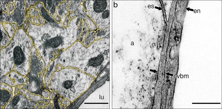
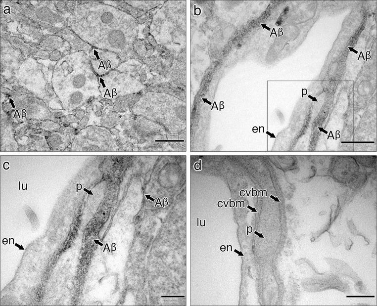
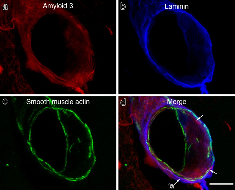
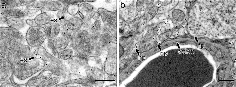
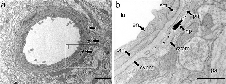
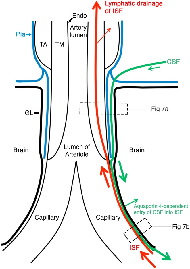
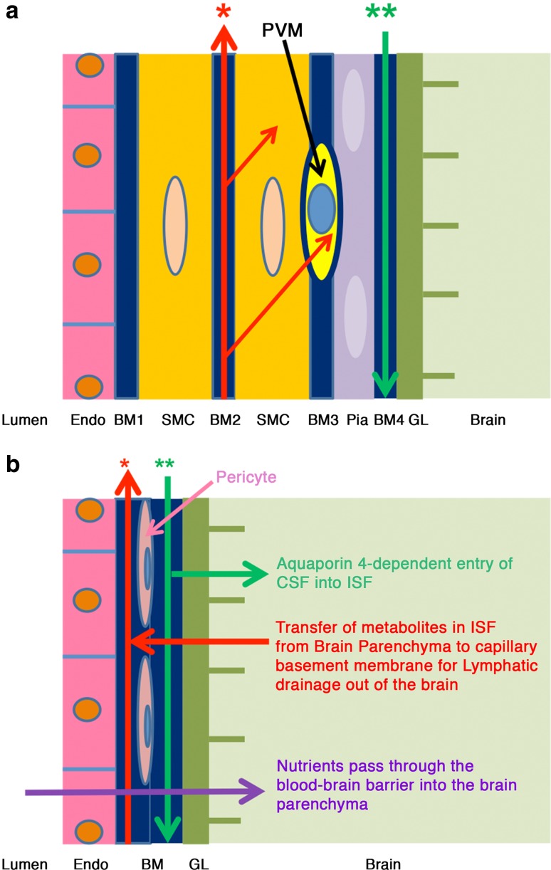
In the absence of conventional lymphatics, drainage of interstitial fluid and solutes from the brain parenchyma to cervical lymph nodes is along basement membranes in the walls of cerebral capillaries and tunica media of arteries. Perivascular pathways are also involved in the entry of CSF into the brain by the convective influx/glymphatic system. The objective of this study is to differentiate the cerebral vascular basement membrane pathways by which fluid passes out of the brain from the pathway by which CSF enters the brain. Experiment 1: 0.5 µl of soluble biotinylated or fluorescent Aβ, or 1 µl 15 nm gold nanoparticles was injected into the mouse hippocampus and their distributions determined at 5 min by transmission electron microscopy. Aβ was distributed within the extracellular spaces of the hippocampus and within basement membranes of capillaries and tunica media of arteries. Nanoparticles did not enter capillary basement membranes from the extracellular spaces. Experiment 2: 2 µl of 15 nm nanoparticles were injected into mouse CSF. Within 5 min, groups of nanoparticles were present in the pial-glial basement membrane on the outer aspect of cortical arteries between the investing layer of pia mater and the glia limitans. The results of this study and previous research suggest that cerebral vascular basement membranes form the pathways by which fluid passes into and out of the brain but that different basement membrane layers are involved. The significance of these findings for neuroimmunology, Alzheimer's disease, drug delivery to the brain and the concept of the Virchow-Robin space are discussed.
Overview
The claims and statistics of this paper haven’t been checked yet.
Run the check — free, no account needed. It reads the open-access text and checks the sources it cites.
Integrity signals
Registry
Cites
Statistics
Figures
Verification
Claims the paper makes
The claims and statistics of this paper haven’t been checked yet.
Run the check — free, no account needed. It reads the open-access text and checks the sources it cites.
Claims other papers make about this one
Each is checked against this paper’s own text: does it say what the citing paper says it does?
The source backs part of the claim, not all of it — or backs it for a different population, condition or comparison.
In the citing paper
“Moreover, animal studies report diverging findings regarding the sites of perivascular brain influx and efflux and direction of perivascular flow (7, 15, 16).”
From Brain-wide glymphatic enhancement and clearance in humans assessed with MRI. (2018) · cites this paper as [16]
Experimental — accuracy not yet measuredIn the citing paper
“For example, in the cortex, a packed barrier of endothelial cells, basement membrane, and glia limitans surrounds penetrating arteries, leaving little to no fluid-filled space, whereas the lenticulostriate branches in the basal ganglia are surrounded by a larger perivascular space (13).”
From The anatomy and immunology of vasculature in the central nervous system (2019) · cites this paper as [13]
In the citing paper
“Movement of ISF from the brain parenchyma occurs primarily between basement membranes in the perivascular spaces of capillaries and arteries. This continues along basement membranes in the tunica media of cerebral and leptomeningeal arteries (13).”
From The anatomy and immunology of vasculature in the central nervous system (2019) · cites this paper as [13]
Self-check
Percentages, sums and reported differences re-computed from the paper’s own numbers.
The self-check needs the paper’s full text.
Citations
Cites
Iliff, J. J., Wang, M., Liao, Y., Plogg, B. A., Peng, W., Gundersen, G. A., Benveniste, H., Vates, G. E., Deane, R., Goldman, S. A., et al. (2012) · Science Translational Medicine · doi:10.1126/scitranslmed.3003748 · Cited by 5,824
Turkevich Method for Gold Nanoparticle Synthesis Revisited
Kimling, J., Maier, M., Okenve, B., Kotaidis, V., Ballot, H., & Plech, A. (2006) · The Journal of Physical Chemistry B · doi:10.1021/jp061667w · Cited by 2,283
Direct brain infusion of glial cell line–derived neurotrophic factor in Parkinson disease
Gill, S. S., Patel, N. K., Hotton, G. R., O'Sullivan, K., McCarter, R., Bunnage, M., Brooks, D. J., Svendsen, C. N., & Heywood, P. (2003) · Nature Medicine · doi:10.1038/nm850 · Cited by 1,290
A new look at cerebrospinal fluid circulation
Brinker, T., Stopa, E., Morrison, J., & Klinge, P. (2014) · Fluids and Barriers of the CNS · doi:10.1186/2045-8118-11-10 · Cited by 819
Evidence for bulk flow of brain interstitial fluid: significance for physiology and pathology
Abbott, N. J. (2004) · Neurochemistry International · doi:10.1016/j.neuint.2003.11.006 · Cited by 786
Rennels, M. L., Gregory, T. F., Blaumanis, O. R., Fujimoto, K., & Grady, P. A. (1985) · Brain Research · doi:10.1016/0006-8993(85)91383-6 · Cited by 752
Carare, R. O., Bernardes‐Silva, M., Newman, T. A., Page, A. M., Nicoll, J. A. R., Perry, V. H., & Weller, R. O. (2008) · Neuropathology and Applied Neurobiology · doi:10.1111/j.1365-2990.2007.00926.x · Cited by 665
Self-citationKida, S., Pantazis, A., & Weller, R. O. (1993) · Neuropathology and Applied Neurobiology · doi:10.1111/j.1365-2990.1993.tb00476.x · Cited by 520
Self-citationDynamics of CNS Barriers: Evolution, Differentiation, and Modulation
Abbott, N. J. (2005) · Cellular and Molecular Neurobiology · doi:10.1007/s10571-004-1374-y · Cited by 471
(1990) · Europe PMC (PubMed Central) · Cited by 463
Self-citation
Cited by
Sorted by citations
The Neurovascular Unit Coming of Age: A Journey through Neurovascular Coupling in Health and Disease
Iadecola, C. (2017) · Neuron · doi:10.1016/j.neuron.2017.07.030 · Cited by 2,480
Flow of cerebrospinal fluid is driven by arterial pulsations and is reduced in hypertension.
Mestre, H., Tithof, J., Du, T., Song, W., Peng, W., Sweeney, A. M., Olveda, G., Thomas, J. H., Nedergaard, M., & Kelley, D. H. (2018) · Nature Communications · doi:10.1038/s41467-018-07318-3 · Cited by 1,110
The movers and shapers in immune privilege of the CNS
Engelhardt, B., Vajkoczy, P., & Weller, R. O. (2017) · Nature Immunology · doi:10.1038/ni.3666 · Cited by 893
Self-citationPerivascular spaces in the brain: anatomy, physiology and pathology
Wardlaw, J. M., Benveniste, H., Nedergaard, M., Zlokovic, B. V., Mestre, H., Lee, H., Doubal, F. N., Brown, R., Ramirez, J., MacIntosh, B. J., et al. (2020) · Nature Reviews Neurology · doi:10.1038/s41582-020-0312-z · Cited by 870
Rasmussen, M. K., Mestre, H., & Nedergaard, M. (2021) · Physiological Reviews · doi:10.1152/physrev.00031.2020 · Cited by 638
The role of brain barriers in fluid movement in the CNS: is there a ‘glymphatic’ system?
Abbott, N. J., Pizzo, M. E., Preston, J. E., Janigro, D., & Thorne, R. G. (2018) · Acta Neuropathologica · doi:10.1007/s00401-018-1812-4 · Cited by 593
Emerging concepts in sporadic cerebral amyloid angiopathy
Charidimou, A., Boulouis, G., Gurol, M. E., Ayata, C., Bacskai, B. J., Frosch, M. P., Viswanathan, A., & Greenberg, S. M. (2017) · Brain · doi:10.1093/brain/awx047 · Cited by 555
Brain-wide glymphatic enhancement and clearance in humans assessed with MRI.
Ringstad, G., Valnes, L. M., Dale, A. M., Pripp, A. H., Vatnehol, S.-A. S., Emblem, K. E., Mardal, K.-A., & Eide, P. K. (2018) · JCI Insight · doi:10.1172/jci.insight.121537 · Cited by 533
“Moreover, animal studies report diverging findings regarding the sites of perivascular brain influx and efflux and direction of perivascular flow (7, 15, 16).”
Neurovascular and Cognitive Dysfunction in Hypertension
Iadecola, C., & Gottesman, R. F. (2019) · Circulation Research · doi:10.1161/circresaha.118.313260 · Cited by 530
The vascular basement membrane in the healthy and pathological brain
Thomsen, M. S., Routhe, L. G., & Moos, T. (2017) · Journal of Cerebral Blood Flow & Metabolism · doi:10.1177/0271678x17722436 · Cited by 512
Blood-Brain Barrier Dysfunction and the Pathogenesis of Alzheimer’s Disease
Yamazaki, Y., & Kanekiyo, T. (2017) · International Journal of Molecular Sciences · doi:10.3390/ijms18091965 · Cited by 451
Targeting the transferrin receptor for brain drug delivery
Johnsen, K. B., Burkhart, A., Thomsen, L. B., Andresen, T. L., & Moos, T. (2019) · Progress in Neurobiology · doi:10.1016/j.pneurobio.2019.101665 · Cited by 379
Vascular, glial, and lymphatic immune gateways of the central nervous system
Engelhardt, B., Carare, R. O., Bechmann, I., Flügel, A., Laman, J. D., & Weller, R. O. (2016) · Acta Neuropathologica · doi:10.1007/s00401-016-1606-5 · Cited by 362
Self-citationSmith, A. J., Yao, X., Dix, J. A., Jin, B.-J., & Verkman, A. S. (2017) · eLife · doi:10.7554/elife.27679 · Cited by 362
Inoue, Y., Shue, F., Bu, G., & Kanekiyo, T. (2023) · Molecular Neurodegeneration · doi:10.1186/s13024-023-00640-5 · Cited by 380
Cerebrovascular Smooth Muscle Cells as the Drivers of Intramural Periarterial Drainage of the Brain
Aldea, R., Weller, R. O., Wilcock, D. M., Carare, R. O., & Richardson, G. W. (2019) · Frontiers in Aging Neuroscience · doi:10.3389/fnagi.2019.00001 · Cited by 340
Self-citationThe anatomy and immunology of vasculature in the central nervous system
Mastorakos, P., & McGavern, D. (2019) · Science Immunology · doi:10.1126/sciimmunol.aav0492 · Cited by 333
Albargothy, N. J., Johnston, D. A., MacGregor-Sharp, M., Weller, R. O., Verma, A., Hawkes, C. A., & Carare, R. O. (2018) · Acta Neuropathologica · doi:10.1007/s00401-018-1862-7 · Cited by 311
Self-citationVascular Cognitive Impairment (VCI)
Rundek, T., Tolea, M. I., Ariko, T., Fagerli, E. A., & Camargo, C. J. (2021) · Neurotherapeutics · doi:10.1007/s13311-021-01170-y · Cited by 347
Interstitial solute transport in 3D reconstructed neuropil occurs by diffusion rather than bulk flow
Holter, K. E., Kehlet, B., Devor, A., Sejnowski, T. J., Dale, A. M., Omholt, S. W., Ottersen, O. P., Nagelhus, E. A., Mardal, K.-A., & Pettersen, K. H. (2017) · Proceedings of the National Academy of Sciences · doi:10.1073/pnas.1706942114 · Cited by 282
Pizzo, M. E., Wolak, D. J., Kumar, N. N., Brunette, E., Brunnquell, C. L., Hannocks, M., Abbott, N. J., Meyerand, M. E., Sorokin, L., Stanimirovic, D. B., et al. (2018) · The Journal of Physiology · doi:10.1113/jp275105 · Cited by 272
Achariyar, T. M., Li, B., Peng, W., Verghese, P. B., Shi, Y., McConnell, E., Benraiss, A., Kasper, T., Song, W., Takano, T., et al. (2016) · Molecular Neurodegeneration · doi:10.1186/s13024-016-0138-8 · Cited by 256
The glymphatic hypothesis: the theory and the evidence
Hladky, S. B., & Barrand, M. A. (2022) · Fluids and Barriers of the CNS · doi:10.1186/s12987-021-00282-z · Cited by 278
The increasing impact of cerebral amyloid angiopathy: essential new insights for clinical practice
Banerjee, G., Carare, R. O., Cordonnier, C., Greenberg, S. M., Schneider, J. A., Smith, E. E., van Buchem, M. A., van der Grond, J., Verbeek, M. M., & Werring, D. J. (2017) · Journal of Neurology Neurosurgery & Psychiatry · doi:10.1136/jnnp-2016-314697 · Cited by 251
Self-citationBrain Microvascular Pericytes in Vascular Cognitive Impairment and Dementia
Uemura, M. T., Maki, T., Ihara, M., Lee, V. M. ‐., & Trojanowski, J. Q. (2020) · Frontiers in Aging Neuroscience · doi:10.3389/fnagi.2020.00080 · Cited by 246
Non-invasive MR imaging of human brain lymphatic networks with connections to cervical lymph nodes
Albayram, S., Smith, G., Tufan, F., Tuna, İ. S., Bostancıklıoğlu, M., Zile, M. R., & Albayram, Ö. (2022) · Nature Communications · doi:10.1038/s41467-021-27887-0 · Cited by 243
Eide, P. K., & Ringstad, G. A. (2018) · Journal of Cerebral Blood Flow & Metabolism · doi:10.1177/0271678x18760974 · Cited by 239
Weller, R. O., Sharp, M. M., Christodoulides, M., Carare, R. O., & Møllgård, K. (2018) · Acta Neuropathologica · doi:10.1007/s00401-018-1809-z · Cited by 222
“5b) [64] and by high-resolution confocal microscopy (Albargothy, Hawkes and Carare submitted paper) and not along periarteriolar “spaces”.”
BackgroundSelf-citationThe “Neuro-Glial-Vascular” Unit: The Role of Glia in Neurovascular Unit Formation and Dysfunction
Kugler, E., Greenwood, J., & MacDonald, R. B. (2021) · Frontiers in Cell and Developmental Biology · doi:10.3389/fcell.2021.732820 · Cited by 228
Clearance of Amyloid Beta and Tau in Alzheimer’s Disease: from Mechanisms to Therapy
Xin, S.-H., Tan, L., Cao, X., Yu, J.-T., & Tan, L. (2018) · Neurotoxicity Research · doi:10.1007/s12640-018-9895-1 · Cited by 222
Hladky, S. B., & Barrand, M. A. (2018) · Fluids and Barriers of the CNS · doi:10.1186/s12987-018-0113-6 · Cited by 206
Molecular characterization of perivascular drainage pathways in the murine brain
Hannocks, M.-J., Pizzo, M. E., Huppert, J., Deshpande, T., Abbott, N. J., Thorne, R. G., & Sorokin, L. (2018) · Journal of Cerebral Blood Flow & Metabolism · doi:10.1177/0271678x17749689 · Cited by 201
Chen, T., Dai, Y., Hu, C., Lin, Z., Wang, S., Yang, J., Zeng, L., Li, S., & Li, W. (2024) · Fluids and Barriers of the CNS · doi:10.1186/s12987-024-00557-1 · Cited by 205
Retinal imaging in Alzheimer's and neurodegenerative diseases
Snyder, P. J., Alber, J., Alt, C., Bain, L. J., Bouma, B. E., Bouwman, F. H., DeBuc, D. C., Campbell, M. C. W., Carrillo, M. C., Chew, E. Y., et al. (2020) · Alzheimer s & Dementia · doi:10.1002/alz.12179 · Cited by 197
Self-citationDysfunction of the blood–brain barrier in Alzheimer's disease: Evidence from human studies
Kurz, C., Walker, L., Rauchmann, B., & Perneczky, R. (2022) · Neuropathology and Applied Neurobiology · doi:10.1111/nan.12782 · Cited by 183
“Aβ via the cerebrovascular system, but its connection to other intracerebral Aβ drainage systems, such as the intramural periarterial drainage (IPAD) pathway, remains to be clarified.(11,12,23) Advancing age and carrying the apolipoprotein (ApoE) ε4 (APOE ε4) genotype, both related to enhanced cerebral amyloid angiopa…”
BackgroundHow Do Meningeal Lymphatic Vessels Drain the CNS?
Raper, D. M. S., Louveau, A., & Kipnis, J. (2016) · Trends in Neurosciences · doi:10.1016/j.tins.2016.07.001 · Cited by 184
Mechanical stress regulates transport in a compliant 3D model of the blood-brain barrier
Partyka, P. P., Godsey, G. A., Galie, J. R., Kosciuk, M. C., Acharya, N. K., Nagele, R. G., & Galie, P. A. (2016) · Biomaterials · doi:10.1016/j.biomaterials.2016.11.012 · Cited by 178
Bacyinski, A., Xu, M., Wang, W., & Hu, J. (2017) · Frontiers in Neuroanatomy · doi:10.3389/fnana.2017.00101 · Cited by 170
Gouveia-Freitas, K., & de Bastos-Leite, A. J. (2021) · Neuroradiology · doi:10.1007/s00234-021-02718-7 · Cited by 166
Hypertension, Neurodegeneration, and Cognitive Decline
Pacholko, A. G., & Iadecola, C. (2024) · Hypertension · doi:10.1161/hypertensionaha.123.21356 · Cited by 170
Chen, S., Shao, L., & Ma, L. (2021) · Frontiers in Cellular Neuroscience · doi:10.3389/fncel.2021.716825 · Cited by 154
Perivascular spaces and their role in neuroinflammation
Ineichen, B. V., Okar, S. V., Proulx, S. T., Engelhardt, B., Lassmann, H., & Reich, D. S. (2022) · Neuron · doi:10.1016/j.neuron.2022.10.024 · Cited by 151
Emerging links between cerebrovascular and neurodegenerative diseases—a special role for pericytes
Lendahl, U., Nilsson, P., & Betsholtz, C. (2019) · EMBO Reports · doi:10.15252/embr.201948070 · Cited by 148
Glymphatic system disruption as a mediator of brain trauma and chronic traumatic encephalopathy
Sullan, M. J., Asken, B. M., Jaffee, M. S., DeKosky, S. T., & Bauer, R. M. (2017) · Neuroscience & Biobehavioral Reviews · doi:10.1016/j.neubiorev.2017.08.016 · Cited by 135
Pathological changes within the cerebral vasculature in Alzheimer’s disease: New perspectives
Fisher, R. A., Miners, J. S., & Love, S. (2022) · Brain Pathology · doi:10.1111/bpa.13061 · Cited by 133
Kakuda, N., Miyasaka, T., Iwasaki, N., Nirasawa, T., Wada‐Kakuda, S., Takahashi‐Fujigasaki, J., Murayama, S., Ihara, Y., & Ikegawa, M. (2017) · Acta Neuropathologica Communications · doi:10.1186/s40478-017-0477-x · Cited by 131
Klostranec, J. M., Vucevic, D., Bhatia, K. D., Kortman, H. G. J., Krings, T., Murphy, K., terBrugge, K. G., & Mikulis, D. J. (2021) · Radiology · doi:10.1148/radiol.2021202043 · Cited by 130
Davies, J., Siebenhandl-Wolff, P., Tranquart, F., Jones, P. A., & Evans, P. M. (2022) · Archives of Toxicology · doi:10.1007/s00204-021-03189-8 · Cited by 123
Enzmann, G., Kargaran, S., & Engelhardt, B. (2018) · Therapeutic Advances in Neurological Disorders · doi:10.1177/1756286418794184 · Cited by 119
Impact of sex and APOE4 on cerebral amyloid angiopathy in Alzheimer’s disease
Shinohara, M., Murray, M. E., Frank, R. D., Shinohara, M., DeTure, M., Yamazaki, Y., Tachibana, M., Atagi, Y., Davis, M. D., Liu, C.-C., et al. (2016) · Acta Neuropathologica · doi:10.1007/s00401-016-1580-y · Cited by 123
Endothelial Barrier Function and Leukocyte Transmigration in Atherosclerosis
Sluiter, T. J., van Buul, J. D., Huveneers, S., Quax, P. H. A., & de Vries, M. R. (2021) · Biomedicines · doi:10.3390/biomedicines9040328 · Cited by 123
Saito, S., Yamamoto, Y., Maki, T., Hattori, Y., Ito, H., Mizuno, K., Harada-Shiba, M., Kalaria, R. N., Fukushima, M., Takahashi, R., et al. (2017) · Acta Neuropathologica Communications · doi:10.1186/s40478-017-0429-5 · Cited by 110
Damisah, E. C., Hill, R., Тонг, Л., Murray, K. N., & Grutzendler, J. (2017) · Nature Neuroscience · doi:10.1038/nn.4564 · Cited by 114
Decreased CSF clearance and increased brain amyloid in Alzheimer’s disease
Li, Y., Rusinek, H., Butler, T., Glodzik, L., Pirraglia, E., Babich, J. W., Mozley, P. D., Nehmeh, S. A., Pahlajani, S. S., Wang, X. H., et al. (2022) · Fluids and Barriers of the CNS · doi:10.1186/s12987-022-00318-y · Cited by 110
Self-citationGlymphatic Cerebrospinal Fluid and Solute Transport Quantified by MRI and PET Imaging
Benveniste, H., Lee, H., Ozturk, B., Chen, X., Koundal, S., Vaska, P., Tannenbaum, A., & Volkow, N. D. (2020) · Neuroscience · doi:10.1016/j.neuroscience.2020.11.014 · Cited by 106
Hecht, M., Krämer, L. M., Arnim, C., Otto, M., & Thal, D. (2018) · Acta Neuropathologica · doi:10.1007/s00401-018-1834-y · Cited by 92
Solar, P., Zamani, A., Lakatosová, K., & Joukal, M. (2022) · Fluids and Barriers of the CNS · doi:10.1186/s12987-022-00312-4 · Cited by 103
The glymphatic system and meningeal lymphatics of the brain: new understanding of brain clearance
Yankova, G., Bogomyakova, O. B., & Тулупов, А. А. (2021) · Reviews in the Neurosciences · doi:10.1515/revneuro-2020-0106 · Cited by 110
Brain perivascular macrophages: current understanding and future prospects
Wen, W., Cheng, J., & Tang, Y. (2023) · Brain · doi:10.1093/brain/awad304 · Cited by 105
Pirici, I., Balsanu, T. A., Cătălin, B., Mărgăritescu, C., Divan, T., Văcăraș, V., Mogoantă, L., Pirici, D. N., Carare, R. O., & Muresanu, D. F. (2017) · International Journal of Molecular Sciences · doi:10.3390/ijms19010046 · Cited by 100
Self-citationMestre, H., Verma, N., Greene, T. D., Lin, L. A., Ladrón-de-Guevara, A., Sweeney, A. M., Liu, G., Thomas, V. K., Galloway, C. A., Bentley, K. L. D. M., et al. (2022) · Nature Communications · doi:10.1038/s41467-022-31257-9 · Cited by 97
Tachibana, M., Yamazaki, Y., Liu, C., Bu, G., & Kanekiyo, T. (2017) · Experimental Neurology · doi:10.1016/j.expneurol.2017.10.023 · Cited by 92
Singhrao, S. K., & Olsen, I. (2018) · Journal of Alzheimer s Disease Reports · doi:10.3233/adr-180080 · Cited by 93
Wu, C., Lirng, J., Ling, Y., Wang, Y.-F., Wu, H., Fuh, J., Lin, P., Wang, S., & Chen, S. (2020) · Annals of Neurology · doi:10.1002/ana.25928 · Cited by 90
Taoka, T., & Naganawa, S. (2020) · Japanese Journal of Radiology · doi:10.1007/s11604-020-01017-0 · Cited by 88
Neural stem cell therapy for neurovascular injury in Alzheimer's disease
Boese, A. C., Hamblin, M. H., & Lee, J. (2019) · Experimental Neurology · doi:10.1016/j.expneurol.2019.113112 · Cited by 89
Sharp, M. K., Carare, R. O., & Martin, B. A. (2019) · Fluids and Barriers of the CNS · doi:10.1186/s12987-019-0132-y · Cited by 87
Self-citationHe, J., Zhao, X., Xu, L., & Mao, C. (2019) · Journal of Alzheimer s Disease · doi:10.3233/jad-190764 · Cited by 88
Golanov, E. V., Bovshik, E. I., Wong, K. K., Pautler, R. G., Foster, C. H., Federley, R., Zhang, J. Y., Mancuso, J. J., Wong, S. T. C., & Britz, G. W. (2017) · Journal of Cerebral Blood Flow & Metabolism · doi:10.1177/0271678x17701157 · Cited by 86
The mechanisms behind perivascular fluid flow
Daversin, C., Vinje, V., Mardal, K., & Rognes, M. E. (2020) · PLoS ONE · doi:10.1371/journal.pone.0244442 · Cited by 82
Structural and Functional Remodeling of the Brain Vasculature Following Stroke
Andrade, M. F., Raman‐Nair, J., & Lacoste, B. (2020) · Frontiers in Physiology · doi:10.3389/fphys.2020.00948 · Cited by 82
Kim, S. H., Ahn, J. H., Yang, H. J., Lee, P., Koh, G. Y., & Jeong, Y. (2020) · Acta Neuropathologica Communications · doi:10.1186/s40478-020-01042-0 · Cited by 79
Basal lamina changes in neurodegenerative disorders
Nguyen, B., Bix, G., & Yao, Y. (2021) · Molecular Neurodegeneration · doi:10.1186/s13024-021-00502-y · Cited by 75
Carare, R. O., Aldea, R., Agarwal, N., Bacskai, B. J., Bechman, I., Boche, D., Bu, G., Bulters, D. O., Alt, C., Counts, S., et al. (2020) · Alzheimer s & Dementia Diagnosis Assessment & Disease Monitoring · doi:10.1002/dad2.12053 · Cited by 77
Self-citationDivecha, Y. A., Rampes, S., Tromp, S., Boyanova, S., Fleckney, A. L., Fidanboylu, M., & Thomas, S. A. (2025) · Pharmacological Reviews · doi:10.1016/j.pharmr.2025.100052 · Cited by 83
Emerging applications of nanotechnology for diagnosis and therapy of disease: a review
Bayford, R. H., Rademacher, T., Roitt, I. M., & Wang, S. X. (2017) · Physiological Measurement · doi:10.1088/1361-6579/aa7182 · Cited by 75
Pulsatile flow drivers in brain parenchyma and perivascular spaces: a resistance network model study
Rey, J. A., & Sarntinoranont, M. (2018) · Fluids and Barriers of the CNS · doi:10.1186/s12987-018-0105-6 · Cited by 74
An immunohistochemical study of lymphatic elements in the human brain
Mezey, É., Szalayova, I., Hogden, C. T., Brady, A., Dósa, A., Sótonyi, P., & Palkovits, M. (2021) · Proceedings of the National Academy of Sciences · doi:10.1073/pnas.2002574118 · Cited by 74
Current understanding of lymphatic vessels in the central nervous system
Tamura, R., Yoshida, K., & Toda, M. (2019) · Neurosurgical Review · doi:10.1007/s10143-019-01133-0 · Cited by 74
Dóra, D., Ferenczi, S., Stavely, R. G., Tóth, V. É., Varga, Z. V., Kovács, T., Bódi, I., Hotta, R., Kovács, K., Goldstein, A. M., et al. (2021) · Cellular and Molecular Gastroenterology and Hepatology · doi:10.1016/j.jcmgh.2021.07.003 · Cited by 74
Lam, M. A., Hemley, S. J., Najafi, E., Vella, N. G. F., Bilston, L. E., & Stoodley, M. A. (2017) · Scientific Reports · doi:10.1038/s41598-017-13455-4 · Cited by 71
Peritumoral Edema in Gliomas: A Review of Mechanisms and Management
Ohmura, K., Tomita, H., & Hara, A. (2023) · Biomedicines · doi:10.3390/biomedicines11102731 · Cited by 71
Inhibition of Aquaporin 4 Decreases Amyloid Aβ40 Drainage Around Cerebral Vessels
Roşu, G. C., Cătălin, B., Bălşeanu, T. A., Mogoantă, L., Mărgăritescu, C., Kumar‐Singh, S., & Pirici, D. N. (2020) · Molecular Neurobiology · doi:10.1007/s12035-020-02044-8 · Cited by 69
Rasschaert, M., Weller, R. O., Schroeder, J. A., Brochhausen, C., & Idée, J. (2020) · Journal of Magnetic Resonance Imaging · doi:10.1002/jmri.27124 · Cited by 65
Self-citationSmall vessels, dementia and chronic diseases – molecular mechanisms and pathophysiology
Horsburgh, K. J., Wardlaw, J. M., Agtmael, T. V., Allan, S. M., Ashford, M. L. J., Bath, P. M., Brown, R., Berwick, J., Cader, M. Z., Carare, R. O., et al. (2018) · Clinical Science · doi:10.1042/cs20171620 · Cited by 65
Self-citationRole of the glymphatic system in ageing and diabetes mellitus impaired cognitive function
Zhang, L., Chopp, M., Jiang, Q., & Zhang, Z. (2019) · Stroke and Vascular Neurology · doi:10.1136/svn-2018-000203 · Cited by 59
A Kinetic Model for Blood Biomarker Levels After Mild Traumatic Brain Injury
Azizi, S., Hier, D. B., Allen, B., Obafemi-Ajayi, T., Olbricht, G. R., Thimgan, M. S., & Wunsch, D. C. (2021) · Frontiers in Neurology · doi:10.3389/fneur.2021.668606 · Cited by 58
Henrich‐Noack, P., Nikitovic, D., Neagu, M. T., Docea, A. O., Engin, A. B., Gelperina, S. E., Штильман, М. И., Mitsias, P. D., Tzanakakis, G. N., Gozes, I., et al. (2019) · Nanomedicine Nanotechnology Biology and Medicine · doi:10.1016/j.nano.2019.01.016 · Cited by 58
The lymphatic drainage systems in the brain: a novel target for ischemic stroke?
Wang, Y.-J., Sun, Y., Pei, Y., Ma, H.-W., Ma, H., Qin, L., & Yan, J.-H. (2022) · Neural Regeneration Research · doi:10.4103/1673-5374.346484 · Cited by 58
The glymphatic system’s role in traumatic brain injury-related neurodegeneration
Peters, M. E., & Lyketsos, C. G. (2023) · Molecular Psychiatry · doi:10.1038/s41380-023-02070-7 · Cited by 57
Pharmacokinetics and pharmacodynamics of antibiotics in central nervous system infections
Nau, R., Seele, J., Djukic, M., & Eiffert, H. (2017) · Current Opinion in Infectious Diseases · doi:10.1097/qco.0000000000000418 · Cited by 53
The Pattern of AQP4 Expression in the Ageing Human Brain and in Cerebral Amyloid Angiopathy
Owasil, R., O’Neill, R., Keable, A., Nimmo, J., MacGregor Sharp, M., Kelly, L., Saito, S., Simpson, J. E., Weller, R. O., Smith, C., et al. (2020) · International Journal of Molecular Sciences · doi:10.3390/ijms21041225 · Cited by 48
Self-citationLi, M., Kitamura, A., Beverley, J., Koudelka, J., Duncombe, J., Lennen, R. J., Jansen, M. A., Marshall, I., Platt, B., Wiegand, U. K., et al. (2022) · Frontiers in Aging Neuroscience · doi:10.3389/fnagi.2021.788519 · Cited by 55
Self-citationXu, M., Xiao, M., Li, S., & Yang, B. (2017) · Advances in experimental medicine and biology · doi:10.1007/978-94-024-1057-0_5 · Cited by 53
Current Advances in Immunotherapy for Glioblastoma
Mende, A., Schulte, J. D., Okada, H., & Clarke, J. L. (2021) · Current Oncology Reports · doi:10.1007/s11912-020-01007-5 · Cited by 53
The Role of Basement Membranes in Cerebral Amyloid Angiopathy
Howe, M. D., McCullough, L. D., & Urayama, A. (2020) · Frontiers in Physiology · doi:10.3389/fphys.2020.601320 · Cited by 50
Mehta, N. H., Suss, R. A., Dyke, J. P., Theise, N. D., Chiang, G. C.-Y., Strauß, S., Saint-Louis, L. A., Li, Y., Pahlajani, S. S., Babaria, V., et al. (2022) · Neurobiology of Disease · doi:10.1016/j.nbd.2022.105776 · Cited by 51
Self-citationPhysiological and Pathological Factors Affecting Drug Delivery to the Brain by Nanoparticles
Islam, Y., Leach, A. G., Smith, J. A., Pluchino, S., Coxon, C. R., Sivakumaran, M., Downing, J. R., Fatokun, A. A., Teixidò, M., & Ehtezazi, T. (2021) · Advanced Science · doi:10.1002/advs.202002085 · Cited by 52
Fluid outflow in the rat spinal cord: the role of perivascular and paravascular pathways
Liu, S., Lam, M. A., Sial, A. W., Hemley, S. J., Bilston, L. E., & Stoodley, M. A. (2018) · Fluids and Barriers of the CNS · doi:10.1186/s12987-018-0098-1 · Cited by 52
Hozumi, I., Kurita, H., Ozawa, K., Furuta, N., Inden, M., Sekine, S.-I., Yamada, M., Hayashi, Y., Kimura, A., Inuzuka, T., et al. (2018) · Journal of the Neurological Sciences · doi:10.1016/j.jns.2018.03.014 · Cited by 49
Metadata
- Authors
- Morris, A. W. J., Sharp, M. M., Albargothy, N. J., Fernandes, R., Hawkes, C. A., Verma, A., Weller, R. O., & Carare, R. O.
- Affiliations
- Southampton General Hospital (GB)
- The Open University (GB)
- Biogen (United States) (US)
- Corresponding author
- Carare, Roxana O.
- ORCID
- Venue
- Acta Neuropathologica
- Published
- 2016-03-14
- Volume
- 131
- Issue
- 5
- Pages
- 725-736
- ISSN
- 0001-6322 (ISSN-L)
- 1432-0533
- PMC
- PMC4835509
- OpenAlex
- W2298591538
- Cited by
- 289
- References
- 29
- Citations by year
- 2026: 14 · 2025: 19 · 2024: 18 · 2023: 22 · 2022: 35 · 2021: 28 · 2020: 48 · 2019: 33 · 2018: 29 · 2017: 34 · 2016: 9
- FWCI
- 19.63
- Type
- journal article
- Language
- en
- Open access
- hybrid
- doi.org
- Locations
- Acta Neuropathologica (publishedVersion, cc-by)
- PubMed Central (publishedVersion, cc-by)
- PubMed (publishedVersion)
- Open Research Online - ORO (The Open University) (acceptedVersion)
- ePrints Soton (University of Southampton) (acceptedVersion)
- Lancaster EPrints (Lancaster University) (submittedVersion)
- Funders
- Biotechnology and Biological Sciences Research Council (BB/K015540/1)
- Age UK (PhD-368)
- Biogen Idec
- Topics
- Health Sciences › Medicine › Clinical Neurology › Cerebrospinal fluid and hydrocephalus
- Life Sciences › Neuroscience › Neurology › Barrier Structure and Function Studies
- Life Sciences › Biochemistry, Genetics and Molecular Biology › Molecular Biology › Ion Transport and Channel Regulation
- Keywords
- vascular basement membrane, cerebrospinal fluid, glymphatic system, interstitial fluid drainage, amyloid-beta, gold nanoparticles, glia limitans, cerebral microvasculature, perivascular spaces, brain drug delivery, neuroimmunology, Alzheimer's disease, hippocampus
- MeSH terms
- Actins, Animals, Basement Membrane, Blood Vessels, Cerebrospinal Fluid, Cisterna Magna, Extracellular Space, Fluorescent Dyes, Hippocampus, Laminin, Male, Mice, Inbred C57BL, Peptide Fragments, Amyloid beta-Peptides, Biotinylation, Microscopy, Electron, Transmission, Mice, Metal Nanoparticles
About these checks
Registry, retraction, Cites and statistics results are computed, not judged, with one exception: a printed reference that no index lists is read once by a model before it counts as a citation error. Claims are extracted by a model and kept only when their quote appears verbatim in the text; each cited source is then checked, with a model’s help, for whether it says what the claim relies on. Those verdicts can be wrong — read the claim’s quote and the cited source before relying on one.
Statistics and arithmetic are recomputed, not judged.
Confidence bands are shown only for fields calibrated on our annotated test set. None is calibrated yet, so no band is shown.
What counts as a claim ›How we measure confidence ›How we check this ›
Are you an author, or do you see a problem with this page? Request a review
