

The DOI matches its Crossref record; 29 of the 29 references it cites were found in a registry or an index.
Vascular basement membranes as pathways for the passage of fluid into and out of the brain
Open accessAbstract
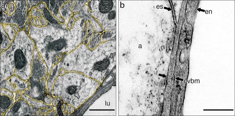
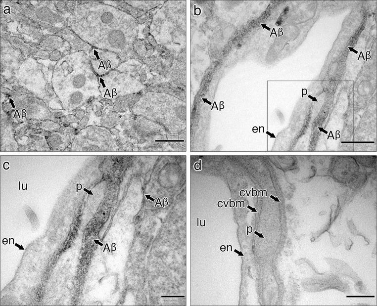
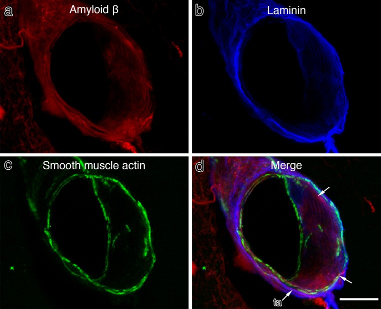
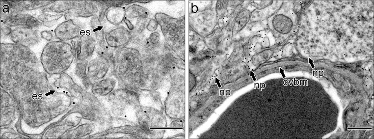
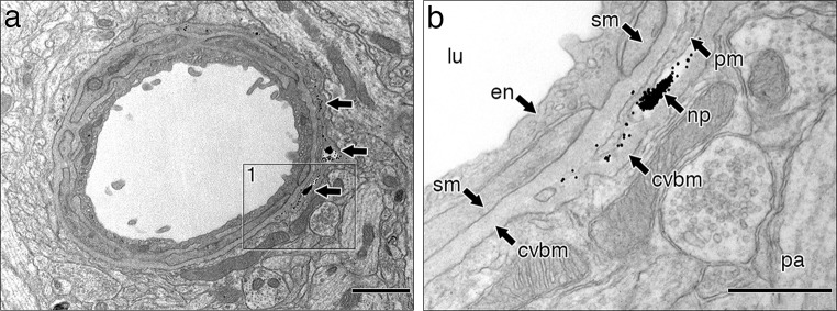
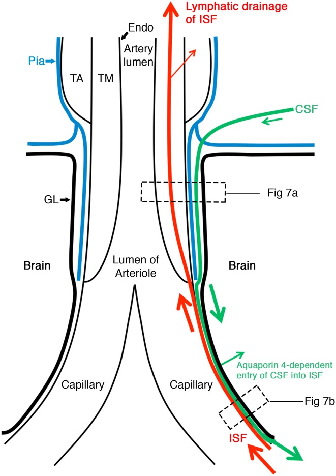
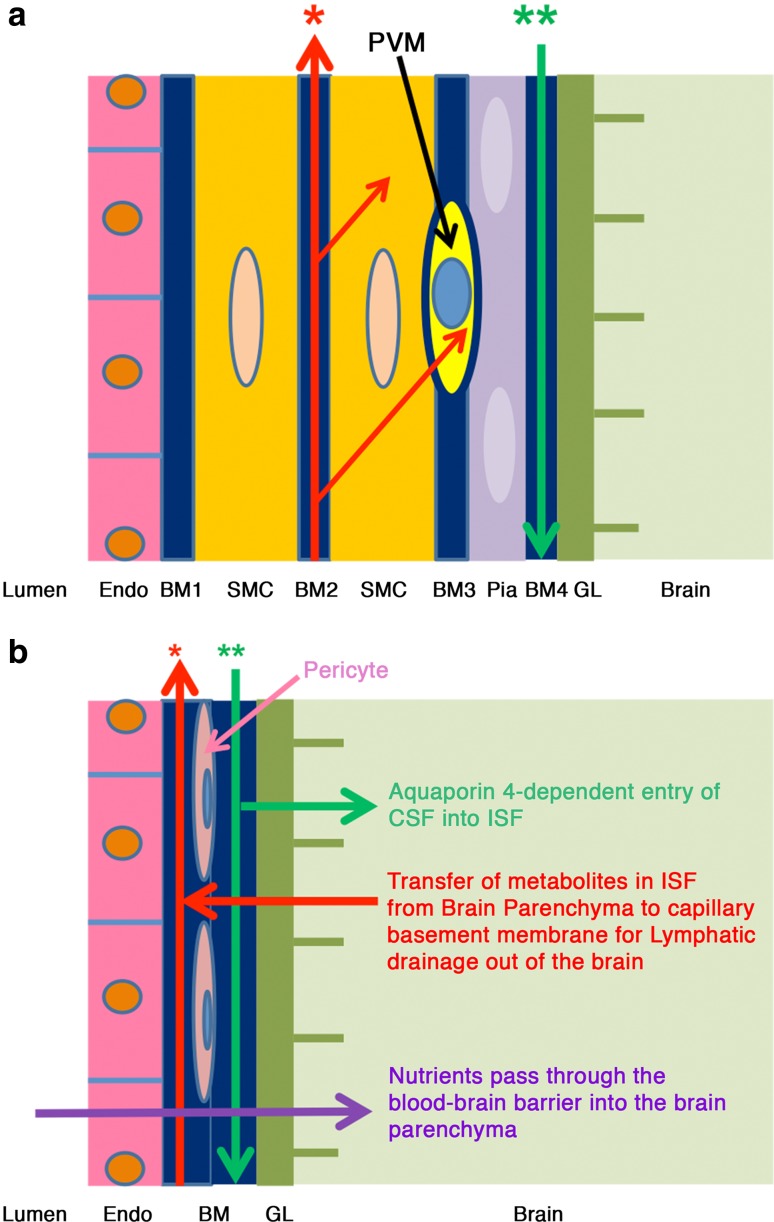
In the absence of conventional lymphatics, drainage of interstitial fluid and solutes from the brain parenchyma to cervical lymph nodes is along basement membranes in the walls of cerebral capillaries and tunica media of arteries. Perivascular pathways are also involved in the entry of CSF into the brain by the convective influx/glymphatic system. The objective of this study is to differentiate the cerebral vascular basement membrane pathways by which fluid passes out of the brain from the pathway by which CSF enters the brain. Experiment 1: 0.5 µl of soluble biotinylated or fluorescent Aβ, or 1 µl 15 nm gold nanoparticles was injected into the mouse hippocampus and their distributions determined at 5 min by transmission electron microscopy. Aβ was distributed within the extracellular spaces of the hippocampus and within basement membranes of capillaries and tunica media of arteries. Nanoparticles did not enter capillary basement membranes from the extracellular spaces. Experiment 2: 2 µl of 15 nm nanoparticles were injected into mouse CSF. Within 5 min, groups of nanoparticles were present in the pial-glial basement membrane on the outer aspect of cortical arteries between the investing layer of pia mater and the glia limitans. The results of this study and previous research suggest that cerebral vascular basement membranes form the pathways by which fluid passes into and out of the brain but that different basement membrane layers are involved. The significance of these findings for neuroimmunology, Alzheimer's disease, drug delivery to the brain and the concept of the Virchow-Robin space are discussed.
Overview
The claims and statistics of this paper haven’t been checked yet.
Run the check — free, no account needed. It reads the open-access text and checks the sources it cites.
Integrity signals
Registry
Cites
Statistics
Figures
Verification
Claims the paper makes
The claims and statistics of this paper haven’t been checked yet.
Run the check — free, no account needed. It reads the open-access text and checks the sources it cites.
Claims other papers make about this one
Each is checked against this paper’s own text: does it say what the citing paper says it does?
The source backs part of the claim, not all of it — or backs it for a different population, condition or comparison.
In the citing paper
“Moreover, animal studies report diverging findings regarding the sites of perivascular brain influx and efflux and direction of perivascular flow (7, 15, 16).”
From Brain-wide glymphatic enhancement and clearance in humans assessed with MRI. (2018) · cites this paper as [16]
Experimental — accuracy not yet measuredIn the citing paper
“For example, in the cortex, a packed barrier of endothelial cells, basement membrane, and glia limitans surrounds penetrating arteries, leaving little to no fluid-filled space, whereas the lenticulostriate branches in the basal ganglia are surrounded by a larger perivascular space (13).”
From The anatomy and immunology of vasculature in the central nervous system (2019) · cites this paper as [13]
In the citing paper
“Movement of ISF from the brain parenchyma occurs primarily between basement membranes in the perivascular spaces of capillaries and arteries. This continues along basement membranes in the tunica media of cerebral and leptomeningeal arteries (13).”
From The anatomy and immunology of vasculature in the central nervous system (2019) · cites this paper as [13]
Self-check
Percentages, sums and reported differences re-computed from the paper’s own numbers.
The self-check needs the paper’s full text.
Citations
Cites
Iliff, J. J., Wang, M., Liao, Y., Plogg, B. A., Peng, W., Gundersen, G. A., Benveniste, H., Vates, G. E., Deane, R., Goldman, S. A., et al. (2012) · Science Translational Medicine · doi:10.1126/scitranslmed.3003748 · Cited by 5,824
Turkevich Method for Gold Nanoparticle Synthesis Revisited
Kimling, J., Maier, M., Okenve, B., Kotaidis, V., Ballot, H., & Plech, A. (2006) · The Journal of Physical Chemistry B · doi:10.1021/jp061667w · Cited by 2,283
Direct brain infusion of glial cell line–derived neurotrophic factor in Parkinson disease
Gill, S. S., Patel, N. K., Hotton, G. R., O'Sullivan, K., McCarter, R., Bunnage, M., Brooks, D. J., Svendsen, C. N., & Heywood, P. (2003) · Nature Medicine · doi:10.1038/nm850 · Cited by 1,290
A new look at cerebrospinal fluid circulation
Brinker, T., Stopa, E., Morrison, J., & Klinge, P. (2014) · Fluids and Barriers of the CNS · doi:10.1186/2045-8118-11-10 · Cited by 819
Evidence for bulk flow of brain interstitial fluid: significance for physiology and pathology
Abbott, N. J. (2004) · Neurochemistry International · doi:10.1016/j.neuint.2003.11.006 · Cited by 786
Rennels, M. L., Gregory, T. F., Blaumanis, O. R., Fujimoto, K., & Grady, P. A. (1985) · Brain Research · doi:10.1016/0006-8993(85)91383-6 · Cited by 752
Carare, R. O., Bernardes‐Silva, M., Newman, T. A., Page, A. M., Nicoll, J. A. R., Perry, V. H., & Weller, R. O. (2008) · Neuropathology and Applied Neurobiology · doi:10.1111/j.1365-2990.2007.00926.x · Cited by 665
Self-citationKida, S., Pantazis, A., & Weller, R. O. (1993) · Neuropathology and Applied Neurobiology · doi:10.1111/j.1365-2990.1993.tb00476.x · Cited by 520
Self-citationDynamics of CNS Barriers: Evolution, Differentiation, and Modulation
Abbott, N. J. (2005) · Cellular and Molecular Neurobiology · doi:10.1007/s10571-004-1374-y · Cited by 471
(1990) · Europe PMC (PubMed Central) · Cited by 463
Self-citation
Cited by
Sorted by citations
Items 101–200 of 289
What SARS-CoV-2 does to our brains
Aschman, T., Mothes, R., Heppner, F. L., & Radbruch, H. (2022) · Immunity · doi:10.1016/j.immuni.2022.06.013 · Cited by 50
Role of Nanomedicine-Based Therapeutics in the Treatment of CNS Disorders
Guo, Z.-H., Khattak, S., Rauf, M. A., Ansari, M. A., Alomary, M. N., Razak, S., Yang, C., Wu, D., & Ji, X. (2023) · Molecules · doi:10.3390/molecules28031283 · Cited by 49
Tanaka, M., Saitô, S., Inoue, T., Satoh‐Asahara, N., & Ihara, M. (2019) · International Journal of Molecular Sciences · doi:10.3390/ijms20092139 · Cited by 48
van Veluw, S. J., Benveniste, H., Bakker, E. N. T. P., Carare, R. O., Greenberg, S. M., Iliff, J. J., Lorthois, S., Nostrand, W. E. V., Petzold, G. C., Shih, A. Y., et al. (2024) · Cellular and Molecular Life Sciences · doi:10.1007/s00018-024-05277-1 · Cited by 50
Self-citationAntigen Presentation After Stroke
Miró‐Mur, F., Urra, X., Gallizioli, M., Chamorro, Á., & Planas, A. M. (2016) · Neurotherapeutics · doi:10.1007/s13311-016-0469-8 · Cited by 47
Sheng, X., Zhang, C., Zhao, J., Xu, J., Zhang, P., Ding, Q., & Zhang, J. (2024) · Cell & Bioscience · doi:10.1186/s13578-024-01269-7 · Cited by 53
Liu, E., Sun, L., Zhang, Y., Wang, A., & Yan, J. (2019) · Acta neurochirurgica. Supplementum · doi:10.1007/978-3-030-04615-6_10 · Cited by 44
A Review of Hematoma Components Clearance Mechanism After Subarachnoid Hemorrhage
Pan, P., Xu, L., Zhang, H., Yuan, L., Lu, X., Chen, G., Tang, H., & Wu, J. (2020) · Frontiers in Neuroscience · doi:10.3389/fnins.2020.00685 · Cited by 49
Endo, Y., Hasegawa, K., Nomura, R., Arishima, H., Kikuta, K., Yamashita, T., Inoue, Y., Ueda, M., Ando, Y., Wilson, M. R., et al. (2019) · Acta Neuropathologica Communications · doi:10.1186/s40478-019-0662-1 · Cited by 42
Howe, M. D., Furr, J. W., Munshi, Y., Roy-O’Reilly, M. A., Maniskas, M. E., Koellhoffer, E. C., d’Aigle, J., Sansing, L. H., McCullough, L. D., & Urayama, A. (2019) · GeroScience · doi:10.1007/s11357-019-00118-7 · Cited by 43
Kassem, L. M., Ibrahim, N., & Farhana, S. A. (2020) · Journal of Nanotechnology · doi:10.1155/2020/8147080 · Cited by 43
McIntee, F. L., Giannoni, P., Blais, S. P., Sommer, G., Neubert, T. A., Rostagno, A. A., & Ghiso, J. A. (2016) · Frontiers in Aging Neuroscience · doi:10.3389/fnagi.2016.00223 · Cited by 42
Taxifolin: A Potential Therapeutic Agent for Cerebral Amyloid Angiopathy
Saitô, S., Tanaka, M., Satoh‐Asahara, N., Carare, R. O., & Ihara, M. (2021) · Frontiers in Pharmacology · doi:10.3389/fphar.2021.643357 · Cited by 45
Self-citationUric acid therapy for vasculoprotection in acute ischemic stroke
Chamorro, Á., Amaro, S., & Jiménez‐Altayó, F. (2019) · Brain Circulation · doi:10.4103/bc.bc_1_19 · Cited by 43
Subpial Hemorrhage in Neonates: What Radiologists Need to Know
Barreto, A. R. F., Carrasco, M., Dabrowski, A. K., Sun, L. R., & Tekes, A. (2021) · American Journal of Roentgenology · doi:10.2214/ajr.20.22999 · Cited by 40
Established, New and Emerging Concepts in Brain Vascular Development
Gupta, A., Rarick, K. R., & Ramchandran, R. (2021) · Frontiers in Physiology · doi:10.3389/fphys.2021.636736 · Cited by 42
A roadmap for potassium buffering/dispersion via the glial network of the CNS
Beckner, M. E. (2020) · Neurochemistry International · doi:10.1016/j.neuint.2020.104727 · Cited by 41
Howe, M. D., Atadja, L. A., Furr, J. W., Maniskas, M. E., Zhu, L., McCullough, L. D., & Urayama, A. (2018) · Neurobiology of Aging · doi:10.1016/j.neurobiolaging.2018.07.019 · Cited by 36
An Update of Nanoparticle-Based Approaches for Glioblastoma Multiforme Immunotherapy
Taiarol, L., Formicola, B., Magro, R. D., Sesana, S., & Re, F. (2020) · Nanomedicine · doi:10.2217/nnm-2020-0132 · Cited by 38
Glymphatic System and Subsidiary Pathways Drive Nanoparticles Away from the Brain
Liu, R., Jia, W., Wang, Y., Hu, C., Yu, W., Huang, Y., Wang, L., & Gao, H. (2022) · Research · doi:10.34133/2022/9847612 · Cited by 36
Ueno, M., Chiba, Y., Murakami, R., Matsumoto, K., Fujihara, R., Uemura, N., Yanase, K., & Kamada, M. (2019) · International Journal of Molecular Sciences · doi:10.3390/ijms20102600 · Cited by 34
Pathophysiology of Cerebral Edema—A Comprehensive Review
Dalby, T., Wohl, E., Dinsmore, M., Unger, Z., Chowdhury, T., & Venkatraghavan, L. (2020) · Journal of Neuroanaesthesiology and Critical Care · doi:10.1055/s-0040-1721165 · Cited by 37
Targeting nanoparticles to malignant tumors
Sharma, N., Bietar, K., & Stochaj, U. (2022) · Biochimica et Biophysica Acta (BBA) - Reviews on Cancer · doi:10.1016/j.bbcan.2022.188703 · Cited by 36
Filippidis, A., Conroy, T. J., Maragkos, G. Α., Holsapple, J. W., & Davies, K. G. (2017) · World Neurosurgery · doi:10.1016/j.wneu.2017.03.131 · Cited by 32
Moura, R. P., Almeida, A., & Sarmento, B. F. C. C. (2017) · Progress in Neurobiology · doi:10.1016/j.pneurobio.2017.09.001 · Cited by 32
Dankner, M., Lam, S., Degenhard, T., Garzia, L., Guiot, M., Petrecca, K., & Siegel, P. M. (2021) · Cancers · doi:10.3390/cancers13040732 · Cited by 32
Metabolite Clearance During Wakefulness and Sleep
Hladky, S. B., & Barrand, M. A. (2017) · Handbook of experimental pharmacology · doi:10.1007/164_2017_37 · Cited by 36
Carotid Stiffness is Associated with Brain Amyloid-β Burden in Amnestic Mild Cognitive Impairment
Pasha, E. P., Rutjes, E., Tomoto, T., Tarumi, T., Stowe, A. M., Claassen, J. A. H. R., Cullum, C. M., Zhu, D. C., & Zhang, R. (2020) · Journal of Alzheimer s Disease · doi:10.3233/jad-191073 · Cited by 32
Nanoparticle systems for nose-to-brain delivery
Liu, Q., & Zhang, Q. (2018) · Elsevier eBooks · doi:10.1016/b978-0-12-814001-7.00010-x · Cited by 30
Kelly, L., Brown, C., Michalik, D., Hawkes, C. A., Aldea, R., Agarwal, N., Salib, R. J., Alzetani, A., Ethell, D. W., Counts, S., et al. (2023) · Alzheimer s & Dementia · doi:10.1002/alz.13512 · Cited by 31
Self-citationDevelopment of a Multicomponent Intervention to Prevent Alzheimer's Disease
Saitô, S., Yamamoto, Y., & Ihara, M. (2019) · Frontiers in Neurology · doi:10.3389/fneur.2019.00490 · Cited by 30
Brain Vascular Health in ALS Is Mediated through Motor Cortex Microvascular Integrity
Schreiber, S., Bernal, J., Arndt, P., Schreiber, F., Müller, P., Morton, L., Braun-Dullaeus, R. C., Hernández, M. V., Duarte, R., Wardlaw, J. M., et al. (2023) · Cells · doi:10.3390/cells12060957 · Cited by 30
Role of the glymphatic system in idiopathic intracranial hypertension
Steinruecke, M., Tiefenbach, J., Park, J. J., & Kaliaperumal, C. (2022) · Clinical Neurology and Neurosurgery · doi:10.1016/j.clineuro.2022.107446 · Cited by 28
Sharp, M. M., Saitô, S., Keable, A., Gatherer, M., Aldea, R., Agarwal, N., Simpson, J. E., Wharton, S. B., Weller, R. O., & Carare, R. O. (2020) · Acta Neuropathologica Communications · doi:10.1186/s40478-020-01009-1 · Cited by 30
Self-citationVascular and Nonvascular Mechanisms of Cognitive Impairment and Dementia
Kara, B., Gordon, M. N., Gifani, M., Dorrance, A. M., & Counts, S. (2022) · Clinics in Geriatric Medicine · doi:10.1016/j.cger.2022.07.006 · Cited by 29
Shear-Induced Amyloid Formation in the Brain: I. Potential Vascular and Parenchymal Processes
Trumbore, C. N. (2016) · Journal of Alzheimer's Disease · doi:10.3233/jad-160027 · Cited by 27
van der Thiel, M. M., Backes, W. H., Ramakers, I. H. G. B., & Jansen, J. F. A. (2022) · Neuroscience & Biobehavioral Reviews · doi:10.1016/j.neubiorev.2022.104999 · Cited by 30
Vasomotion Drives Periarterial Drainage of Aβ from the Brain
Carare, R. O., Aldea, R., Bulters, D. O., Alzetani, A., Birch, A. A., Richardson, G. W., & Weller, R. O. (2020) · Neuron · doi:10.1016/j.neuron.2020.01.011 · Cited by 27
Self-citationNimmo, J., Johnston, D. A., Dodart, J., Sharp, M. M., Weller, R. O., Nicoll, J. A. R., Verma, A. K., & Carare, R. O. (2020) · Alzheimer s & Dementia Diagnosis Assessment & Disease Monitoring · doi:10.1002/dad2.12070 · Cited by 27
Self-citationTissue-Engineered Models of the Human Brain: State-of-the-Art Analysis and Challenges
Tarricone, G., Carmagnola, I., & Chiono, V. (2022) · Journal of Functional Biomaterials · doi:10.3390/jfb13030146 · Cited by 30
N-terminally truncated Aβ4-x proteoforms and their relevance for Alzheimer’s pathophysiology
Rostagno, A. A., Cabrera, E., Lashley, T., & Ghiso, J. A. (2022) · Translational Neurodegeneration · doi:10.1186/s40035-022-00303-3 · Cited by 23
Cerebral microvascular abnormalities in patients with idiopathic intracranial hypertension
Eidsvaag, V. A., Hansson, H., Heuser, K., Nagelhus, E. A., & Eide, P. K. (2018) · Brain Research · doi:10.1016/j.brainres.2018.02.017 · Cited by 24
Aberrant waste disposal in neurodegeneration: why improved sleep could be the solution
Wafford, K. A. (2021) · Cerebral Circulation - Cognition and Behavior · doi:10.1016/j.cccb.2021.100025 · Cited by 25
Mass Spectrometry Imaging in Alzheimer's Disease
Ikegawa, M., Kakuda, N., Miyasaka, T., Toyama, Y., Nirasawa, T., Minta, K., & Hanrieder, J. (2023) · Brain Connectivity · doi:10.1089/brain.2022.0057 · Cited by 26
Seasonal adaptations of the hypothalamo-neurohypophyseal system of the dromedary camel
Alim, F. Z. D., Romanova, E. V., Tay, Y.-L., Rahman, A. Y. B. A., Chan, K., Hong, K., Rogers, M. F., Southey, B. R., Greenwood, M. P., Mecawi, A. S., et al. (2019) · PLoS ONE · doi:10.1371/journal.pone.0216679 · Cited by 23
The perivascular pathways for influx of cerebrospinal fluid are most efficient in the midbrain
Dobson, H., Sharp, M. M., Cumpsty, R., Criswell, T. P., Wellman, T., Finucane, C., Sullivan, J. M., Weller, R. O., Verma, A., & Carare, R. O. (2017) · Clinical Science · doi:10.1042/cs20171265 · Cited by 27
Self-citationPlog, B. A., Kim, K., Verhaege, D., Kim, M. W., Papadopoulos, Z., Dikranian, K. T., Dykstra, T., Cao, J., Perrin, R. J., Schwetye, K. E., et al. (2025) · Nature Neuroscience · doi:10.1038/s41593-025-01977-4 · Cited by 22
Cell type-targeting nanoparticles in treating central nervous system diseases: Challenges and hopes
Guo, S., & Yi, C. (2023) · Nanotechnology Reviews · doi:10.1515/ntrev-2023-0158 · Cited by 25
Hu, X., Wang, Y., Du, W., Liang, L., Wang, W., & Jin, X. (2022) · Oxidative Medicine and Cellular Longevity · doi:10.1155/2022/7762078 · Cited by 21
RetractedIkegawa, M., Nirasawa, T., Kakuda, N., Miyasaka, T., Kuzuhara, Y., Murayama, S., & Ihara, Y. (2019) · Journal of Visualized Experiments · doi:10.3791/57645 · Cited by 21
Current Management and Therapeutic Strategies for Cerebral Amyloid Angiopathy
Inoue, Y., Ando, Y., Misumi, Y., & Ueda, M. (2021) · International Journal of Molecular Sciences · doi:10.3390/ijms22083869 · Cited by 23
The blood-brain interface: a culture change
Galea, I., & Perry, V. H. (2017) · Brain Behavior and Immunity · doi:10.1016/j.bbi.2017.10.014 · Cited by 20
Soontornniyomkij, V., Moore, D. J., Gouaux, B. D., Soontornniyomkij, B., Sinsheimer, J. S., & Levine, A. J. (2019) · Journal of NeuroVirology · doi:10.1007/s13365-019-00761-y · Cited by 21
γ-Secretase Activity Is Associated with Braak Senile Plaque Stages
Kakuda, N., Yamaguchi, H., Akazawa, K., Hata, S., Suzuki, T., Hatsuta, H., Murayama, S., Funamoto, S., & Ihara, Y. (2020) · American Journal Of Pathology · doi:10.1016/j.ajpath.2020.02.009 · Cited by 20
Ramaglia, V., Florescu, A., Zuo, M., Sheikh‐Mohamed, S., & Gommerman, J. L. (2021) · The Journal of Immunology · doi:10.4049/jimmunol.2000833 · Cited by 23
Take, Y., Chikai, Y., Shimamori, K., Kuragano, M., Kurita, H., & Tokuraku, K. (2021) · Biochemistry and Biophysics Reports · doi:10.1016/j.bbrep.2021.101189 · Cited by 20
The elusive brain perivascular fibroblast: a potential role in vascular stability and homeostasis
Sosa, M. J., Shih, A. Y., & Bonney, S. K. (2023) · Frontiers in Cardiovascular Medicine · doi:10.3389/fcvm.2023.1283434 · Cited by 19
Effect of extradural constriction on CSF flow in rat spinal cord
Berliner, J. A., Woodcock, T., Najafi, E., Hemley, S. J., Lam, M. A., Cheng, S., Bilston, L. E., & Stoodley, M. A. (2019) · Fluids and Barriers of the CNS · doi:10.1186/s12987-019-0127-8 · Cited by 23
Hu, H., Wan, S., Hu, Y., Wang, Q., Li, H., & Zhang, N. (2025) · Annals of Medicine · doi:10.1080/07853890.2024.2445194 · Cited by 20
Mănescu, M. D., Cătălin, B., Bâldea, I., Mateescu, V. O., Roşu, G. C., Boboc, I. K. Ş., Istrate-Ofiţeru, A.-M., Liliac, I. M., Streba, C. T., Kumar‐Singh, S., et al. (2025) · Alzheimer s & Dementia · doi:10.1002/alz.70164 · Cited by 17
Abikenari, M. A., Sjöholm, M., Liu, J., Nageeb, G., Ha, J. H., Wu, J. Y., Ren, A. L., Sayadi, J. J., Lim, J., Cho, K. B., et al. (2025) · Frontiers in Physics · doi:10.3389/fphy.2025.1723329 · Cited by 20
High-resolution 3D demonstration of regional heterogeneity in the glymphatic system
He, X.-Z., Li, X., Li, Z.-H., Meng, J., Mao, R.-T., Zhang, X.-K., Zhang, R., Huang, H.-L., Gui, Q., Xu, G., et al. (2022) · Journal of Cerebral Blood Flow & Metabolism · doi:10.1177/0271678x221109997 · Cited by 18
The brain’sglymphatic system:physiological anatomy and clinical perspectives
Nikolenko, V. N., Oganesyan, M. V., Yakhno, N. N., Orlov, E. A., Porubayeva, E. E., & Popova, E. (2018) · Neurology neuropsychiatry Psychosomatics · doi:10.14412/2074-2711-2018-4-94-100 · Cited by 18
Umlauf, A., Soontornniyomkij, B., Sundermann, E. E., Gouaux, B. D., Ellis, R. J., Levine, A. J., Moore, D. J., & Soontornniyomkij, V. (2019) · AIDS · doi:10.1097/qad.0000000000002336 · Cited by 18
Hu, N., Shi, X., Zhang, Q., Liu, W., Zhu, Y., Wang, Y., Hou, Y., Ji, Y., Cao, Y., Zeng, Q., et al. (2019) · Nano Research · doi:10.1007/s12274-019-2510-9 · Cited by 18
Proteostasis in Cerebral Small Vessel Disease
Haffner, C. (2019) · Frontiers in Neuroscience · doi:10.3389/fnins.2019.01142 · Cited by 17
Zhou, Y., & Tan, G. Z. (2020) · Macromolecular Materials and Engineering · doi:10.1002/mame.202000180 · Cited by 18
Mapping the Brain’s Glymphatic System
Voumvourakis, K. I., Thomaidis, N. S., & Tsiodras, S. (2026) · Biomedicines · doi:10.3390/biomedicines14020409 · Cited by 17
Xiao, M., Hou, J., Xu, M., Li, S., & Yang, B. (2023) · Advances in experimental medicine and biology · doi:10.1007/978-981-19-7415-1_7 · Cited by 17
Imaging of brain barrier inflammation and brain fluid drainage in human neurological diseases
Okar, S. V., Fagiani, F., Absinta, M., & Reich, D. S. (2024) · Cellular and Molecular Life Sciences · doi:10.1007/s00018-023-05073-3 · Cited by 20
Cerebrospinal fluid flow increases from newborn to adult stages
Palma, C. D., Goulay, R., Chagnot, S., de Lizarrondo, S. M., Anfray, A., Salaün, J., Maubert, E., Lechapt, E., Andreiuolo, F. D. M., Gakuba, C., et al. (2018) · Developmental Neurobiology · doi:10.1002/dneu.22622 · Cited by 19
Kelly, L., Sharp, M. M., Thomas, I., Brown, C., Schrag, M., Antunes, L. V., Solopova, E., Martínez‐González, J., Rodrı́guez, C., & Carare, R. O. (2023) · Cerebral Circulation - Cognition and Behavior · doi:10.1016/j.cccb.2023.100171 · Cited by 14
Self-citationKeable, A., O’Neill, R., MacGregor Sharp, M., Gatherer, M., Yuen, H. M., Johnston, D. A., Weller, R. O., & Carare, R. O. (2020) · International Journal of Molecular Sciences · doi:10.3390/ijms21124371 · Cited by 15
Self-citationRelationship between comprehensive geriatric assessment and amyloid PET in older persons with MCI
Lauretani, F., Ruffini, L., Scarlattei, M., & Maggio, M. G. (2020) · BMC Geriatrics · doi:10.1186/s12877-020-01746-x · Cited by 16
Follicle on the Roof: Tertiary Lymphoid Structures in Central Nervous System Autoimmunity
Zuo, M., Wang, A. A., & Gommerman, J. L. (2025) · Immunological Reviews · doi:10.1111/imr.70045 · Cited by 16
Meningeal Immunity, Drainage, and Tertiary Lymphoid Structure Formation
Louveau, A. (2018) · Methods in molecular biology · doi:10.1007/978-1-4939-8709-2_3 · Cited by 14
Periventricular magnetisation transfer abnormalities in early multiple sclerosis
Pirpamer, L., Kincses, B., Kincses, Z. T., Kiss, C., Damulina, A., Khalil, M., Stollberger, R., Schmidt, R., Enzinger, C., & Ropele, S. (2022) · NeuroImage Clinical · doi:10.1016/j.nicl.2022.103012 · Cited by 15
Cognitive behavioral therapy (CBT) for preventing Alzheimer’s disease
Reid, L. D., Avens, F. E., & Walf, A. A. (2017) · Behavioural Brain Research · doi:10.1016/j.bbr.2017.07.024 · Cited by 16
Zhu, H., Li, S., Wang, Y., Song, B., Gao, Y., Xu, Y., & Li, Y. (2023) · Neurobiology of Disease · doi:10.1016/j.nbd.2023.106347 · Cited by 14
Ghali, M. G. Z., Marchenko, V., Yasargil, M. G., & Ghali, G. Z. (2020) · Neurobiology of Disease · doi:10.1016/j.nbd.2020.105022 · Cited by 15
Cerebral microbleeds in vascular dementia from clinical aspects to host-microbial interaction
Tonomura, S., & Gyanwali, B. (2021) · Neurochemistry International · doi:10.1016/j.neuint.2021.105073 · Cited by 13
Neurofluid as Assessed by Diffusion-Weighted Imaging
Taoka, T. (2021) · Magnetic Resonance Imaging Clinics of North America · doi:10.1016/j.mric.2021.01.002 · Cited by 13
Ludwig, H., Reitemeyer, M., Bock, H., & Sigler, M. (2019) · Child s Nervous System · doi:10.1007/s00381-019-04467-8 · Cited by 13
Review of Alterations in Perlecan-Associated Vascular Risk Factors in Dementia
Trout, A. L., Rutkai, I., Biose, I. J., & Bix, G. J. (2020) · International Journal of Molecular Sciences · doi:10.3390/ijms21020679 · Cited by 14
Zaldua, S., Damen, F. C., Pisharody, R., Thomas, R., Fan, K. D., Ekkurthi, G. K., Scheinman, S. B., Alahmadi, S., Marottoli, F. M., Alford, S. T., et al. (2020) · Heliyon · doi:10.1016/j.heliyon.2020.e03919 · Cited by 13
Therapeutic targeting of neuroimmune mechanisms in neurodegeneration
Monroe, K. M., Hong, S. Y., Lewcock, J. W., & Yang, A. C. (2026) · Nature Reviews Drug Discovery · doi:10.1038/s41573-025-01370-7 · Cited by 16
Krause, M., Peukert, N., Härtig, W., Emmer, A., Mahr, C. V., Richter, C., Dieckow, J., Puchta, J., Pirlich, M., Hoffmann, K., et al. (2018) · Molecular Neurobiology · doi:10.1007/s12035-018-1247-x · Cited by 13
Dilated Perivascular Spaces in the Centrum Semiovale Begin to Develop in Middle Age
Ishikawa, M., Yamada, S., & Yamamoto, K. (2018) · Journal of Alzheimer s Disease · doi:10.3233/jad-170755 · Cited by 11
Boundary waves in a microfluidic device as a model for intramural periarterial drainage
Coloma, M., Schaffer, J. D., Huang, P., & Chiarot, P. R. (2019) · Biomicrofluidics · doi:10.1063/1.5080446 · Cited by 11
Vascular α1A Adrenergic Receptors as a Potential Therapeutic Target for IPAD in Alzheimer’s Disease
Frost, M., Keable, A., Baseley, D., Sealy, A., Zbarcea, D. A., Gatherer, M., Yuen, H. M., Sharp, M. M., Weller, R. O., Attems, J., et al. (2020) · Pharmaceuticals · doi:10.3390/ph13090261 · Cited by 11
Self-citationFabbri, R., Spennato, D., Conte, G., Konstantoulaki, A., Lazzarini, C., Saracino, E., Nicchia, G. P., Frigeri, A., Zamboni, R., Spray, D. C., et al. (2022) · Pharmacology & Therapeutics · doi:10.1016/j.pharmthera.2023.108403 · Cited by 10
Schob, S., Weiß, A., Surov, A., Dieckow, J., Richter, C., Pirlich, M., Horvath-Rizea, D., Härtig, W., Hoffmann, K., Krause, M., et al. (2017) · Molecular Neurobiology · doi:10.1007/s12035-017-0835-5 · Cited by 10
Weber, V., Olzscha, H., Längrich, T., Hartmann, C., Jung, M., Hofmann, B., Horstkorte, R., & Bork, K. (2020) · Journal of Clinical Medicine · doi:10.3390/jcm9113672 · Cited by 10
Mehta, R. I., & Mehta, R. I. (2024) · NMR in Biomedicine · doi:10.1002/nbm.5149 · Cited by 9
Infant critical head injury could be a remote cause of middle-aged cerebral amyloid angiopathy
Tachiyama, K., Nakamori, M., Hayashi, Y., Matsushima, H., Imamura, E., Wakabayashi, S., & Urakami, K. (2020) · Interdisciplinary Neurosurgery · doi:10.1016/j.inat.2020.100794 · Cited by 9
Rey, J. A., Farid, U. M., Najjoum, C. M., Brown, A., Magdoom, K. N., Mareci, T. H., & Sarntinoranont, M. (2023) · Scientific Reports · doi:10.1038/s41598-023-34850-0 · Cited by 8
Segeroth, M., Wachsmuth, L., Gagel, M., Albers, F., Heß, A., & Faber, C. (2023) · Fluids and Barriers of the CNS · doi:10.1186/s12987-023-00443-2 · Cited by 9
The Haptoglobin Response after Aneurysmal Subarachnoid Haemorrhage
Bandyopadhyay, S., Garland, P., Gaastra, B., Zolnourian, A. H., Bulters, D. O., & Galea, I. (2023) · International Journal of Molecular Sciences · doi:10.3390/ijms242316922 · Cited by 9
Weber, V., Bork, K., Horstkorte, R., & Olzscha, H. (2020) · Journal of Visualized Experiments · doi:10.3791/60692 · Cited by 8
Должиков, А. А., Бобынцев, И. И., Белых, А. Е., Shevchenko, O. A., Победа, А. С., Dolzhikova, I. N., & Bibik, P. I. (2020) · Человек и его здоровье · doi:10.21626/vestnik/2020-1/06 · Cited by 7
Metadata
- Authors
- Morris, A. W. J., Sharp, M. M., Albargothy, N. J., Fernandes, R., Hawkes, C. A., Verma, A., Weller, R. O., & Carare, R. O.
- Affiliations
- Southampton General Hospital (GB)
- The Open University (GB)
- Biogen (United States) (US)
- Corresponding author
- Carare, Roxana O.
- ORCID
- Venue
- Acta Neuropathologica
- Published
- 2016-03-14
- Volume
- 131
- Issue
- 5
- Pages
- 725-736
- ISSN
- 0001-6322 (ISSN-L)
- 1432-0533
- PMC
- PMC4835509
- OpenAlex
- W2298591538
- Cited by
- 289
- References
- 29
- Citations by year
- 2026: 14 · 2025: 19 · 2024: 18 · 2023: 22 · 2022: 35 · 2021: 28 · 2020: 48 · 2019: 33 · 2018: 29 · 2017: 34 · 2016: 9
- FWCI
- 19.63
- Type
- journal article
- Language
- en
- Open access
- hybrid
- doi.org
- Locations
- Acta Neuropathologica (publishedVersion, cc-by)
- PubMed Central (publishedVersion, cc-by)
- PubMed (publishedVersion)
- Open Research Online - ORO (The Open University) (acceptedVersion)
- ePrints Soton (University of Southampton) (acceptedVersion)
- Lancaster EPrints (Lancaster University) (submittedVersion)
- Funders
- Biotechnology and Biological Sciences Research Council (BB/K015540/1)
- Age UK (PhD-368)
- Biogen Idec
- Topics
- Health Sciences › Medicine › Clinical Neurology › Cerebrospinal fluid and hydrocephalus
- Life Sciences › Neuroscience › Neurology › Barrier Structure and Function Studies
- Life Sciences › Biochemistry, Genetics and Molecular Biology › Molecular Biology › Ion Transport and Channel Regulation
- Keywords
- vascular basement membrane, cerebrospinal fluid, glymphatic system, interstitial fluid drainage, amyloid-beta, gold nanoparticles, glia limitans, cerebral microvasculature, perivascular spaces, brain drug delivery, neuroimmunology, Alzheimer's disease, hippocampus
- MeSH terms
- Actins, Animals, Basement Membrane, Blood Vessels, Cerebrospinal Fluid, Cisterna Magna, Extracellular Space, Fluorescent Dyes, Hippocampus, Laminin, Male, Mice, Inbred C57BL, Peptide Fragments, Amyloid beta-Peptides, Biotinylation, Microscopy, Electron, Transmission, Mice, Metal Nanoparticles
About these checks
Registry, retraction, Cites and statistics results are computed, not judged, with one exception: a printed reference that no index lists is read once by a model before it counts as a citation error. Claims are extracted by a model and kept only when their quote appears verbatim in the text; each cited source is then checked, with a model’s help, for whether it says what the claim relies on. Those verdicts can be wrong — read the claim’s quote and the cited source before relying on one.
Statistics and arithmetic are recomputed, not judged.
Confidence bands are shown only for fields calibrated on our annotated test set. None is calibrated yet, so no band is shown.
What counts as a claim ›How we measure confidence ›How we check this ›
Are you an author, or do you see a problem with this page? Request a review
