

The DOI matches its Crossref record; 12 of the 12 references it cites were found in a registry or an index.
Mutation-specific neuropathologic signatures in MAPT-associated frontotemporal lobar degeneration
Overview
The claims and statistics of this paper haven’t been checked yet.
Run the check — free, no account needed. It reads the open-access text and checks the sources it cites.
Integrity signals
Registry
Cites
Statistics
Figures
Verification
Claims the paper makes
The claims and statistics of this paper haven’t been checked yet.
Run the check — free, no account needed. It reads the open-access text and checks the sources it cites.
Claims other papers make about this one
Each is checked against this paper’s own text: does it say what the citing paper says it does?
In the citing paper
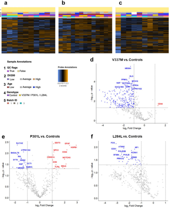
“A recent study on the differences in pathology between MAPT coding-variant patients included gene expression profiling on ~700 genes associated with neurodegeneration 41 .”
From Tau is an m 6 A-associated RNA binding protein that causes RNA dysregulation in disease (2026) · cites this paper as [41]
In the citing paper
“This is further corroborated by the significant overlap between transcriptomic changes in our MAPT V337M iNeurons and MAPT V337M patient brain samples 41 ( Figure S6A ).”
From Tau is an m 6 A-associated RNA binding protein that causes RNA dysregulation in disease (2026) · cites this paper as [41]
In the citing paper
“While the authors admit that their dataset is underpowered, the most differentially expressed genes were identified in the MAPT V337M patient tissue.”
From Tau is an m 6 A-associated RNA binding protein that causes RNA dysregulation in disease (2026) · cites this paper as [41]
Self-check
Percentages, sums and reported differences re-computed from the paper’s own numbers.
The self-check needs the paper’s full text.
Citations
Cites
Neuropathological stageing of Alzheimer-related changes.
Braak, H., & Braak, E. (1991) · Acta Neuropathologica · doi:10.1007/bf00308809 · Cited by 16,123
Accurate structure prediction of biomolecular interactions with AlphaFold 3
Abramson, J., Adler, J., Dunger, J., Evans, R., Green, T., Pritzel, A., Ronneberger, O., Willmore, L., Ballard, A. J., Bambrick, J., et al. (2024) · Nature · doi:10.1038/s41586-024-07487-w · Cited by 15,861 · cited 1×
“Predicted structural models of wild-type and mutant tau were generated using alphafold 3 [1].”
Cairns, N. J., Bigio, E. H., Mackenzie, I. R. A., Neumann, M., Lee, V. M.-Y., Hatanpaa, K. J., White, C. L., Schneider, J. A., Grinberg, L. T., Halliday, G., et al. (2007) · Acta Neuropathologica · doi:10.1007/s00401-007-0237-2 · Cited by 1,110
Identification of region-specific astrocyte subtypes at single cell resolution
Batiuk, M. Y., Martirosyan, A., Wahis, J., de Vin, F., Marneffe, C., Kusserow, C., Koeppen, J., Viana, J. F., Oliveira, J. F., Voet, T., et al. (2020) · Nature Communications · doi:10.1038/s41467-019-14198-8 · Cited by 805
the Arizona Parkinson’s Disease Consortium, Beach, T. G., Adler, C. H., Lue, L., Sue, L. I., Bachalakuri, J., Henry-Watson, J., Sasse, J., Boyer, S., Shirohi, S., et al. (2009) · Acta Neuropathologica · doi:10.1007/s00401-009-0538-8 · Cited by 698
Aging-related tau astrogliopathy (ARTAG): Harmonized evaluation strategy
Kovacs, G. G., Ferrer, I., Grinberg, L. T., Alafuzoff, I., Attems, J., Budka, H., Cairns, N. J., Crary, J. F., Duyckaerts, C., Ghetti, B., et al. (2016) · Acta Neuropathologica · doi:10.1007/s00401-015-1509-x · Cited by 532 · cited 1×
“Additional neuropathological analyses included the presence of hippocampal sclerosis (HS), the presence and distribution of beta amyloid pathology, pTDP-43 proteinopathy, age-related tau astrogliopathy (ARTAG), and alpha-synuclein pathology following established guidelines [3, 6, 41, 53, 69].”
Progressive Supranuclear Palsy: Pathology and Genetics
Dickson, D. W., Rademakers, R., & Hutton, M. L. (2007) · Brain Pathology · doi:10.1111/j.1750-3639.2007.00054.x · Cited by 314
Hippocampal sclerosis and TDP ‐43 pathology in aging and Alzheimer disease
Nag, S., Yu, L., Capuano, A. W., Wilson, R. S., Leurgans, S. E., Bennett, D. A., & Schneider, J. A. (2015) · Annals of Neurology · doi:10.1002/ana.24388 · Cited by 305
Transcriptomic cytoarchitecture reveals principles of human neocortex organization
Jorstad, N. L., Close, J., Johansen, N., Yanny, A. M., Barkan, E. R., Travaglini, K. J., Bertagnolli, D., Campos, J., Casper, T., Crichton, K., et al. (2023) · Science · doi:10.1126/science.adf6812 · Cited by 209
Self-citationTau binding protein CAPON induces tau aggregation and neurodegeneration
Hashimoto, S., Matsuba, Y., Kamano, N., Mihira, N., Sahara, N., Takano, J., Muramatsu, S.-I., Saido, T. C., & Saito, T. (2019) · Nature Communications · doi:10.1038/s41467-019-10278-x · Cited by 111
Research works printed but not linked: 84
- Ahmed Z, Bigio EH, Budka H, Dickson DW, Ferrer I, Ghetti B et al (2013) Globular glial tauopathies (GGT): consensus recommendations. Acta Neuropathol (Berl) 126:537–544. https://doi.org/10.1007/s00401-013-1171-0 · DOI
- Attems J, Toledo JB, Walker L, Gelpi E, Gentleman S, Halliday G et al (2021) Neuropathological consensus criteria for the evaluation of Lewy pathology in post-mortem brains: a multi-centre study. Acta Neuropathol (Berl) 141:159–172. https://doi.org/10.1007/s00401-020-02255-2 · DOI
- Badihian N, Tosakulwong N, Weigand SD, Ali F, Clark HM, Stierwalt J et al (2024) Relationships between regional burden of tau pathology and age at death and disease duration in PSP. Parkinsonism Relat Disord 127:107109. https://doi.org/10.1016/j.parkreldis.2024.107109 · DOI
- Bird TD, Nochlin D, Poorkaj P, Cherrier M, Kaye J, Payami H et al (1999) A clinical pathological comparison of three families with frontotemporal dementia and identical mutations in the tau gene (P301L). Brain 122:741–756. https://doi.org/10.1093/brain/122.4.741 · DOI
- Bird TD, Wijsman EM, Nochlin D, Leehey M, Sumi SM, Payami H et al (1997) Chromosome 17 and hereditary dementia: linkage studies in three non-Alzheimer families and kindreds with late-onset FAD. Neurology 48:949–954. https://doi.org/10.1212/wnl.48.4.949 · DOI
- Borrego-Écija S, Morgado J, Palencia-Madrid L, Grau-Rivera O, Reñé R, Hernández I et al (2017) Frontotemporal dementia caused by the P301L mutation in the MAPT gene: clinicopathological features of 13 cases from the same geographical origin in Barcelona, Spain. Dement Geriatr Cogn Disord 44:213–221. https://doi.org/10.1159/000480077 · DOI
- Carlos AF, Tosakulwong N, Weigand SD, Buciuc M, Ali F, Clark HM et al (2022) Histologic lesion type correlates of magnetic resonance imaging biomarkers in four-repeat tauopathies. Brain Commun 4:fcac108. https://doi.org/10.1093/braincomms/fcac108 · DOI
- Chambraud B, Daguinot C, Guillemeau K, Genet M, Dounane O, Meduri G et al (2021) Decrease of neuronal FKBP4/FKBP52 modulates perinuclear lysosomal positioning and MAPT/Tau behavior during MAPT/Tau-induced proteotoxic stress. Autophagy 17:3491–3510. https://doi.org/10.1080/15548627.2021.1875611 · DOI
- Chung D-EC, Roemer S, Petrucelli L, Dickson DW (2021) Cellular and pathological heterogeneity of primary tauopathies. Mol Neurodegener 16:57. https://doi.org/10.1186/s13024-021-00476-x · DOI
- Crowther RA, Goedert M (2000) Abnormal tau-containing filaments in neurodegenerative diseases. J Struct Biol 130:271–279. https://doi.org/10.1006/jsbi.2000.4270 · DOI
- Cudaback E, Yang Y, Montine TJ, Keene CD (2015) APOE genotype-dependent modulation of astrocyte chemokine CCL3 production. Glia 63:51–65. https://doi.org/10.1002/glia.22732 · DOI
- Dickson DW, Bergeron C, Chin SS, Duyckaerts C, Horoupian D, Ikeda K et al (2002) Office of rare diseases neuropathologic criteria for corticobasal degeneration. J Neuropathol Exp Neurol 61:935–946. https://doi.org/10.1093/jnen/61.11.935 · DOI · Paper page
- Dickson DW, Kouri N, Murray ME, Josephs KA (2011) Neuropathology of frontotemporal lobar degeneration-tau (FTLD-Tau). J Mol Neurosci 45:384–389. https://doi.org/10.1007/s12031-011-9589-0 · DOI · Paper page
- Domoto-Reilly K, Davis MY, Keene CD, Bird TD (2017) Unusually long duration and delayed penetrance in a family with FTD and mutation in MAPT (V337M). Am J Med Genet B Neuropsychiatr Genet 174:70–74. https://doi.org/10.1002/ajmg.b.32443 · DOI
- D’Souza I, Poorkaj P, Hong M, Nochlin D, Lee VM-Y, Bird TD et al (1999) Missense and silent tau gene mutations cause frontotemporal dementia with parkinsonism-chromosome 17 type, by affecting multiple alternative RNA splicing regulatory elements. Proc Natl Acad Sci USA 96:5598–5603. https://doi.org/10.1073/pnas.96.10.5598 · DOI
- Dugger BN, Dickson DW (2017) Pathology of neurodegenerative diseases. Cold Spring Harb Perspect Biol 9:a028035. https://doi.org/10.1101/cshperspect.a028035 · DOI · Paper page
- Ferrer I, Andrés-Benito P, Zelaya MV, Aguirre MEE, Carmona M, Ausín K et al (2020) Familial globular glial tauopathy linked to MAPT mutations: molecular neuropathology and seeding capacity of a prototypical mixed neuronal and glial tauopathy. Acta Neuropathol (Berl) 139:735–771. https://doi.org/10.1007/s00401-019-02122-9 · DOI
- Ferrer I, Lopez-Gonzalez I, Carmona M, Arregui L, Dalfo E, Torrejon-Escribano B et al (2014) Glial and neuronal tau pathology in tauopathies: characterization of disease-specific phenotypes and tau pathology progression. J Neuropathol Exp Neurol. https://doi.org/10.1097/nen.0000000000000030 · DOI · Paper page
- Forrest SL, Kril JJ, Halliday GM (2019) Cellular and regional vulnerability in frontotemporal tauopathies. Acta Neuropathol (Berl) 138:705–727. https://doi.org/10.1007/s00401-019-02035-7 · DOI
- Forrest SL, Kril JJ, Stevens CH, Kwok JB, Hallupp M, Kim WS et al (2018) Retiring the term FTDP-17 as MAPT mutations are genetic forms of sporadic frontotemporal tauopathies. Brain J Neurol 141:521–534. https://doi.org/10.1093/brain/awx328 · DOI
- Ghetti B, Oblak AL, Boeve BF, Johnson KA, Dickerson BC, Goedert M (2015) Invited review: frontotemporal dementia caused by microtubule-associated protein tau gene (MAPT) mutations: a chameleon for neuropathology and neuroimaging. Neuropathol Appl Neurobiol 41:24–46. https://doi.org/10.1111/nan.12213 · DOI
- Giannini LA, Mol MO, Rajicic A, van Buuren R, Sarkar L, Arezoumandan S et al (2023) Presymptomatic and early pathological features of MAPT-associated frontotemporal lobar degeneration. Acta Neuropathol Commun 11:126. https://doi.org/10.1186/s40478-023-01588-9 · DOI
- Giannini LAA, Ohm DT, Rozemuller AJM, Dratch L, Suh E, van Deerlin VM et al (2022) Isoform-specific patterns of tau burden and neuronal degeneration in MAPT-associated frontotemporal lobar degeneration. Acta Neuropathol (Berl) 144:1065–1084. https://doi.org/10.1007/s00401-022-02487-4 · DOI
- Gibbons GS, Banks RA, Kim B, Changolkar L, Riddle DM, Leight SN et al (2018) Detection of Alzheimer disease (AD)-specific tau pathology in AD and NonAD Tauopathies by immunohistochemistry with novel conformation-selective tau antibodies. J Neuropathol Exp Neurol 77:216–228. https://doi.org/10.1093/jnen/nly010 · DOI · Paper page
- Goedert M (2005) Tau gene mutations and their effects. Mov Disord Off J Mov Disord Soc 20(Suppl 12):S45-52. https://doi.org/10.1002/mds.20539 · DOI
- Goedert M, Crowther RA, Scheres SHW, Spillantini MG (2024) Tau and neurodegeneration. Cytoskeleton 81:95–102. https://doi.org/10.1002/cm.21812 · DOI
- Goedert M, Spillantini MG, Falcon B, Zhang W, Newell KL, Hasegawa M et al (2021) Tau protein and frontotemporal dementias. Adv Exp Med Biol 1281:177–199. https://doi.org/10.1007/978-3-030-51140-1_12 · DOI
- Guo T, Noble W, Hanger DP (2017) Roles of tau protein in health and disease. Acta Neuropathol (Berl) 133:665–704. https://doi.org/10.1007/s00401-017-1707-9 · DOI · Paper page
- Hauw JJ, Daniel SE, Dickson D, Horoupian DS, Jellinger K, Lantos PL et al (1994) Preliminary NINDS neuropathologic criteria for Steele-Richardson-Olszewski syndrome (progressive supranuclear palsy). Neurology 44:2015–2019. https://doi.org/10.1212/wnl.44.11.2015 · DOI
- Howat WJ, Wilson BA (2014) Tissue fixation and the effect of molecular fixatives on downstream staining procedures. Methods San Diego Calif 70:12–19. https://doi.org/10.1016/j.ymeth.2014.01.022 · DOI
- Howe S, McCann H, Xiong Z, Nguyen Q, Steyn FJ, Ngo ST et al (2025) Long-term fixation impact on archived human nervous tissues for sequencing-based transcriptomics. Brain Commun 7:fcaf428. https://doi.org/10.1093/braincomms/fcaf428 · DOI
- Hyman BT, Phelps CH, Beach TG, Bigio EH, Cairns NJ, Carrillo MC et al (2012) National Institute on Aging–Alzheimer’s Association guidelines for the neuropathologic assessment of Alzheimer’s disease. Alzheimers Dement 8:1–13. https://doi.org/10.1016/j.jalz.2011.10.007 · DOI · Paper page
- Irwin DJ, Cairns NJ, Grossman M, McMillan CT, Lee EB, Van Deerlin VM et al (2015) Frontotemporal lobar degeneration: defining phenotypic diversity through personalized medicine. Acta Neuropathol (Berl) 129:469–491. https://doi.org/10.1007/s00401-014-1380-1 · DOI
- Jicha GA, Nelson PT (2019) Hippocampal sclerosis, argyrophilic grain disease, and primary age-related tauopathy. Continuum 25:208–233. https://doi.org/10.1212/CON.0000000000000697 · DOI
- Josephs KA, Dickson DW, Tosakulwong N, Weigand SD, Murray ME, Petrucelli L et al (2017) Rates of hippocampal atrophy and post-mortem TDP-43 in Alzheimer’s disease: a longitudinal retrospective study. Lancet Neurol 16:917–924. https://doi.org/10.1016/S1474-4422(17)30284-3 · DOI · Paper page
- Josephs KA, Hodges JR, Snowden JS, Mackenzie IR, Neumann M, Mann DM et al (2011) Neuropathological background of phenotypical variability in frontotemporal dementia. Acta Neuropathol (Berl) 122:137–153. https://doi.org/10.1007/s00401-011-0839-6 · DOI
- Josephs KA, Whitwell JL, Knopman DS, Hu WT, Stroh DA, Baker M et al (2008) Abnormal TDP-43 immunoreactivity in AD modifies clinicopathologic and radiologic phenotype. Neurology 70:1850–1857. https://doi.org/10.1212/01.wnl.0000304041.09418.b1 · DOI
- Komuro Y, Xu G, Bhaskar K, Lamb BT (2015) Human tau expression reduces adult neurogenesis in a mouse model of tauopathy. Neurobiol Aging 36:2034–2042. https://doi.org/10.1016/j.neurobiolaging.2015.03.002 · DOI
- Koriath C, Lashley T, Taylor W, Druyeh R, Dimitriadis A, Denning N et al (2019) ApoE4 lowers age at onset in patients with frontotemporal dementia and tauopathy independent of amyloid-β copathology. Alzheimers Dement Diagn Assess Dis Monit 11:277–280. https://doi.org/10.1016/j.dadm.2019.01.010 · DOI
- Kouri N, Murray ME, Reddy JS, Serie DJ, Soto-Beasley A, Allen M et al (2021) Latent trait modeling of tau neuropathology in progressive supranuclear palsy. Acta Neuropathol (Berl) 141:667–680. https://doi.org/10.1007/s00401-021-02289-0 · DOI
- Kovacs GG (2015) Invited review: neuropathology of tauopathies: principles and practice. Neuropathol Appl Neurobiol 41:3–23. https://doi.org/10.1111/nan.12208 · DOI · Paper page
- Kovacs GG (2019) Molecular pathology of neurodegenerative diseases: principles and practice. J Clin Pathol 72:725–735. https://doi.org/10.1136/jclinpath-2019-205952 · DOI
- Kovacs GG, Lee VM, Trojanowski JQ (2017) Protein astrogliopathies in human neurodegenerative diseases and aging. Brain Pathol 27:675–690. https://doi.org/10.1111/bpa.12536 · DOI
- Kovacs GG, Lukic MJ, Irwin DJ, Arzberger T, Respondek G, Lee EB et al (2020) Distribution patterns of tau pathology in progressive supranuclear palsy. Acta Neuropathol (Berl) 140:99–119. https://doi.org/10.1007/s00401-020-02158-2 · DOI · Paper page
- Latimer CS, Burke BT, Liachko NF, Currey HN, Kilgore MD, Gibbons LE et al (2019) Resistance and resilience to Alzheimer’s disease pathology are associated with reduced cortical pTau and absence of limbic-predominant age-related TDP-43 encephalopathy in a community-based cohort. Acta Neuropathol Commun 7:91. https://doi.org/10.1186/s40478-019-0743-1 · DOI
- Latimer CS, Liachko NF (2021) Tau and TDP-43 synergy: a novel therapeutic target for sporadic late-onset Alzheimer’s disease. GeroScience 43:1627–1634. https://doi.org/10.1007/s11357-021-00407-0 · DOI
- Latimer CS, Melief EJ, Ariza-Torres J, Howard K, Keen AR, Keene LM et al (2023) Protocol for the systematic fixation, circuit-based sampling, and qualitative and quantitative neuropathological analysis of human brain tissue. Methods Mol Biol Clifton NJ 2561:3–30. https://doi.org/10.1007/978-1-0716-2655-9_1 · DOI
- Lester E, Ooi FK, Bakkar N, Ayers J, Woerman AL, Wheeler J et al (2021) Tau aggregates are RNA-protein assemblies that mislocalize multiple nuclear speckle components. Neuron 109:1675-1691.e9. https://doi.org/10.1016/j.neuron.2021.03.026 · DOI · Paper page
- Li D, Zand MS, Dye TD, Goniewicz ML, Rahman I, Xie Z (2022) An evaluation of RNA-seq differential analysis methods. PLoS ONE 17:e0264246. https://doi.org/10.1371/journal.pone.0264246 · DOI
- Lin L-C, Nana AL, Hepker M, Hwang J-HL, Gaus SE, Spina S et al (2019) Preferential tau aggregation in von Economo neurons and fork cells in frontotemporal lobar degeneration with specific MAPT variants. Acta Neuropathol Commun 7:159. https://doi.org/10.1186/s40478-019-0809-0 · DOI
- Mackenzie IRA, Neumann M (2016) Molecular neuropathology of frontotemporal dementia: insights into disease mechanisms from postmortem studies. J Neurochem 138:54–70. https://doi.org/10.1111/jnc.13588 · DOI
- McMillan PJ, Benbow SJ, Uhrich R, Saxton A, Baum M, Strovas T et al (2023) Tau–RNA complexes inhibit microtubule polymerization and drive disease-relevant conformation change. Brain 146:3206–3220. https://doi.org/10.1093/brain/awad032 · DOI · Paper page
- Miki T, Yokota O, Takenoshita S, Mori Y, Yamazaki K, Ozaki Y et al (2018) Frontotemporal lobar degeneration due to P301L tau mutation showing apathy and severe frontal atrophy but lacking other behavioral changes: a case report and literature review. Neuropathol Off J Jpn Soc Neuropathol 38:268–280. https://doi.org/10.1111/neup.12441 · DOI
- Mirra SS, Murrell JR, Gearing M, Spillantini MG, Goedert M, Crowther RA et al (1999) Tau pathology in a family with dementia and a P301L mutation in tau. J Neuropathol Exp Neurol 58:335–345. https://doi.org/10.1097/00005072-199904000-00004 · DOI · Paper page
- Mishra A, Ferrari R, Heutink P, Hardy J, Pijnenburg Y, Posthuma D et al (2017) Gene-based association studies report genetic links for clinical subtypes of frontotemporal dementia. Brain 140:1437–1446. https://doi.org/10.1093/brain/awx066 · DOI
- Nelson PT, Dickson DW, Trojanowski JQ, Jack CR, Boyle PA, Arfanakis K et al (2019) Limbic-predominant age-related TDP-43 encephalopathy (LATE): consensus working group report. Brain 142:1503–1527. https://doi.org/10.1093/brain/awz099 · DOI · Paper page
- Neumann M, Sampathu DM, Kwong LK, Truax AC, Micsenyi MC, Chou TT et al (2006) Ubiquitinated TDP-43 in frontotemporal lobar degeneration and amyotrophic lateral sclerosis. Science 314:130–133. https://doi.org/10.1126/science.1134108 · DOI · Paper page
- Ohm DT, Cousins KAQ, Xie SX, Peterson C, McMillan CT, Massimo L et al (2022) Signature laminar distributions of pathology in frontotemporal lobar degeneration. Acta Neuropathol (Berl) 143:363–382. https://doi.org/10.1007/s00401-021-02402-3 · DOI
- Ohm DT, Xie SX, Capp N, Arezoumandan S, Cousins KAQ, Rascovsky K et al (2025) Cytoarchitectonic gradients of laminar degeneration in behavioural variant frontotemporal dementia. Brain 148:102–118. https://doi.org/10.1093/brain/awae263 · DOI
- Poorkaj P, Bird TD, Wijsman E, Nemens E, Garruto RM, Anderson L et al (1998) Tau is a candidate gene for chromosome 17 frontotemporal dementia. Ann Neurol 43:815–825. https://doi.org/10.1002/ana.410430617 · DOI · Paper page
- Poorkaj P, Grossman M, Steinbart E, Payami H, Sadovnick A, Nochlin D et al (2001) Frequency of Tau gene mutations in familial and sporadic cases of non-Alzheimer dementia. Arch Neurol 58:383–387. https://doi.org/10.1001/archneur.58.3.383 · DOI · Paper page
- Qi C, Lövestam S, Murzin AG, Peak-Chew S, Franco C, Bogdani M et al (2025) Tau filaments with the Alzheimer fold in human MAPT mutants V337M and R406W. Nat Struct Mol Biol 32:1297–1304. https://doi.org/10.1038/s41594-025-01498-5 · DOI
- Rademakers R, Cruts M, van Broeckhoven C (2004) The role of tau (MAPT) in frontotemporal dementia and related tauopathies. Hum Mutat 24:277–295. https://doi.org/10.1002/humu.20086 · DOI
- Rockland KS (2017) Anatomy of the cerebral cortex. Neuropsychiatric disorders. Academic Press, pp 3–36
- Schweighauser M, Shi Y, Murzin AG, Garringer HJ, Vidal R, Murrell JR et al (2025) Distinct tau filament folds in human MAPT mutants P301L and P301T. Nat Struct Mol Biol 32:1470–1478. https://doi.org/10.1038/s41594-025-01575-9 · DOI
- Seeley WW (2008) Selective functional, regional, and neuronal vulnerability in frontotemporal dementia. Curr Opin Neurol 21:701–707. https://doi.org/10.1097/WCO.0b013e3283168e2d · DOI
- Shi Y, Yamada K, Liddelow SA, Smith ST, Zhao L, Luo W et al (2017) ApoE4 markedly exacerbates tau-mediated neurodegeneration in a mouse model of tauopathy. Nature 549:523–527. https://doi.org/10.1038/nature24016 · DOI · Paper page
- Shiarli A-M, Jennings R, Shi J, Bailey K, Davidson Y, Tian J et al (2006) Comparison of extent of tau pathology in patients with frontotemporal dementia with Parkinsonism linked to chromosome 17 (FTDP-17), frontotemporal lobar degeneration with Pick bodies and early onset Alzheimer’s disease. Neuropathol Appl Neurobiol 32:374–387. https://doi.org/10.1111/j.1365-2990.2006.00736.x · DOI
- Siletti K, Hodge R, Mossi Albiach A, Lee KW, Ding S-L, Hu L et al (2023) Transcriptomic diversity of cell types across the adult human brain. Science 382:eadd7046. https://doi.org/10.1126/science.add7046 · DOI · Paper page
- Spillantini MG, Bird TD, Ghetti B (1998) Frontotemporal dementia and Parkinsonism linked to chromosome 17: a new group of tauopathies. Brain Pathol 8:387–402. https://doi.org/10.1111/j.1750-3639.1998.tb00162.x · DOI
- Spillantini MG, Goedert M (2013) Tau pathology and neurodegeneration. Lancet Neurol 12:609–622. https://doi.org/10.1016/S1474-4422(13)70090-5 · DOI · Paper page
- Spillantini MG, Van Swieten JC, Goedert M (2000) Tau gene mutations in frontotemporal dementia and Parkinsonism linked to chromosome 17 (FTDP-17). Neurogenetics 2:193–205. https://doi.org/10.1007/pl00022972 · DOI
- Spina S, Schonhaut DR, Boeve BF, Seeley WW, Ossenkoppele R, O’Neil JP et al (2017) Frontotemporal dementia with the V337M MAPT mutation. Neurology 88:758–766. https://doi.org/10.1212/WNL.0000000000003636 · DOI · Paper page
- Strang KH, Golde TE, Giasson BI (2019) MAPT mutations, tauopathy, and mechanisms of neurodegeneration. Lab Invest 99:912–928. https://doi.org/10.1038/s41374-019-0197-x · DOI · Paper page
- Strittmatter WJ, Weisgraber KH, Goedert M, Saunders AM, Huang D, Corder EH et al (1994) Hypothesis: microtubule instability and paired helical filament formation in the Alzheimer disease brain are related to apolipoprotein E genotype. Exp Neurol 125:163–171. https://doi.org/10.1006/exnr.1994.1019. (discussion 172–174) · DOI
- Sumi SM, Bird TD, Nochlin D, Raskind MA (1992) Familial presenile dementia with psychosis associated with cortical neurofibrillary tangles and degeneration of the amygdala. Neurology 42:120–120. https://doi.org/10.1212/WNL.42.1.120 · DOI · Paper page
- Tacik P, Sanchez-Contreras M, DeTure M, Murray ME, Rademakers R, Ross OA et al (2017) Clinicopathologic heterogeneity in frontotemporal dementia and parkinsonism linked to chromosome 17 (FTDP‐17) due to microtubule‐associated protein tau (MAPT) p.P301L mutation, including a patient with globular glial tauopathy. Neuropathol Appl Neurobiol 43(3):200–214. https://doi.org/10.1111/nan.12367 · DOI
- Taylor LM, McMillan PJ, Liachko NF, Strovas TJ, Ghetti B, Bird TD et al (2018) Pathological phosphorylation of tau and TDP-43 by TTBK1 and TTBK2 drives neurodegeneration. Mol Neurodegener 13:7. https://doi.org/10.1186/s13024-018-0237-9 · DOI
- Tomé SO, Tsaka G, Ronisz A, Ospitalieri S, Gawor K, Gomes LA et al (2023) TDP-43 pathology is associated with increased tau burdens and seeding. Mol Neurodegener 18:71. https://doi.org/10.1186/s13024-023-00653-0 · DOI
- Trabzuni D, Wray S, Vandrovcova J, Ramasamy A, Walker R, Smith C et al (2012) MAPT expression and splicing is differentially regulated by brain region: relation to genotype and implication for tauopathies. Hum Mol Genet 21:4094–4103. https://doi.org/10.1093/hmg/dds238 · DOI · Paper page
- Wang Y, Mandelkow E (2016) Tau in physiology and pathology. Nat Rev Neurosci 17:5–21. https://doi.org/10.1038/nrn.2015.1 · DOI · Paper page
- Wolfe MS (2009) Tau mutations in neurodegenerative diseases. J Biol Chem 284:6021–6025. https://doi.org/10.1074/jbc.R800013200 · DOI
- Woodworth DC, Lou JJ, Yong WH, Head E, Corrada MM, Nelson PT et al (2025) Common neuropathologic change drivers of hippocampal sclerosis of ageing. Brain 148:2400–2411. https://doi.org/10.1093/brain/awaf158 · DOI
- Xia Y, Sorrentino ZA, Kim JD, Strang KH, Riffe CJ, Giasson BI (2019) Impaired tau-microtubule interactions are prevalent among pathogenic tau variants arising from missense mutations. J Biol Chem 294:18488–18503. https://doi.org/10.1074/jbc.RA119.010178 · DOI
Cited by
Tau is an m 6 A-associated RNA binding protein that causes RNA dysregulation in disease
Dixon, G., Keys, A., Rothamel, K., McKetney, J., Jiang, Y. X., Gordon, L. A., Page, M. L., Liu, C., Hou, K., Flanagan, K., et al. (2026) · doi:10.64898/2026.10.26.762095
Metadata
- Authors
- Bogdani, M., Jadhav, V. S., Kraemer, B. C., Grabowski, T. J., Jayadev, S., Domoto-Reilly, K., Liachko, N. F., Bird, T. D., Dirk Keene, C., & Latimer, C. S.
- ORCID
- Latimer, Caitlin S.
- Venue
- Acta Neuropathologica
- Published
- 2026-07-03
- Volume
- 152
- Issue
- 1
- ISSN
- 1432-0533
- PMC
- PMC13331856
- Cited by
- 1
- References
- 12
- Language
- en
- Licence
- Funders
- U.S. Department of Veterans Affairs (IK6BX006467, I01BX005762)
- National Institutes of Health (RF1AG078374, P30 AG066509, U19AG063911, R01AG066729, U19 AG066567, R01AG090551)
- U.S. Department of Defense (W81XWH-21-S-TBIPH2)
About these checks
Registry, retraction, Cites and statistics results are computed, not judged, with one exception: a printed reference that no index lists is read once by a model before it counts as a citation error. Claims are extracted by a model and kept only when their quote appears verbatim in the text; each cited source is then checked, with a model’s help, for whether it says what the claim relies on. Those verdicts can be wrong — read the claim’s quote and the cited source before relying on one.
Statistics and arithmetic are recomputed, not judged.
Confidence bands are shown only for fields calibrated on our annotated test set. None is calibrated yet, so no band is shown.
What counts as a claim ›How we measure confidence ›How we check this ›
Are you an author, or do you see a problem with this page? Request a review
Data sources
Bibliographic data: Semantic Scholar, OpenAlex, Crossref.
Contains information from the Semantic Scholar Academic Graph Datasets, made available under the ODC Attribution License (ODC-BY 1.0).
Semantic Scholar’s data is described in: Kinney, R., et al. (2023). The Semantic Scholar Open Data Platform. arXiv:2301.10140.
