

The DOI matches its Crossref record; 72 of the 72 references it cites were found in a registry or an index.
Dysfunction of NMDA receptors in Alzheimer’s disease
Open accessAbstract
N-methyl-D-aspartate receptors (NMDARs) play a pivotal role in the synaptic transmission and synaptic plasticity thought to underlie learning and memory. NMDARs activation has been recently implicated in Alzheimer's disease (AD) related to synaptic dysfunction. Synaptic NMDARs are neuroprotective, whereas overactivation of NMDARs located outside of the synapse cause loss of mitochondrial membrane potential and cell death. NMDARs dysfunction in the glutamatergic tripartite synapse, comprising presynaptic and postsynaptic neurons and glial cells, is directly involved in AD. This review discusses that both beta-amyloid (Aβ) and tau perturb synaptic functioning of the tripartite synapse, including alterations in glutamate release, astrocytic uptake, and receptor signaling. Particular emphasis is given to the role of NMDARs as a possible convergence point for Aβ and tau toxicity and possible reversible stages of the AD through preventive and/or disease-modifying therapeutic strategies.
Overview
The claims and statistics of this paper haven’t been checked yet.
Run the check — free, no account needed. It reads the open-access text and checks the sources it cites.
Integrity signals
Registry
Cites
Statistics
Figures
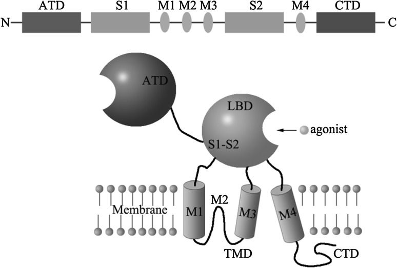
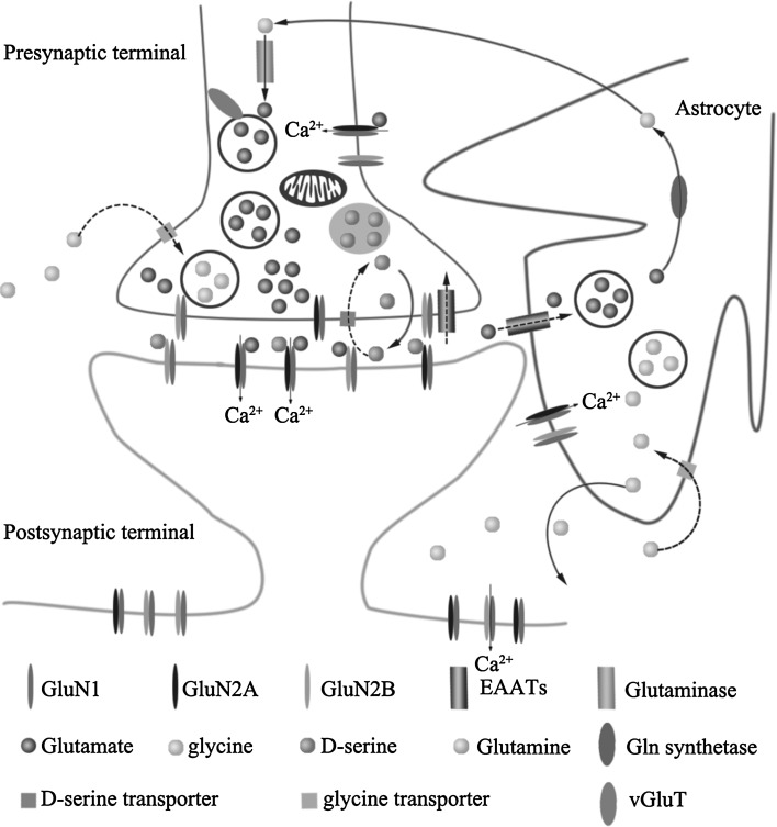
Verification
Claims the paper makes
The claims and statistics of this paper haven’t been checked yet.
Run the check — free, no account needed. It reads the open-access text and checks the sources it cites.
Claims other papers make about this one
Each is checked against this paper’s own text: does it say what the citing paper says it does?
In the citing paper
“The relevance of NMDAR in AD lends support from studies showing that partial blockade of NMDAR overstimulation with NMDAR antagonists rescues Aβ-induced LTP impairment and cognitive function in various animal models [69].”
From Molecular and cellular mechanisms underlying the pathogenesis of Alzheimer’s disease (2020) · cites this paper as [69]
Self-check
Percentages, sums and reported differences re-computed from the paper’s own numbers.
The self-check needs the paper’s full text.
Citations
Cites
1 retracted
Disturbed Neurotransmitter Transporter Expression in Alzheimer’s Disease Brain
Chen, K. H., Reese, E. A., Kim, H., Rapoport, S. I., & Rao, J. S. (2011) · Journal of Alzheimer s Disease · doi:10.3233/jad-2011-110002 · Cited by 122 · cited 1×
“The expression of glutamate transporters is reduced significantly both in human tissue from AD patients [54] and animal models of AD [55].”
BackgroundRetractedAmyloid oligomers exacerbate tau pathology in a mouse model of tauopathy
Selenica, M.-L. B., Brownlow, M., Jimenez, J. P., Lee, D. C., Pena, G., Dickey, C. A., Gordon, M. N., & Morgan, D. (2013) · Neurodegenerative Diseases · doi:10.1159/000337230 · Cited by 10,359
Shrestha, S., & Offer, S. M. (2016) · Medical Epigenetics · doi:10.1159/000444713 · Cited by 8,951
Südhof, T. C. (2004) · Annual Review of Neuroscience · doi:10.1146/annurev.neuro.26.041002.131412 · Cited by 2,491 · cited 1×
“Following presynaptic neuronal depolarization, calcium channels open, permitting the influx of calcium and triggering the fusion of vesicles with the membrane, resulting in the exocytosis of glutamate into the synapse [15].”
BackgroundDendritic Function of Tau Mediates Amyloid-β Toxicity in Alzheimer's Disease Mouse Models
Ittner, L. M., Ke, Y. D., Delerue, F., Bi, M., Gladbach, A., van Eersel, J., Wölfing, H., Chieng, B. C., Christie, M. J., Napier, I. A., et al. (2010) · Cell · doi:10.1016/j.cell.2010.06.036 · Cited by 1,821 · cited 1×
“Tau reductions cannot transport Fyn to dendritic spines, prevent the memory deficits and excitability caused by A b [58].”
BackgroundSynaptic versus extrasynaptic NMDA receptor signalling: implications for neurodegenerative disorders
Hardingham, G. E., & Bading, H. (2010) · Nature Reviews Neuroscience · doi:10.1038/nrn2911 · Cited by 1,644 · cited 1×
“…can also be taken up by the presynaptic terminal or astrocytes by their respective transporters Activation of synaptic NMDARs mediates synaptic plasticity and is thought to be beneficial for neurons, whereas prolonged activation of NMDARs with subsequent overload of Ca 2 ? entry is deleterious [32].”
BackgroundShankar, G. M., Bloodgood, B. L., Townsend, M., Walsh, D. M., Selkoe, D. J., & Sabatini, B. L. (2007) · The Journal of Neuroscience · doi:10.1523/jneurosci.4970-06.2007 · Cited by 1,592 · cited 2×
“More-over, treatment of rat organotypic slices containing pyramidal neurons with A b oligomers decrease dendritic spine density and reduce NMDAR-mediated calcium influx into active spines [50].”
BackgroundRegulation of NMDA receptor trafficking by amyloid-β
Snyder, E. M., Nong, Y., Almeida, C. G., Paul, S., Moran, T., Choi, E. Y., Nairn, A. C., Salter, M. W., Lombroso, P. J., Gouras, G. K., et al. (2005) · Nature Neuroscience · doi:10.1038/nn1503 · Cited by 1,561 · cited 1×
“When applied to cultured cortical neurons, Ab enhances endocytosis of NMDARs and decreases expression of NMDARs [53].”
Long-term potentiation depends on release of d-serine from astrocytes
Henneberger, C., Papouin, T., Oliet, S. H. R., & Rusakov, D. A. (2010) · Nature · doi:10.1038/nature08673 · Cited by 1,389 · cited 1×
“The increased influx of calcium transform into chemical signals which in turn, leading to the establishment of LTP, a process believed to underlie learning and memory [18].”
BackgroundHoover, B. R., Reed, M. N., Su, J., Penrod, R. D., Kotilinek, L. A., Grant, M. K., Pitstick, R., Carlson, G. A., Lanier, L. M., Yuan, L.-L., et al. (2010) · Neuron · doi:10.1016/j.neuron.2010.11.030 · Cited by 1,118 · cited 1×
“Tau phosphorylation results in mislocalization of tau from axons to dendritic spines and decreased expression of AMPA receptors and LTP deficits, and subsequently synaptic impairment [56].”
Background
Cited by
Sorted by citations
Molecular and cellular mechanisms underlying the pathogenesis of Alzheimer’s disease
Guo, T., Zhang, D., Zeng, Y., Huang, T. Y., Xu, H., & Zhao, Y. (2020) · Molecular Neurodegeneration · doi:10.1186/s13024-020-00391-7 · Cited by 994
“The relevance of NMDAR in AD lends support from studies showing that partial blockade of NMDAR overstimulation with NMDAR antagonists rescues Aβinduced LTP impairment and cognitive function in various animal models [69].”
BackgroundAlzheimer’s disease: Recent treatment strategies
Vaz, M., & Silvestre, S. (2020) · European Journal of Pharmacology · doi:10.1016/j.ejphar.2020.173554 · Cited by 547
Costa, L. G., Cole, T. B., Dao, K., Chang, Y. C., Coburn, J. L., & Garrick, J. M. (2020) · Pharmacology & Therapeutics · doi:10.1016/j.pharmthera.2020.107523 · Cited by 511
Calcium signaling and molecular mechanisms underlying neurodegenerative diseases
Pchitskaya, E. I., Александровна, П. Е., & Борисович, Б. И. (2017) · Cell Calcium · doi:10.1016/j.ceca.2017.06.008 · Cited by 355
“In case of hippocampus, TRPC6 and Orai2 were recently identified as SOCE-forming channels in hippocampal neurons (Zhang et al., 2016a).”
BackgroundInfluentialSharma, P., Srivastava, P., Seth, A., Tripathi, P. N., Banerjee, A. G., & Shrivastava, S. K. (2019) · Progress in Neurobiology · doi:10.1016/j.pneurobio.2018.12.006 · Cited by 331
Phytochemicals as inhibitors of NF-κB for treatment of Alzheimer’s disease
Seo, E.-J., Fischer, N., & Efferth, T. (2018) · Pharmacological Research · doi:10.1016/j.phrs.2017.11.030 · Cited by 265
“The loss of mitochondrial membrane potential causing cell death, which is directly involved in AD pathogenesis, is attenuated [86].”
BackgroundDepression in neurodegenerative diseases: Common mechanisms and current treatment options
Galts, C., Bettio, L. E. B., Jewett, D. C., Yang, C. C., Brocardo, P. S., Rodrigues, A. L. S., Thacker, J. S., & Gil‐Mohapel, J. (2019) · Neuroscience & Biobehavioral Reviews · doi:10.1016/j.neubiorev.2019.04.002 · Cited by 278
Safiri, S., Jolfayi, A. G., Fazlollahi, A., Morsali, S., Sarkesh, A., Sorkhabi, A. D., Golabi, B., Aletaha, R., Asghari, K. M., Hamidi, S., et al. (2024) · Frontiers in Medicine · doi:10.3389/fmed.2024.1474043 · Cited by 273
“NMDA receptor dysfunction, involving pre-and postsynaptic neurons and glial cells, directly affects AD (375).”
Temido‐Ferreira, M., Ferreira, D. G., Batalha, V. L., Marques-Morgado, I., Coelho, J. E., Pereira, P., Gomes, R., Pinto, A., Carvalho, S., Canas, P. M., et al. (2018) · Molecular Psychiatry · doi:10.1038/s41380-018-0110-9 · Cited by 194
Yu, S. P., Jiang, M. Q., Shim, S. S., Pourkhodadad, S., & Wei, L. (2023) · Molecular Neurodegeneration · doi:10.1186/s13024-023-00636-1 · Cited by 178
The basis of cellular and regional vulnerability in Alzheimer’s disease
Mrdjen, D., Fox, E. J., Bukhari, S. A., Montine, K. S., Bendall, S. C., & Montine, T. J. (2019) · Acta Neuropathologica · doi:10.1007/s00401-019-02054-4 · Cited by 162
NMDA and AMPA receptors dysregulation in Alzheimer's disease
Babaei, P. (2021) · European Journal of Pharmacology · doi:10.1016/j.ejphar.2021.174310 · Cited by 155
Pál, B. Z. (2018) · Cellular and Molecular Life Sciences · doi:10.1007/s00018-018-2837-5 · Cited by 140
Findley, C. A., Bartke, A., Hascup, K. N., & Hascup, E. R. (2019) · ASN NEURO · doi:10.1177/1759091419855541 · Cited by 133
“(Hardingham et al., 2002; Hardingham and Bading, 2003; Bezprozvanny and Mattson, 2008; Zhang et al., 2016).”
InfluentialKabir, M. T., Sufian, M. A., Uddin, M. S., Begum, M. M., Akhter, S., Islam, A., Mathew, B., Islam, M. S., Amran, M. S., & Ashraf, G. M. (2019) · Current Pharmaceutical Design · doi:10.2174/1381612825666191011102444 · Cited by 122
Neuroinflammation in Neurodegenerative Disorders: Current Knowledge and Therapeutic Implications
Giri, P. M., Banerjee, A., Ghosal, A., & Layek, B. (2024) · International Journal of Molecular Sciences · doi:10.3390/ijms25073995 · Cited by 129
“Overactivation of NMDA receptors has been observed in AD patients, leading to a higher level of glutamate in the brain [28].”
BackgroundCalcium-Handling Defects and Neurodegenerative DiseasePreprint
Schrank, S., Barrington, N. M., & Stutzmann, G. E. (2019) · Cold Spring Harbor Perspectives in Biology · doi:10.1101/cshperspect.a035212 · Cited by 115
Dysregulation of Intracellular Calcium Signaling in Alzheimer's Disease
Александровна, П. Е., Pchitskaya, E. I., & Борисович, Б. И. (2018) · Antioxidants and Redox Signaling · doi:10.1089/ars.2018.7506 · Cited by 106
“It was reported that in early disease stages, Ab activates NMDAR and induces rapid Ca elevation in neurons (40, 87, 99, 134).”
BackgroundFani, G., Mannini, B., Vecchi, G., Cascella, R., Cecchi, C., Dobson, C. M., Vendruscolo, M., & Chiti, F. (2021) · ACS Chemical Neuroscience · doi:10.1021/acschemneuro.0c00811 · Cited by 106
Torres, L., Robinson, S.-A., Kim, D.-G., Yan, A., Cleland, T. A., & Bynoe, M. S. (2018) · Journal of Neuroinflammation · doi:10.1186/s12974-018-1086-8 · Cited by 97
“The potent downregulation or loss of NMDAR is strongly indicative of the Aβ-induced synaptic depression and impaired synaptic plasticity that is seen in AD [41, 42].”
BackgroundWang, X., & Zheng, W. (2019) · The FASEB Journal · doi:10.1096/fj.201801751r · Cited by 97
“Food and Drug Administration– approved extrasynaptic NMDAR antagonist memantine (107, 108), more therapeutic strategies for AD may also be developed by regulating the NMDAR system.”
BackgroundViel, T., Toricelli, M., Pereira, A. R., Souza Abrao, G., Malerba, H., Maia, J., & Buck, H. (2021) · Neural Regeneration Research · doi:10.4103/1673-5374.286952 · Cited by 102
“…high densities of β-amyloid plaques in the hippocampus are known to induce increased replacement of NMDA Glu-N2A subunits with Glu-N2B (via calpains) facilitating receptor binding to SAP-102, which presents high mobility for extra-synaptic regions (Parsons and Raymond, 2014; Zhang et al., 2016).”
Mira, R. G., & Cerpa, W. F. (2020) · Cellular and Molecular Neurobiology · doi:10.1007/s10571-020-00924-0 · Cited by 98
Jadiya, P., Garbincius, J. F., & Elrod, J. W. (2021) · Acta Neuropathologica Communications · doi:10.1186/s40478-021-01224-4 · Cited by 94
Obesity as a Risk Factor for Alzheimer’s Disease: Implication of Leptin and Glutamate
Lloret, A., Monllor, P., Esteve, D., Cervera‐Ferri, A., & Lloret, M.-Á. (2019) · Frontiers in Neuroscience · doi:10.3389/fnins.2019.00508 · Cited by 94
“Interestingly, in AD the aforementioned overactivation is produced in extrasynaptic NMDA receptors rather than in synaptic NMDA receptors (Zhang et al., 2016).”
BackgroundCurrent Pharmacotherapy and Multi-Target Approaches for Alzheimer’s Disease
Cheong, S. L., Tiew, J. K., Fong, Y. H., Leong, H. W., Chan, Y. M., Chan, Z. L., & Kong, E. W. J. (2022) · Pharmaceuticals · doi:10.3390/ph15121560 · Cited by 94
“Thus, free ROS production is favored, subsequently causing an increase in oxidative stress and altered synapse function [24].”
BackgroundDrug development for Alzheimer’s disease: Review
Lao, K., Ji, N., Zhang, X., Qiao, W., Tang, Z., & Gou, X. (2019) · Journal of drug targeting · doi:10.1080/1061186x.2018.1474361 · Cited by 86
“The NMDARs are cationic channels gated by the neurotransmitter glutamate which play an essential role in excitatory transmission, synaptic integration, learning and memory [13].”
BackgroundGSK-3β, FYN, and DYRK1A: Master Regulators in Neurodegenerative Pathways
Demuro, S., Martino, R. M. C. D., Ortega, J. A., & Cavalli, A. (2021) · International Journal of Molecular Sciences · doi:10.3390/ijms22169098 · Cited by 88
“nMDARs are the primary channel that mediates Ca2+ signals in hippocampal neurons and contribute to the expression of long-term potentiation (LTP) and long-term depression (LTD), which are two major forms of long-lasting synaptic plasticity, by employing both NR2A and NR2B subunits [88].”
BackgroundNaturally Occurring Antioxidant Therapy in Alzheimer’s Disease
Collins, A. E., Saleh, T. M., & Kalisch, B. E. (2022) · Antioxidants · doi:10.3390/antiox11020213 · Cited by 93
Could Alzheimer’s Disease Originate in the Periphery and If So How So?
Morris, G., Berk, M., Maes, M. H. J., & Puri, B. K. (2018) · Molecular Neurobiology · doi:10.1007/s12035-018-1092-y · Cited by 86
“tate (NMDA) receptors, which has been repeatedly reported in AD patients [254], via oxidation of cysteine groups on key”
BackgroundRatan, Y., Rajput, A., Maleysm, S., Pareek, A., Jain, V. K., Pareek, A., Kaur, R., & Singh, G. (2023) · Biomedicines · doi:10.3390/biomedicines11051398 · Cited by 89
“These findings demonstrate that the partial inhibition of NMDAR jaded stimulation with antagonists of NMDAR restores A β -initiated long-term potentiation (LTP) deficits and cognitive performance in distinct animal models, suggesting the importance of NMDAR in AD [99].”
BackgroundCalcium signalling: A common target in neurological disorders and neurogenesis
Glaser, T., Arnaud-Sampaio, V. F., Lameu, C., & Ulrich, H. (2018) · Seminars in Cell and Developmental Biology · doi:10.1016/j.semcdb.2018.12.002 · Cited by 77
Olajide, O. J., Suvanto, M. E., & Chapman, C. A. (2021) · Biology Open · doi:10.1242/bio.056796 · Cited by 77
Gadhave, K., Kumar, D., Uversky, V. N., & Giri, R. (2020) · Medicinal Research Reviews · doi:10.1002/med.21719 · Cited by 71
“Here, synaptic NMDA receptors promote calcium entry leading to the induction of the CREB phosphorylation, whereas extrasynaptic NMDA receptors show the opposite effects and initiate the CREB shut‐off pathway.364 Transcriptional activity of CREB and associated LTP generation are decreased in AD mostly due to the Aβ‐ind…”
BackgroundInfluentialJakaria, M., Park, S., Haque, M. E., Karthivashan, G., Kim, I., Ganesan, P., & Choi, D. (2018) · Frontiers in Molecular Neuroscience · doi:10.3389/fnmol.2018.00307 · Cited by 71
A Novel NMDA Receptor Antagonist Protects against Cognitive Decline Presented by Senescent Mice
Companys‐Alemany, J., Turcu, A. L., Bellver‐Sanchís, A., Loza, M. I., Brea, J. M., Canudas, A. M., Leiva, R., Vázquez, S., Pallàs, M., & Griñán‐Ferré, C. (2020) · Pharmaceutics · doi:10.3390/pharmaceutics12030284 · Cited by 63
“On the other hand, there is accumulating evidence that several neurotransmitter pathways, such as acetylcholine, dopamine, glutamate, and serotonin [12] are involved in the pathological alterations of AD [13,14].”
BackgroundSouza, C. D. S., Grangeiro, M. S., Pereira, É. P. L., dos Santos, C. C., da Silva, A. B., Sampaio, G. P., Figueiredo, D. D. R., David, J. M., David, J. P., da Silva, V. D. A., et al. (2018) · NeuroToxicology · doi:10.1016/j.neuro.2018.02.001 · Cited by 64
The Role of D-Amino Acids in Alzheimer’s Disease
Piubelli, L., Murtas, G., Rabattoni, V., & Pollegioni, L. (2021) · Journal of Alzheimer s Disease · doi:10.3233/jad-201217 · Cited by 63
“As stated above, dysfunctional glutamatergic transmission mediated by altered NMDA receptor activity has been proposed to contribute to the pathogenesis of AD [41, 76, 117].”
Memantine Derivatives as Multitarget Agents in Alzheimer’s Disease
Marotta, G., Basagni, F., Rosini, M., & Minarini, A. (2020) · Molecules · doi:10.3390/molecules25174005 · Cited by 61
Homocysteine and Age-Related Central Nervous System Diseases: Role of Inflammation
Tawfik, A. M., Elsherbiny, N. M., Zaidi, Y., & Rajpurohit, P. (2021) · International Journal of Molecular Sciences · doi:10.3390/ijms22126259 · Cited by 61
“These changes are concomitant with NMDAR activation and oxidative stress, ultimately resulting in AD pathology [30].”
BackgroundRole of GPCR signaling and calcium dysregulation in Alzheimer's disease
Sushma, & Mondal, A. C. (2019) · Molecular and Cellular Neuroscience · doi:10.1016/j.mcn.2019.103414 · Cited by 56
Therapeutic Strategies to Target Calcium Dysregulation in Alzheimer’s Disease
Calvo-Rodríguez, M., Kharitonova, E. K., & Bacskai, B. J. (2020) · Cells · doi:10.3390/cells9112513 · Cited by 60
“Aβ42 peptides interact with NMDARs, potentiating their activity and leading to increased Ca2+ influx, thus contributing to the synapse loss observed in AD [44,45].”
BackgroundChennakesavan, K., Saravanan, N., Essa, M. M., Guillemin, G. J., Jayachandran, S. K., & Anusuyadevi, M. (2018) · Neurological Research · doi:10.1080/01616412.2018.1544745 · Cited by 58
“regulates the NMDARs and disturbs the ionic balance between synaptic and extrasynaptic NMDAR signaling [13,14], but the exact mechanisms underlying Aβ-”
BackgroundRegulation of Melatonin and Neurotransmission in Alzheimer’s Disease
Roy, J., Tsui, K. C., Ng, J., Fung, M., & Lim, L. W. (2021) · International Journal of Molecular Sciences · doi:10.3390/ijms22136841 · Cited by 55
Understanding the Modulatory Effects of Cannabidiol on Alzheimer’s Disease
Xiong, Y., & Lim, C. (2021) · Brain Sciences · doi:10.3390/brainsci11091211 · Cited by 60
“Increased extracellular glutamate excessively activates both synaptic and extrasynaptic N-methyl-D-aspartate (NMDA) receptors, leading to intense transient influx of Ca2+ and activation of signaling pathways responsible for neuronal dysfunction and cell death [6,7].”
BackgroundOlajide, O. J., Gbadamosi, I., Yawson, E. O., Arogundade, T. T., Lewu, F. S., Ogunrinola, K. Y., Adigun, O. O., Bamisi, O., Lambe, E., Arietarhire, L. O., et al. (2021) · Journal of Molecular Neuroscience · doi:10.1007/s12031-020-01747-w · Cited by 58
Role of Receptors in Relation to Plaques and Tangles in Alzheimer’s Disease Pathology
Sharma, K., Pradhan, S., Duffy, L. K., Yeasmin, S., Bhattarai, N., & Schulte, M. K. (2021) · International Journal of Molecular Sciences · doi:10.3390/ijms222312987 · Cited by 59
“The synapse receptor proteins modulate the extracellular glutamate levels and are considered deregulated in AD pathology [64].”
BackgroundMaffioli, E., Murtas, G., Rabattoni, V., Badone, B., Tripodi, F., Iannuzzi, F., Licastro, D., Nonnis, S., Rinaldi, A. M., Motta, Z., et al. (2022) · Cell Reports · doi:10.1016/j.celrep.2022.111271 · Cited by 56
NMDA Receptors: Distribution, Role, and Insights into Neuropsychiatric Disorders
Beaurain, M., Salabert, A., Payoux, P., Gras, E., & Talmont, F. (2024) · Pharmaceuticals · doi:10.3390/ph17101265 · Cited by 57
“The pathophysiology of AD encompasses both structural and functional abnormalities, particularly in the hippocampus and neocortex, which are particularly involved in memory and cognition [143].”
BackgroundIn Vivo Chimeric Alzheimer’s Disease Modeling of Apolipoprotein E4 Toxicity in Human Neurons
Najm, R., Zalocusky, K. A., Zilberter, M., Yoon, S. Y., Hao, Y., Koutsodendris, N., Nelson, M., Rao, A., Taubes, A., Jones, E. A., et al. (2020) · Cell Reports · doi:10.1016/j.celrep.2020.107962 · Cited by 52
Peineau, S., Rabiant, K., Pierrefiche, O., & Potier, B. (2018) · Pharmacological Research · doi:10.1016/j.phrs.2018.01.018 · Cited by 56
Jeon, S. G., Yoo, A., Chun, D. W., Hong, S. B., Chung, H., Kim, J.-I., & Moon, M. (2020) · Aging and Disease · doi:10.14336/ad.2019.0718 · Cited by 57
“Furthermore, under AD conditions, there are other pathways that cause cognitive dysfunction and memory decline, such as abnormal activity of the NMDA receptor [160, 161].”
BackgroundHampel, H. J., Vergallo, A., Caraci, F., Cuello, A. C., Lemercier, P., Vellas, B. J., Giudici, K. V., Baldacci, F., Hänisch, B., Haberkamp, M., et al. (2020) · Neuropharmacology · doi:10.1016/j.neuropharm.2020.108081 · Cited by 52
Binder, J., Ursu, O., Bologa, C., Jiang, S., Maphis, N., Dadras, S., Chisholm, D., Weick, J., Myers, O., Kumar, P., et al. (2022) · Communications Biology · doi:10.1038/s42003-022-03068-7 · Cited by 49
DNA damage and its links to neuronal aging and degeneration
Delint‐Ramírez, I., & Madabhushi, R. (2025) · Neuron · doi:10.1016/j.neuron.2024.12.001 · Cited by 57
Oxidative Stress in Alzheimer’s Disease: Molecular Hallmarks of Underlying Vulnerability
Uddin, M. S., & Kabir, M. T. (2019) · Springer eBooks · doi:10.1007/978-981-13-9636-6_5 · Cited by 50
Martín‐Belmonte, A., Aguado, C., Alfaro‐Ruiz, R., Moreno‐Martínez, A. E., de la Ossa, L., Hernández, J. M., Buisson, A., Früh, S., Bettler, B., Shigemoto, R., et al. (2019) · Brain Pathology · doi:10.1111/bpa.12802 · Cited by 50
Emerging potential of cannabidiol in reversing proteinopathies
Dash, R., Ali, M. C., Jahan, I., Munni, Y. A., Mitra, S., Hannan, M. A., Timalsina, B., Oktaviani, D. F., Choi, H. J., & Moon, I. S. (2020) · Ageing Research Reviews · doi:10.1016/j.arr.2020.101209 · Cited by 50
Jha, S. K., Jha, N. K., Kumar, D., Sharma, R., Shrivastava, A., Ambasta, R. K., & Kumar, P. (2016) · Journal of Alzheimer s Disease · doi:10.3233/jad-160623 · Cited by 48
Gomaa, A. A., Makboul, R. M., El-Mokhtar, M. A., Abdel-Rahman, E. A., Ahmed, I. A., & Nicola, M. A. (2019) · Cytokine · doi:10.1016/j.cyto.2018.10.017 · Cited by 45
Kaur, S., DasGupta, G., & Singh, S. (2019) · Neurophysiology · doi:10.1007/s11062-019-09823-7 · Cited by 45
Xu, L., Zhou, Y., Hu, L., Jiang, H., Dong, Y.-B., Shen, H., Lou, Z., Yang, S., Ji, Y., Ruan, L., et al. (2021) · Frontiers in Aging Neuroscience · doi:10.3389/fnagi.2021.772980 · Cited by 43
Liu, W., Li, Y., Zhao, T., Gong, M., Wang, X., Zhang, Y., Xu, L., Li, W., Li, Y., & Jia, J. (2023) · Progress in Neurobiology · doi:10.1016/j.pneurobio.2023.102534 · Cited by 46
Self-citationGutti, G., Ramakrishna, K., Kumar, D., Beohar, M., Ganeshpurkar, A., Kumar, A., Krishnamurthy, S., & Singh, S. K. (2019) · European Journal of Medicinal Chemistry · doi:10.1016/j.ejmech.2019.111613 · Cited by 42
Yang, H., Yan, H., Li, X., Liu, J., Cao, S., Huang, B., Huang, D., & Wu, L. (2018) · Frontiers in Cellular Neuroscience · doi:10.3389/fncel.2018.00129 · Cited by 41
“Both Cx43 and NMDARs are responsible for the function of tripartite synapse through glutamate release and activation of Ca2+ signaling (Kekesi et al., 2015; Zhang et al., 2016).”
BackgroundHuang, Y., Huang, X., Zhang, L., Han, F., Pang, K., Li, X., & Shen, J. (2017) · CNS Neuroscience & Therapeutics · doi:10.1111/cns.12775 · Cited by 39
nNOS – CAPON interaction mediates amyloid‐β‐induced neurotoxicity, especially in the early stages
Zhang, Y., Zhu, Z., Liang, H., Zhang, L., Zhou, Q., Ni, H., Luo, C., & Zhu, D. (2018) · Aging Cell · doi:10.1111/acel.12754 · Cited by 39
“Extrasynaptic NMDARs promote inactivation of ERK, dephosphorylation of CREB, and phosphorylation of p38, while synaptic NMDARs provide a neuroprotective effect by inducing the expression of survival genes and inhibiting the expression of apoptotic genes (Zhang et al., 2016).”
BackgroundSelf-citationBashir, S., Uzair, M., Abualait, T., Arshad, M. M. U. D., Khallaf, R. A., Niaz, A., Thani, Z., Yoo, W., Túnez, I., Demirtaş-Tatlıdede, A., et al. (2022) · Molecular Medicine Reports · doi:10.3892/mmr.2022.12625 · Cited by 42
Paudel, Y. N., Angelopoulou, E., Piperi, C., Othman, I., & Shaikh, M. F. (2020) · Biology · doi:10.3390/biology9060122 · Cited by 44
“Furthermore, Aβ regulates the NMDA receptors (NMDARs) and disrupts the ionic balance between synaptic and extra synaptic NMDAR signaling [72,73]; however, the precise mechanism behind Aβ-mediated excitotoxicity stills remains obscure.”
BackgroundJurcău, A., & Simion, A. (2020) · CNS & Neurological Disorders - Drug Targets · doi:10.2174/1871527319666200303121016 · Cited by 38
Companys‐Alemany, J., Turcu, A. L., Schneider, M., Müller, C. E., Vázquez, S., Griñán‐Ferré, C., & Pallàs, M. (2022) · Cellular and Molecular Life Sciences · doi:10.1007/s00018-022-04438-4 · Cited by 43
“Nevertheless, the overactivation of NMDARs is a crucial factor that promotes the increase of intracellular Ca2+ levels leading to synaptic dysfunction and neuronal loss [2].”
BackgroundPuranik, N., & Song, M. (2024) · Molecules · doi:10.3390/molecules29235744 · Cited by 49
Neurobiological mechanisms in the kynurenine pathway and major depressive disorder
Bertollo, A. G., Mingoti, M. E. D., & Ignácio, Z. M. (2024) · Reviews in the Neurosciences · doi:10.1515/revneuro-2024-0065 · Cited by 37
Stem Cell Therapy: A Prospective Treatment for Alzheimer's Disease
Lee, J. H., Oh, I., & Lim, H. K. (2016) · Psychiatry Investigation · doi:10.4306/pi.2016.13.6.583 · Cited by 35
MicroRNA-181c Ameliorates Cognitive Impairment Induced by Chronic Cerebral Hypoperfusion in Rats
Chen, F., Li, Q., Min, G., Liu, M., Cui, J., Sun, J., & Li, L. (2016) · Molecular Neurobiology · doi:10.1007/s12035-016-0268-6 · Cited by 36
Liu, Z., Qiu, X., Mak, S., Guo, B., Hu, S., Wang, J., Luo, F., Xu, D., Sun, Y., Zhang, G., et al. (2020) · Chemico-Biological Interactions · doi:10.1016/j.cbi.2020.109020 · Cited by 35
Medawar, E., Benway, T. A., Liu, W., Hanan, T. A., Haslehurst, P., James, O. T., Yap, K., Muessig, L., Moroni, F., Nahaboo Solim, M. A., et al. (2019) · EBioMedicine · doi:10.1016/j.ebiom.2018.12.006 · Cited by 26
Toffa, D. H., Magnerou, M. A., Kassab, A., Djibo, F. H., & Sow, A. D. (2019) · Neurochemistry International · doi:10.1016/j.neuint.2019.03.014 · Cited by 33
“Indeed, Aβ accumulation has membrane prooxidative effects due to an increased production of hydrogen peroxide and hydroxyl radicals (Butterfield et al., 2010; Mota et al., 2014; Zhang et al., 2016).”
InfluentialThe neurotransmitter puzzle of Alzheimer's: Dissecting mechanisms and exploring therapeutic horizons
Sharma, M., Pal, P., & Gupta, S. K. (2024) · Brain Research · doi:10.1016/j.brainres.2024.148797 · Cited by 34
Reversal of Calcium Dysregulation as Potential Approach for Treating Alzheimer's Disease
Александровна, П. Е., Chernyuk, D., & Борисович, Б. И. (2020) · Current Alzheimer Research · doi:10.2174/1567205017666200528162046 · Cited by 34
Ailioaie, L. M., Ailioaie, C., & Litscher, G. (2023) · Pharmaceutics · doi:10.3390/pharmaceutics15030916 · Cited by 40
Go, J., Ha, T. K. Q., Seo, J. Y., Park, T., Ryu, Y., Park, H., Noh, J., Kim, Y., Hwang, J. H., Choi, D., et al. (2018) · Journal of Functional Foods · doi:10.1016/j.jff.2018.02.002 · Cited by 32
Yang, W., Shen, Z., Wen, S., Wang, W., & Hu, M. (2018) · Lipids in Health and Disease · doi:10.1186/s12944-018-0660-5 · Cited by 32
“is closely related to a variety of neurodegenerative diseases [46, 47], in which the balance of amino acids and”
Khalid, S., Zahid, M., Ali, H., Kim, Y. S., & Khan, S. (2018) · BMC Neuroscience · doi:10.1186/s12868-018-0472-6 · Cited by 31
“Such multiple target drugs developed for AD have targeted two or more of known targets (cholinesterases, BACE1, MAO, NMDA) or have disease progression retarding properties, such as metal chelation, reduce oxidative stress or have anti-inflammatory potential, or can prevent Aβ or tau aggregation [14].”
BackgroundRafe, M. R., Saha, P., & Bello, S. T. (2024) · Behavioural Brain Research · doi:10.1016/j.bbr.2024.115173 · Cited by 29
Exploring the Role of P2X Receptors in Alzheimer’s Disease
Godoy, P. A., Ramírez-Molina, O., & Fuentealba, J. (2019) · Frontiers in Pharmacology · doi:10.3389/fphar.2019.01330 · Cited by 31
PCDH7 interacts with GluN1 and regulates dendritic spine morphology and synaptic function
Wang, Y., Campbell, M. K., Tom, I., Foreman, O., Hanson, J. E., & Sheng, M. (2020) · Scientific Reports · doi:10.1038/s41598-020-67831-8 · Cited by 27
Marcantoni, A., Cerullo, M. S., Buxeda, P., Tomagra, G., Giustetto, M., Chiantia, G., Carabelli, V., & Carbone, E. (2020) · The Journal of Physiology · doi:10.1113/jp279345 · Cited by 29
“Specifically, Abeta oligomers affect Ca 2+ entry through NMDARs (Snyder et al. , 2005; Zhang et al. , 2016) and/or voltage-gated calcium channels (VGCCs) (Nimmrich et al. , 2008; Thibault et al. , 2012; Wang & Mattson, 2013).”
Li, W., Marta Kutas, A., Gray, J. A., Hagerman, R. J., & Olichney, J. (2020) · Neuroscience & Biobehavioral Reviews · doi:10.1016/j.neubiorev.2020.09.023 · Cited by 27
“(Bading, 2017; Zhang et al., 2016) Increased intracellular Ca2+ also directly promotes genomic death responses through cytosol to nucleus translocation of Forkhead transcription factor FoxO3a (Dick and Bading, 2010) and promotes beta-secretase activity and cleavage of amyloid precursor protein (APP) into Aβ, creating…”
BackgroundXie, J., Wu, S., Szadowski, H., Min, S., Yang, Y., Bowman, A. B., Rochet, J., Freeman, J. L., & Yuan, C. (2023) · Journal of Biological Chemistry · doi:10.1016/j.jbc.2023.105023 · Cited by 27
“Neurons with developmental Pb exposure have increased NMDAR, CaMKII, and intracellular Ca concentrations.”
InfluentialHamidi, N., Nozad, A., Milan, H. S., & Amani, M. (2019) · Neurobiology of Learning and Memory · doi:10.1016/j.nlm.2019.01.007 · Cited by 28
Kirsebom, B., Nordengen, K., Selnes, P., Waterloo, K. K., Torsetnes, S. B., Gísladóttir, B., Brix, B., Vanmechelen, E., Bråthen, G., Hessen, E., et al. (2018) · Alzheimer s & Dementia Translational Research & Clinical Interventions · doi:10.1016/j.trci.2018.10.003 · Cited by 27
Zhu, L., Yang, L., Zhao, X., Liu, D., Guo, X., Liu, P., Chi, T., Ji, X., & Zou, L. (2017) · Psychopharmacology · doi:10.1007/s00213-017-4775-6 · Cited by 27
Sugandhi, V. V., Gadhave, D. G., Ugale, A. R., Kulkarni, N. N., Nangare, S., Patil, H. P., Rath, S., Saxena, R., Lavate, A., Patel, A. T., et al. (2025) · Ageing Research Reviews · doi:10.1016/j.arr.2025.102775 · Cited by 32
“Over-activation of NMDARs is proven to cause injury to neurons, which can lead to both acute and long-term disorders of the nervous system, such as Alzheimer ’ s dementia (Liu et al., 2019; Zhang et al., 2016).”
Glutamate Metabolism in Mitochondria is Closely Related to Alzheimer’s Disease
Song, J., Yang, X., Zhang, M., Wang, C., & Chen, L. (2021) · Journal of Alzheimer s Disease · doi:10.3233/jad-210595 · Cited by 25
“AsanNMDAreceptor antagonist,memantineisbelievedtoreducetheexci-tatoryneurotoxicityofglutamate[210–212]andhas shownmoderateefficacyandsafetyinthetreatment ofmoderatetosevereAD[213].”
Klotho protein: Its role in aging and central nervous system pathology
Бокша, И. С., Prokhorova, T. A., Savushkina, O. K., & Терешкина, Е. Б. (2017) · Biochemistry (Moscow) · doi:10.1134/s0006297917090024 · Cited by 27
Prabha, S., Choudhury, A., Islam, A., Thakur, S. C., & Hassan, M. I. (2025) · Ageing Research Reviews · doi:10.1016/j.arr.2025.102680 · Cited by 26
Gao, R., Chen, C., Zhao, Q., Li, M., Wang, Q., Zhou, L., Chen, E., Chen, H., Zhang, Y., Cai, X., et al. (2020) · Frontiers in Aging Neuroscience · doi:10.3389/fnagi.2020.00165 · Cited by 25
Self-citationBenwood, C., Walters-Shumka, J., Scheck, K., & Willerth, S. (2023) · Bioelectronics in Medicine · doi:10.1186/s42234-023-00112-7 · Cited by 26
“Another type of treatment - N-methyl-D-aspartic acid (NMDA) receptor agonists - target overly active glutamate receptors and prevent neural degeneration by decreasing the buildup of phosphorylated tau (Zhang et al. 2016).”
BackgroundAnimal Models of Alzheimer’s Disease
Newman, M., Kretzschmar, D., Khan, I., Chen, M., Verdile, G., & Lardelli, M. T. (2017) · Elsevier eBooks · doi:10.1016/b978-0-12-809468-6.00040-1 · Cited by 25
Metadata
- Authors
- Zhang, Y., Li, P., Feng, J., & Wu, M.
- Affiliations
- Central South University (CN)
- Corresponding author
- Wu, Minghua
- Venue
- Neurological Sciences
- Published
- 2016-03-12
- Volume
- 37
- Issue
- 7
- Pages
- 1039-1047
- ISSN
- 1590-1874 (ISSN-L)
- 1590-3478
- PMC
- PMC4917574
- OpenAlex
- W2293608890
- Cited by
- 282
- References
- 72
- Citations by year
- 2026: 15 · 2025: 30 · 2024: 36 · 2023: 29 · 2022: 29 · 2021: 27 · 2020: 39 · 2019: 23 · 2018: 35 · 2017: 15 · 2016: 3
- FWCI
- 2.97
- Type
- journal article
- Language
- en
- Open access
- hybrid
- doi.org
- Locations
- Neurological Sciences (publishedVersion, cc-by)
- PubMed Central (publishedVersion, cc-by)
- PubMed (publishedVersion)
- Topics
- Life Sciences › Neuroscience › Cellular and Molecular Neuroscience › Alzheimer's disease research and treatments
- Life Sciences › Neuroscience › Cellular and Molecular Neuroscience › Neuroscience and Neuropharmacology Research
- Life Sciences › Neuroscience › Neurology › Neuroinflammation and Neurodegeneration Mechanisms
- Keywords
- NMDA receptor, Alzheimer's disease, synaptic dysfunction, tripartite synapse, amyloid-beta, tau protein, glutamate release, astrocytes, synaptic plasticity, mitochondrial membrane potential, neuroprotection, disease-modifying therapy
- MeSH terms
- Alzheimer Disease, Humans, Synapses, Receptors, N-Methyl-D-Aspartate, Amyloid beta-Peptides, tau Proteins
About these checks
Registry, retraction, Cites and statistics results are computed, not judged, with one exception: a printed reference that no index lists is read once by a model before it counts as a citation error. Claims are extracted by a model and kept only when their quote appears verbatim in the text; each cited source is then checked, with a model’s help, for whether it says what the claim relies on. Those verdicts can be wrong — read the claim’s quote and the cited source before relying on one.
Statistics and arithmetic are recomputed, not judged.
Confidence bands are shown only for fields calibrated on our annotated test set. None is calibrated yet, so no band is shown.
What counts as a claim ›How we measure confidence ›How we check this ›
Are you an author, or do you see a problem with this page? Request a review
