

The DOI matches its Crossref record; 13 of the 13 references it cites were found in a registry or an index.
Harnessing strategies for enhancing diabetic wound healing from the perspective of spatial inflammation patterns
Overview
The claims and statistics of this paper haven’t been checked yet.
Run the check — free, no account needed. It reads the open-access text and checks the sources it cites.
Integrity signals
Registry
Cites
Statistics
Figures
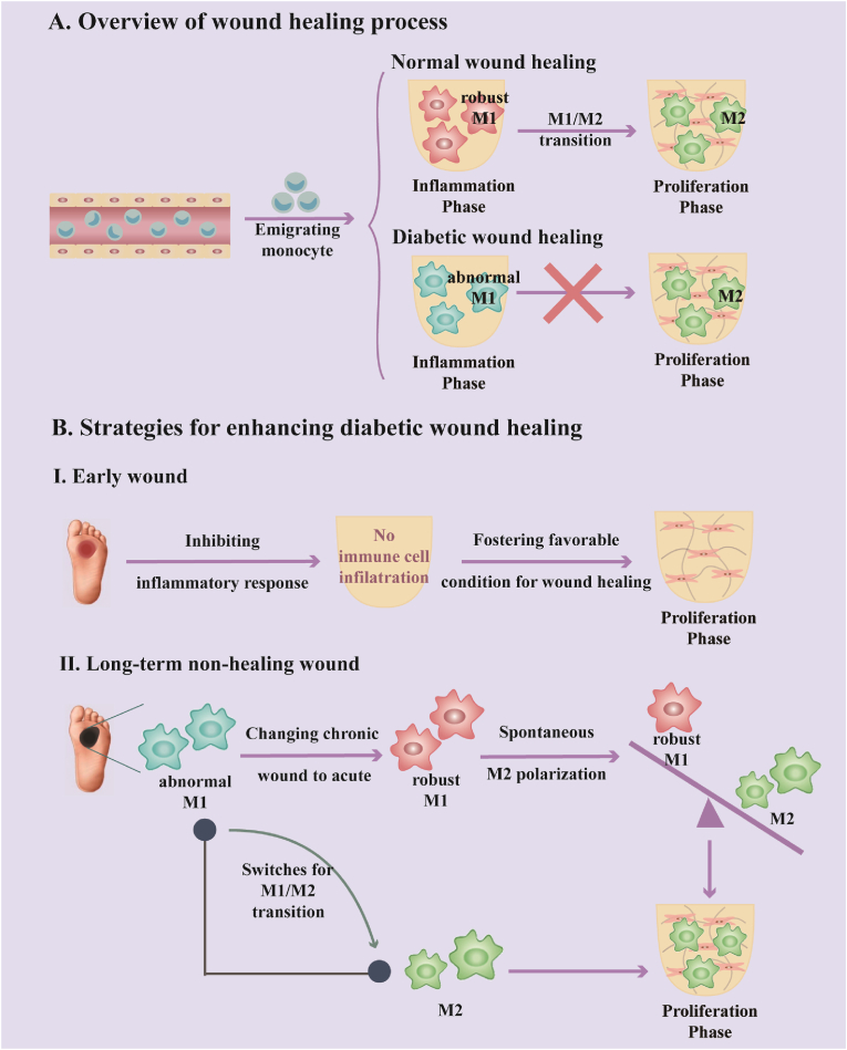
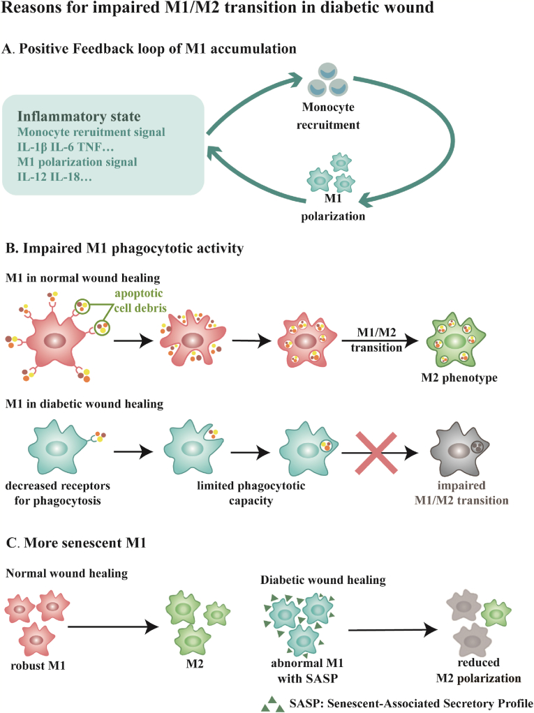
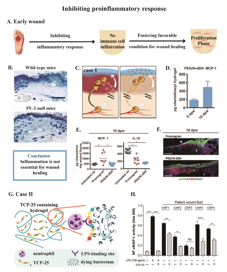
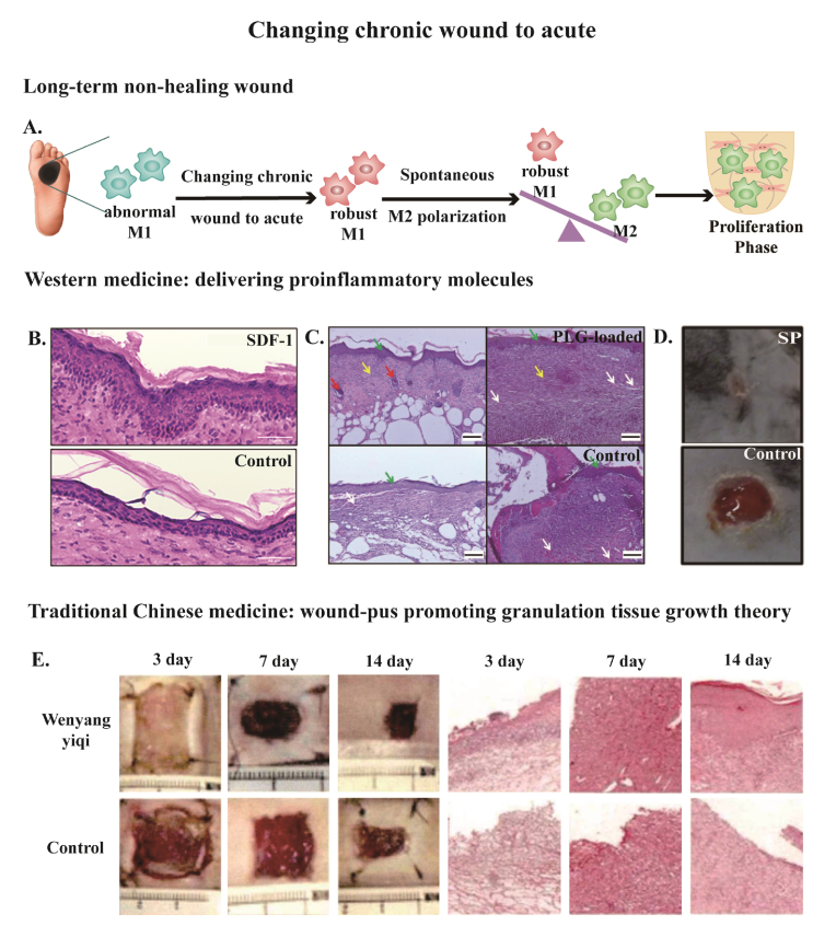
Verification
Claims the paper makes
The claims and statistics of this paper haven’t been checked yet.
Run the check — free, no account needed. It reads the open-access text and checks the sources it cites.
Claims other papers make about this one
Each is checked against this paper’s own text: does it say what the citing paper says it does?
In the citing paper
“The pathological microenvironment in DM bone defect areas is characterized by hyperglycemia, chronic inflammation, vascular dysfunction, and immune dysregulation4–6.”
From Microenvironment-responsive multifunctional enzyme-linked hydrogel for diabetic bone defect regeneration. (2025) · cites this paper as [6]
Self-check
Percentages, sums and reported differences re-computed from the paper’s own numbers.
The self-check needs the paper’s full text.
Citations
Cites
Macrophage Cytokines: Involvement in Immunity and Infectious Diseases
Arango Duque, G., & Descoteaux, A. (2014) · Frontiers in Immunology · doi:10.3389/fimmu.2014.00491 · Cited by 2,708
Transition from inflammation to proliferation: a critical step during wound healing
Landén, N. X., Li, D., & Ståhle, M. (2016) · Cellular and Molecular Life Sciences · doi:10.1007/s00018-016-2268-0 · Cited by 1,980 · cited 1×
“after injury), inflammatory phase (1–3 days), proliferation phase (4–21 days), and remodeling phase (21days-1year) [6,7].”
BackgroundReinke, J. M., & Sorg, H. (2012) · European Surgical Research · doi:10.1159/000339613 · Cited by 1,898
Neutrophil: A Cell with Many Roles in Inflammation or Several Cell Types?
Rosales, C. (2018) · Frontiers in Physiology · doi:10.3389/fphys.2018.00113 · Cited by 1,334
Macrophage Dysfunction Impairs Resolution of Inflammation in the Wounds of Diabetic Mice
Khanna, S., Biswas, S., Shang, Y., Collard, E., Azad, A., Kauh, C., Bhasker, V., Gordillo, G. M., Sen, C. K., & Roy, S. (2010) · PLoS ONE · doi:10.1371/journal.pone.0009539 · Cited by 694
Ellis, S., Lin, E. J., & Tartar, D. (2018) · Current Dermatology Reports · doi:10.1007/s13671-018-0234-9 · Cited by 625 · cited 5×
“247 initiating re-epithelialization by producing epidermal growth factor (EGF) and keratinocyte growth factor (KGF), M1 macrophages hinder keratinocyte migration with over-expressed TNF-α [60,61].”
BackgroundInfluentialAccelerated wound closure in neutrophil-depleted mice
Dovi, J. V., He, L.-K., & DiPietro, L. A. (2003) · Journal of Leukocyte Biology · doi:10.1189/jlb.0802406 · Cited by 525
Garg, K., Pullen, N. A., Oskeritzian, C. A., Ryan, J. J., & Bowlin, G. L. (2013) · Biomaterials · doi:10.1016/j.biomaterials.2013.02.065 · Cited by 457
Substance P Promotes Wound Healing in Diabetes by Modulating Inflammation and Macrophage Phenotype
Leal, E. C., Carvalho, E., Tellechea, A., Kafanas, A., Tecilazich, F., Kearney, C., Kuchibhotla, S., Auster, M. E., Kokkotou, E., Mooney, D. J., et al. (2015) · The American Journal of Pathology · doi:10.1016/j.ajpath.2015.02.011 · Cited by 223
Previtera, M. L., & Sengupta, A. (2015) · PLOS ONE · doi:10.1371/journal.pone.0145813 · Cited by 176
Goren, I., Müller, E., Schiefelbein, D., Christen, U., Pfeilschifter, J., Mühl, H., & Frank, S. (2007) · Journal of Investigative Dermatology · doi:10.1038/sj.jid.5700842 · Cited by 151
Zhuang, Z., Zhang, Y., Sun, S., Li, Q., Chen, K., An, C., Wang, L., van den Beucken, J. J. J. P., & Wang, H. (2020) · ACS Biomaterials Science & Engineering · doi:10.1021/acsbiomaterials.0c00295 · Cited by 126
Self-citationTRPV4 plays a role in matrix stiffness-induced macrophage polarization
Dutta, B., Goswami, R., & Rahaman, S. O. (2020) · Frontiers in Immunology · doi:10.3389/fimmu.2020.570195 · Cited by 124
Research works printed but not linked: 110
- Ouyang, 2021, Risk factors of diabetic foot ulcer in patients with type 2 diabetes: a retrospective cohort study, Am. J. Tourism Res., 13 8, 9554
- Fridman, 2020, Treatment of hard-to-heal diabetic foot ulcers with a hepatic-derived wound matrix, Wounds : a compendium of clinical research and practice, 32, 244
- Thanapaul, 2021, An insight into aging, senescence, and their impacts on wound healing, Advances in geriatric medicine and research, 3
- Gould, 2013, Wound healing in older adults, R. I. Med. J., 99, 34
- Pinto, 2020, Bacteriophages for chronic wound treatment: from traditional to novel delivery systems, Viruses, 12, 235, 10.3390/v12020235 · DOI
- Li, 2021, Macrophage related chronic inflammation in non-healing wounds, Front. Immunol., 12
- Zhu, 2023, Smart bioadhesives for wound healing and closure, Bioact. Mater., 19, 360
- Matoori, 2021, Advanced bandages for diabetic wound healing, Sci. Transl. Med., 13, 10.1126/scitranslmed.abe4839 · DOI
- Sawaya, 2020, Deregulated immune cell recruitment orchestrated by FOXM1 impairs human diabetic wound healing, Nat. Commun., 11, 4678, 10.1038/s41467-020-18276-0 · DOI
- Wood, 2014, Pro-inflammatory chemokine CCL2 (MCP-1) promotes healing in diabetic wounds by restoring the macrophage response, PLoS One, 9, 10.1371/journal.pone.0091574 · DOI
- Louiselle, 2021, Macrophage polarization and diabetic wound healing, Transl. Res. : J. Lab. Clin. Med., 236, 109, 10.1016/j.trsl.2021.05.006 · DOI
- Mirza, 2013, Blocking interleukin-42 induces a healing-associated wound macrophage phenotype and improves healing in type 2 diabetes, Diabetes, 62, 2579, 10.2337/db12-1450 · DOI
- Rehak, 2022, The immune-centric revolution in the diabetic foot: monocytes and lymphocytes role in wound healing and tissue regeneration-A narrative review, J. Clin. Med., 11, 889, 10.3390/jcm11030889 · DOI
- Patel, 2019, Mechanistic insight into diabetic wounds: pathogenesis, molecular targets and treatment strategies to pace wound healing, Biomed. Pharmacother., 112, 10.1016/j.biopha.2019.108615 · DOI
- Lv, 2017, A conducive bioceramic/polymer composite biomaterial for diabetic wound healing, Acta Biomater., 60, 128, 10.1016/j.actbio.2017.07.020 · DOI
- Smith, 2015, How it all starts: initiation of the clotting cascade, Crit. Rev. Biochem. Mol. Biol., 50, 326, 10.3109/10409238.2015.1050550 · DOI · Paper page
- Kerris, 2020, Platelets and platelet extracellular vesicles in hemostasis and sepsis, J. Invest. Med., 68, 813, 10.1136/jim-2019-001195 · DOI
- Locatelli, 2021, Platelets in wound healing: what happens in space?, Front. Bioeng. Biotechnol., 9, 10.3389/fbioe.2021.716184 · DOI
- Kim, 2008, Dynamics of neutrophil infiltration during cutaneous wound healing and infection using fluorescence imaging, J. Invest. Dermatol., 128, 1812, 10.1038/sj.jid.5701223 · DOI
- Soehnlein, 2008, Neutrophil secretion products pave the way for inflammatory monocytes, Blood, 112, 1461, 10.1182/blood-2008-02-139634 · DOI
- Soehnlein, 2009, Mechanisms underlying neutrophil-mediated monocyte recruitment, Blood, 114, 4613, 10.1182/blood-2009-06-221630 · DOI
- Wei, 2012, Keratinocyte expression of inflammatory mediators plays a crucial role in substance P-induced acute and chronic pain, J. Neuroinflammation, 9, 10.1186/1742-2094-9-181 · DOI
- Luger, 1998, Neuropeptides: role in inflammatory skin diseases, J. Eur. Acad. Dermatol. Venereol. : JEADV, 10, 207, 10.1111/j.1468-3083.1998.tb00736.x · DOI
- Wernersson, 2014, Mast cell secretory granules: armed for battle, Nat. Rev. Immunol., 14, 478, 10.1038/nri3690 · DOI
- Barman, 2020, Macrophage dysregulation and impaired skin wound healing in diabetes, Front. Cell Dev. Biol., 8, 10.3389/fcell.2020.00528 · DOI
- Xie, 2016, Effects of IRF1 and IFN-β interaction on the M1 polarization of macrophages and its antitumor function, Int. J. Mol. Med., 38, 148, 10.3892/ijmm.2016.2583 · DOI
- Kotwal, 2017, Macrophage differentiation in normal and accelerated wound healing, Results Probl. Cell Differ., 62, 353, 10.1007/978-3-319-54090-0_14 · DOI
- Brown, 2014, Chapter 25 - the role of the host immune response in tissue engineering and regenerative medicine, 497
- Kim, 2018, Promotion of keratinocyte proliferation by tracheloside through ERK1/2 stimulation, Evid. base Compl. Alternative Med., 2018
- Guillamat-Prats, 2021, The role of MSC in wound healing, scarring and regeneration, Cells, 10, 10.3390/cells10071729 · DOI · Paper page
- Tsalamandris, 2019, The role of inflammation in diabetes: current concepts and future perspectives, European cardiology, 14, 50, 10.15420/ecr.2018.33.1 · DOI · Paper page
- Hicks, 2019, Epidemiology of peripheral neuropathy and lower extremity disease in diabetes, Curr. Diabetes Rep., 19, 10.1007/s11892-019-1212-8 · DOI
- Yagihashi, 2011, Mechanism of diabetic neuropathy: where are we now and where to go?, J Diabetes Investig, 2, 18, 10.1111/j.2040-1124.2010.00070.x · DOI · Paper page
- Theocharidis, 2020, Autonomic nerve dysfunction and impaired diabetic wound healing: the role of neuropeptides, Auton. Neurosci., 223, 10.1016/j.autneu.2019.102610 · DOI
- Tellechea, 2016, Mast cells regulate wound healing in diabetes, Diabetes, 65, 2006, 10.2337/db15-0340 · DOI
- Pradhan, 2009, Inflammation and neuropeptides: the connection in diabetic wound healing, Expet Rev. Mol. Med., 11
- Wu, 2016, Hyperglycaemia inhibits REG3A expression to exacerbate TLR3-mediated skin inflammation in diabetes, Nat. Commun., 7, 10.1038/ncomms13393 · DOI
- Burgess, 2021, Diabetic wound-healing science, Medicina (Kaunas), 57, 1072, 10.3390/medicina57101072 · DOI
- Louiselle, 2021, Macrophage polarization and diabetic wound healing, Transl. Res., 236, 109, 10.1016/j.trsl.2021.05.006 · DOI
- Carmi, 2009, The role of macrophage-derived IL-1 in induction and maintenance of angiogenesis, J. Immunol., 183, 4705, 10.4049/jimmunol.0901511 · DOI · Paper page
- Aitcheson, 2021, Skin wound healing: normal macrophage function and macrophage dysfunction in diabetic wounds, Molecules, 26, 4917, 10.3390/molecules26164917 · DOI
- Morey, 2019, Hyperglycemia acts in synergy with hypoxia to maintain the pro-inflammatory phenotype of macrophages, PLoS One, 14, 10.1371/journal.pone.0220577 · DOI
- Zhong, 2018, Myc-nick promotes efferocytosis through M2 macrophage polarization during resolution of inflammation, Faseb. J. : official publication of the Federation of American Societies for Experimental Biology, 32, 5312, 10.1096/fj.201800223R · DOI
- Wilkinson, 2019, Elevated local senescence in diabetic wound healing is linked to pathological repair via CXCR2, J. Invest. Dermatol., 139, 1171, 10.1016/j.jid.2019.01.005 · DOI
- Jetten, 2014, Anti-inflammatory M2, but not pro-inflammatory M1 macrophages promote angiogenesis in vivo, Angiogenesis, 17, 109, 10.1007/s10456-013-9381-6 · DOI
- Hassanshahi, 2022, Macrophage-mediated inflammation in skin wound healing, Cells, 11, 10.3390/cells11192953 · DOI
- Okonkwo, 2017, Diabetes and wound angiogenesis, Int. J. Mol. Sci., 18, 10.3390/ijms18071419 · DOI
- Ellis, 2018, Immunology of wound healing, Current dermatology reports, 7, 350, 10.1007/s13671-018-0234-9 · DOI · Paper page
- Huang, 2019, High glucose environment induces M1 macrophage polarization that impairs keratinocyte migration via TNF-α: an important mechanism to delay the diabetic wound healing, J. Dermatol. Sci., 96, 159, 10.1016/j.jdermsci.2019.11.004 · DOI
- Abas, 2020, Chapter 19 - collagen in diabetic wound healing, 393
- Joorabloo, 2022, Recent advances in nanomedicines for regulation of macrophages in wound healing, J. Nanobiotechnol., 20, 407, 10.1186/s12951-022-01616-1 · DOI
- Zhou, 2022, The signaling pathways of traditional Chinese medicine in promoting diabetic wound healing, J. Ethnopharmacol., 282, 10.1016/j.jep.2021.114662 · DOI
- Martin, 2003, Wound healing in the PU.1 null mouse--tissue repair is not dependent on inflammatory cells, Curr. Biol. : CB, 13, 1122, 10.1016/S0960-9822(03)00396-8 · DOI
- Lohmann, 2017, Glycosaminoglycan-based hydrogels capture inflammatory chemokines and rescue defective wound healing in mice, Sci. Transl. Med., 9, 10.1126/scitranslmed.aai9044 · DOI
- Puthia, 2020, A dual-action peptide-containing hydrogel targets wound infection and inflammation, Sci. Transl. Med., 12, 10.1126/scitranslmed.aax6601 · DOI
- Wilgus, 2007, Regenerative healing in fetal skin: a review of the literature, Ostomy/Wound Manag., 53, 16
- Wilgus, 2019, Vascular endothelial growth factor and cutaneous scarring, Adv. Wound Care, 8, 671, 10.1089/wound.2018.0796 · DOI
- Tripathi, 2020, Hypertrophic scars and keloids: a review and current treatment modalities, Biomedical Dermatology, 4, 11, 10.1186/s41702-020-00063-8 · DOI
- Lech, 2013, Macrophages and fibrosis: how resident and infiltrating mononuclear phagocytes orchestrate all phases of tissue injury and repair, Biochim. Biophys. Acta, 1832, 989, 10.1016/j.bbadis.2012.12.001 · DOI
- Lin, 1995, Exogenous transforming growth factor-beta amplifies its own expression and induces scar formation in a model of human fetal skin repair, Ann. Surg., 222, 146, 10.1097/00000658-199508000-00006 · DOI
- Wilgus, 2008, Regulation of scar formation by vascular endothelial growth factor, Lab. Invest., 88, 579, 10.1038/labinvest.2008.36 · DOI
- Martin, 1993, Rapid induction and clearance of TGFβ1 is an early response to wounding in the mouse embryo, Dev. Genet., 14, 225, 10.1002/dvg.1020140309 · DOI
- Julier, 2017, Promoting tissue regeneration by modulating the immune system, Acta Biomater., 53, 13, 10.1016/j.actbio.2017.01.056 · DOI · Paper page
- Cheng Jiajun, 2019, A randomized controlled trial of external treatment of traditional Chinese medicine to promote wound healing under the theory of simmering pus and growing meat: a systematic evaluation, Journal of Yunnan University of Traditional Chinese Medicine, 42, 26
- Salcedo, 2003, Role of chemokines in angiogenesis: CXCL12/SDF-1 and CXCR4 interaction, a key regulator of endothelial cell responses, Microcirculation, 10, 359
- Ogłodek, 2015, The MCP-1, CCL-5 and SDF-1 chemokines as pro-inflammatory markers in generalized anxiety disorder and personality disorders, Pharmacol. Rep. : PR, 67, 85, 10.1016/j.pharep.2014.08.006 · DOI
- Gallagher, 2007, Diabetic impairments in NO-mediated endothelial progenitor cell mobilization and homing are reversed by hyperoxia and SDF-1 alpha, J. Clin. Investig., 117, 1249, 10.1172/JCI29710 · DOI
- Bermudez, 2011, Inhibition of stromal cell-derived factor-1α further impairs diabetic wound healing, J. Vasc. Surg., 53, 774, 10.1016/j.jvs.2010.10.056 · DOI
- Kim, 2017, Enhancement of wound closure by modifying dual release patterns of stromal-derived cell factor-1 and a macrophage recruitment agent from gelatin hydrogels, Journal of tissue engineering and regenerative medicine, 11, 2999, 10.1002/term.2202 · DOI
- Yu, 2021, Liposomal SDF-1 alpha delivery in nanocomposite hydrogels promotes macrophage phenotype changes and skin tissue regeneration, ACS Biomater. Sci. Eng., 7, 5230, 10.1021/acsbiomaterials.1c01140 · DOI
- Shen, 2012, Plasminogen is a key proinflammatory regulator that accelerates the healing of acute and diabetic wounds, Blood, 119, 5879, 10.1182/blood-2012-01-407825 · DOI
- Chan, 2001, Accelerated skin wound healing in plasminogen activator inhibitor-1-deficient mice, Am. J. Pathol., 159, 1681, 10.1016/S0002-9440(10)63015-5 · DOI
- Fallah, 2020, Plasminogen is a master regulator and a potential drug candidate for the healing of radiation wounds, Cell Death Dis., 11, 10.1038/s41419-020-2397-0 · DOI
- Das, 2014, Plasminogen promotes macrophage phagocytosis in mice, Blood, 124, 679, 10.1182/blood-2014-01-549659 · DOI
- Al Kayal, 2022, Plasminogen-loaded fibrin scaffold as drug delivery system for wound healing applications, Pharmaceutics, 14, 10.3390/pharmaceutics14020251 · DOI
- Pradhan, 2011, Gene expression of pro-inflammatory cytokines and neuropeptides in diabetic wound healing, J. Surg. Res., 167, 336, 10.1016/j.jss.2009.09.012 · DOI
- Bao Yiru, 2022, Discussion on the mechanism of medicine-wound interaction under the guidance of wound-pus promoting granulation tissue growth theory, Acta Chinese Medicine, 37, 516
- Jiang Yu-feng, 2019, Improving the healing microenvironment is the core concept in wound treatment for both traditional Chinese and Western medicine, Journal of Traumatic Surgery, 21, 241
- Zhu Chao-jun, 2020, Effect of weinongzhangrou method on expression of VEGF, Notch1 and TGFβ1 in granulation tissue of rats with chronic ulcer and its histopathology, Chinese Journal of Surgery of Integrated, 26, 8
- Liu Xian-zhou, 2017, Discussion on simmer pus and cuddle pus promoting the regeneration, Academic Journal of Shanghai University of Traditional Chinese Medicine, 31, 7
- Chen Jia, 2021, vol. 37, 1055
- Kivimäki, 2021, Pinosylvin shifts macrophage polarization to support resolution of inflammation, Molecules, 26, 2772, 10.3390/molecules26092772 · DOI
- Ganesh, 2020, Macrophage mediation in normal and diabetic wound healing responses, Inflamm. Res., 69, 347, 10.1007/s00011-020-01328-y · DOI
- Doyle, 2019, Overview of extracellular vesicles, their origin, composition, purpose, and methods for exosome isolation and analysis, Cells, 8, 727, 10.3390/cells8070727 · DOI
- Bartel, 2020, Extracellular vesicles as mediators of cellular cross talk in the lung microenvironment, Front. Med., 7, 10.3389/fmed.2020.00326 · DOI
- Stremersch, 2016, Therapeutic and diagnostic applications of extracellular vesicles, J. Contr. Release : official journal of the Controlled Release Society, 244, 167, 10.1016/j.jconrel.2016.07.054 · DOI
- Diaz-Garrido, 2021, Cell-to-Cell communication by host-released extracellular vesicles in the gut: implications in health and disease, Int. J. Mol. Sci., 22, 2213, 10.3390/ijms22042213 · DOI
- Hu, 2021, Extracellular vesicle activities regulating macrophage- and tissue-mediated injury and repair responses, Acta Pharm. Sin. B, 11, 1493, 10.1016/j.apsb.2020.12.014 · DOI
- Zheng, 2021, Apoptotic vesicles restore liver macrophage homeostasis to counteract type 2 diabetes, J. Extracell. Vesicles, 10, 10.1002/jev2.12109 · DOI · Paper page
- Dou, 2020, Chimeric apoptotic bodies functionalized with natural membrane and modular delivery system for inflammation modulation, Sci. Adv., 6, 10.1126/sciadv.aba2987 · DOI
- Liu, 2020, Melatonin-stimulated MSC-derived exosomes improve diabetic wound healing through regulating macrophage M1 and M2 polarization by targeting the PTEN/AKT pathway, Stem Cell Res. Ther., 11, 259, 10.1186/s13287-020-01756-x · DOI
- Zhou, 2021, Biomaterial-based physical regulation of macrophage behaviour, J. Mater. Chem. B, 9, 3608, 10.1039/D1TB00107H · DOI · Paper page
- Liu, 2022, Physical confinement in alginate cryogels determines macrophage polarization to a M2 phenotype by regulating a STAT-related mRNA transcription pathway, Biomater. Sci., 10, 2315, 10.1039/D1BM01719E · DOI
- Jiang, 2019, Cryoprotectant enables structural control of porous scaffolds for exploration of cellular mechano-responsiveness in 3D, Nat. Commun., 10, 3491, 10.1038/s41467-019-11397-1 · DOI
- Yin, 2020, S Pore size-mediated macrophage M1-to-M2 transition influences new vessel formation within the compartment of a scaffold, Appl. Mater. Today, 18
- Johnson, 2023, Hyperglycemic conditions enhance the mechanosensitivity of proinflammatory RAW264.7 macrophages, Tissue Eng., 29, 172, 10.1089/ten.tea.2022.0151 · DOI
- Sridharan, 2019, Material stiffness influences the polarization state, function and migration mode of macrophages, Acta Biomater., 89, 47, 10.1016/j.actbio.2019.02.048 · DOI · Paper page
- Yuan, 2022, NIR-triggered hydrogel with dynamic stiffness via ion chelation to modulate macrophage phenotypes, J. Polym. Sci., 60, 3176, 10.1002/pol.20220349 · DOI
- Yang, 2021, Micro/nano-net guides M2-pattern macrophage cytoskeleton distribution via Src-ROCK signalling for enhanced angiogenesis, Biomater. Sci., 9, 3334, 10.1039/D1BM00116G · DOI
- Zheng, 2019, Near-infrared-triggered dynamic surface topography for sequential modulation of macrophage phenotypes, ACS Appl. Mater. Interfaces, 11, 43689, 10.1021/acsami.9b14808 · DOI
- McWhorter Frances, 2013, Modulation of macrophage phenotype by cell shape, Proc. Natl. Acad. Sci. USA, 110, 17253, 10.1073/pnas.1308887110 · DOI · Paper page
- Schoenenberger, 2020, Macromechanics and polycaprolactone fiber organization drive macrophage polarization and regulate inflammatory activation of tendon in vitro and in vivo, Biomaterials, 249, 10.1016/j.biomaterials.2020.120034 · DOI · Paper page
- Barth, 2013, The effect of surface roughness on RAW 264.7 macrophage phenotype, J. Biomed. Mater. Res., Part A, 101, 2679, 10.1002/jbm.a.34562 · DOI · Paper page
- Lee, 2022, High-performance implantable bioelectrodes with immunocompatible topography for modulation of macrophage responses, ACS Nano, 16, 7471, 10.1021/acsnano.1c10506 · DOI
- Milan, 2016, Accelerated wound healing in a diabetic rat model using decellularized dermal matrix and human umbilical cord perivascular cells, Acta Biomater., 45, 234, 10.1016/j.actbio.2016.08.053 · DOI · Paper page
- Zhou, 2022, Glucose and MMP-9 dual-responsive hydrogel with temperature sensitive self-adaptive shape and controlled drug release accelerates diabetic wound healing, Bioact. Mater., 17, 1
- Jia, 2023, Green tea derivative-based hydrogel with ROS-scavenging property for accelerating diabetic wound healing, Mater. Des., 225, 10.1016/j.matdes.2022.111452 · DOI
- Farhan, 2023, Spontaneous NETosis in diabetes: a role of hyperglycemia mediated ROS and autophagy, Front. Med., 10, 10.3389/fmed.2023.1076690 · DOI
- Hicks, 2019, Real-time bacterial detection with an intracellular ROS sensing platform, Biosens. Bioelectron., 141, 10.1016/j.bios.2019.111430 · DOI
- Yang, 2021, Promote anti-inflammatory and angiogenesis using a hyaluronic acid-based hydrogel with miRNA-laden nanoparticles for chronic diabetic wound treatment, Int. J. Biol. Macromol., 166, 166, 10.1016/j.ijbiomac.2020.10.129 · DOI
Cited by
Sorted by citations
Fu, X., Luo, Z., Guo, Y., Meng, W., Jin, S., Chen, J., Cai, Y., Luo, Z., Huang, C., Chen, A., et al. (2025) · Nature Communications · doi:10.1038/s41467-025-65165-5 · Cited by 44
Self-citation
Metadata
- Authors
- Cai, Y., Chen, K., Liu, C., & Qu, X.
- ORCID
- Qu, Xue
- Venue
- Bioactive Materials
- Publisher
- Elsevier BV
- Published
- 2023-10
- Volume
- 28
- Pages
- 243-254
- ISSN
- 2452-199X
- PMC
- PMC10245071
- Cited by
- 112
- References
- 13
- Language
- en
- Licence
- Funders
- Higher Education Discipline Innovation Project (B14018)
- Science and Technology Project of Nantong City
- National Natural Science Foundation of China National Outstanding Youth Science Fund Project (11932012, 31922041, 32171341)
- National Key Research and Development Program of China (2021YFB3800800)
- National Natural Science Foundation of China
About these checks
Registry, retraction, Cites and statistics results are computed, not judged, with one exception: a printed reference that no index lists is read once by a model before it counts as a citation error. Claims are extracted by a model and kept only when their quote appears verbatim in the text; each cited source is then checked, with a model’s help, for whether it says what the claim relies on. Those verdicts can be wrong — read the claim’s quote and the cited source before relying on one.
Statistics and arithmetic are recomputed, not judged.
Confidence bands are shown only for fields calibrated on our annotated test set. None is calibrated yet, so no band is shown.
What counts as a claim ›How we measure confidence ›How we check this ›
Are you an author, or do you see a problem with this page? Request a review
