

The DOI matches its Crossref record; 43 of the 44 references it cites were found in a registry or an index.
The Resting Human Brain and Motor Learning
Open accessAbstract
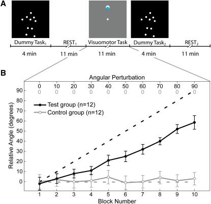
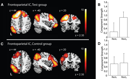
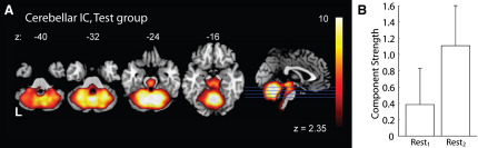
Functionally related brain networks are engaged even in the absence of an overt behavior. The role of this resting state activity, evident as low-frequency fluctuations of BOLD (see [1] for review, [2-4]) or electrical [5, 6] signals, is unclear. Two major proposals are that resting state activity supports introspective thought or supports responses to future events [7]. An alternative perspective is that the resting brain actively and selectively processes previous experiences [8]. Here we show that motor learning can modulate subsequent activity within resting networks. BOLD signal was recorded during rest periods before and after an 11 min visuomotor training session. Motor learning but not motor performance modulated a fronto-parietal resting state network (RSN). Along with the fronto-parietal network, a cerebellar network not previously reported as an RSN was also specifically altered by learning. Both of these networks are engaged during learning of similar visuomotor tasks [9-22]. Thus, we provide the first description of the modulation of specific RSNs by prior learning--but not by prior performance--revealing a novel connection between the neuroplastic mechanisms of learning and resting state activity. Our approach may provide a powerful tool for exploration of the systems involved in memory consolidation.
Overview
The claims and statistics of this paper haven’t been checked yet.
Run the check — free, no account needed. It reads the open-access text and checks the sources it cites.
Integrity signals
Registry
Cites
Statistics
Figures
Verification
Claims the paper makes
The claims and statistics of this paper haven’t been checked yet.
Run the check — free, no account needed. It reads the open-access text and checks the sources it cites.
Claims other papers make about this one
Each is checked against this paper’s own text: does it say what the citing paper says it does?
In the citing paper
“Clusters of brain regions involved in motor learning can be recognized, both at rest (Albert, Robertson, & Miall, 2009;Biswal, Yetkin, Haughton, & Hyde, 1995;Wu, Srinivasan, Kaur, & Cramer, 2014) and in-task (Hasan et al., 2013).”
From Optimizing performance through intrinsic motivation and attention for learning: The OPTIMAL theory of motor learning (2016) · cites this paper as Albert, Robertson, & Miall, 2009
In the citing paper
“Importantly, functional connectivity is related to motor skill learning (Albert et al., 2009;Song, Gotts, Dayan, & Cohen, 2015) and to higher levels of motor skill (Di, Zhu, Jin, Wang, Ye, Zhou et al., 2012;Ito, Matsuda, & Shimojo, 2015;Kim, Han, Kim, & Han, 2015;Milton, Solodkin, Hluštík, & Small, 2007;Li, He, Huang, Zhang, Lu, Lai et al., 2015).”
From Optimizing performance through intrinsic motivation and attention for learning: The OPTIMAL theory of motor learning (2016) · cites this paper as Albert et al., 2009
Self-check
Percentages, sums and reported differences re-computed from the paper’s own numbers.
The self-check needs the paper’s full text.
Citations
Cites
Tzourio-Mazoyer, N., Landeau, B., Papathanassiou, D., Crivello, F., Etard, O., Delcroix, N., Mazoyer, B., & Joliot, M. (2002) · NeuroImage · doi:10.1006/nimg.2001.0978 · Cited by 16,319
A default mode of brain function
Raichle, M. E., MacLeod, A. M., Snyder, A. Z., Powers, W. J., Gusnard, D. A., & Shulman, G. L. (2001) · Proceedings of the National Academy of Sciences · doi:10.1073/pnas.98.2.676 · Cited by 12,190
Jenkinson, M., Bannister, P., Brady, M., & Smith, S. (2002) · NeuroImage · doi:10.1006/nimg.2002.1132 · Cited by 11,888 · cited 2×
“Each EPI volume was motion-corrected using MCFLIRT [3], high-pass filtered (0.01HZ cutoff), masked to eliminate non-brain voxels, spatially-smoothed using a 5mm FWHM filter, demeaned on a voxel-by-voxel basis, whitened, and projected into a 48-dimensional subspace using PICA.”
Fast robust automated brain extraction
Smith, S. M. (2002) · Human Brain Mapping · doi:10.1002/hbm.10062 · Cited by 10,496 · cited 2×
“The role of this resting state activity, evident as low-frequency fluctuations of BOLD (see[1]forreview,[2‐4])orelectrical[ 5 ,6]signals,isunclear.”
The human brain is intrinsically organized into dynamic, anticorrelated functional networks
Fox, M. D., Snyder, A. Z., Vincent, J. L., Corbetta, M., Van Essen, D. C., & Raichle, M. E. (2005) · Proceedings of the National Academy of Sciences · doi:10.1073/pnas.0504136102 · Cited by 8,553
Fast and robust fixed-point algorithms for independent component analysis
Hyvärinen, A. (1999) · IEEE Transactions on Neural Networks · doi:10.1109/72.761722 · Cited by 6,304 · cited 2×
“The role of this resting state activity, evident as low-frequency fluctuations of BOLD (see[1]forreview,[2‐4])orelectrical[5, 6 ]signals,isunclear.”
Consistent resting-state networks across healthy subjects
Damoiseaux, J. S., Rombouts, S. A. R. B., Barkhof, F., Scheltens, P., Stam, C. J., Smith, S. M., & Beckmann, C. F. (2006) · Proceedings of the National Academy of Sciences · doi:10.1073/pnas.0601417103 · Cited by 4,326 · cited 1×
“We identified six previously reported RSNs (see Figures 2A–2E and 2H of [4]).”
BackgroundProbabilistic Independent Component Analysis for Functional Magnetic Resonance Imaging
Beckmann, C. F., & Smith, S. M. (2004) · IEEE Transactions on Medical Imaging · doi:10.1109/tmi.2003.822821 · Cited by 2,707 · cited 7×
“The change in strength of the two RSN components identified by PICA across participants within the test group (Figure 2, main paper) was not linearly correlated with behavioral measures of learning, but this does not imply there is no relationship.”
InfluentialMazziotta, J. C., Toga, A. W., Evans, A. C., Fox, P. T., Lancaster, J. L., Zilles, K., Woods, R. P., Paus, T., Simpson, G. V., Pike, G. B., et al. (2001) · Philosophical Transactions of the Royal Society B Biological Sciences · doi:10.1098/rstb.2001.0915 · Cited by 2,417 · cited 3×
“Non-brain structures were removed from the high-resolution structural image using BET [5] and the transformation matrix used for the affine registration of this image to the ICBM152 brain [2] was applied to the PICA output from each session.”
A default mode of brain function: a brief history of an evolving idea
Raichle, M. E., & Snyder, A. Z. (2007) · NeuroImage · doi:10.1016/j.neuroimage.2007.02.041 · Cited by 2,173
Cited by
Sorted by citations
Dynamic functional connectivity: Promise, issues, and interpretations
Hutchison, R. M., Womelsdorf, T., Allen, E. A., Bandettini, P. A., Calhoun, V. D., Corbetta, M., Della Penna, S., Duyn, J. H., Glover, G. H., Gonzalez-Castillo, J., et al. (2013) · NeuroImage · doi:10.1016/j.neuroimage.2013.05.079 · Cited by 2,962
“, 2007), learning (Albert et al., 2009; Bassett et al., 2011; Lewis et al., 2009; Tambini et al., 2010), and large state transitions such as sleep (Horovitz et al.”
BackgroundToward discovery science of human brain function
Biswal, B. B., Mennes, M., Zuo, X.-N., Gohel, S., Kelly, C., Smith, S. M., Beckmann, C. F., Adelstein, J. S., Buckner, R. L., Colcombe, S., et al. (2010) · Proceedings of the National Academy of Sciences · doi:10.1073/pnas.0911855107 · Cited by 2,970
“For instance, functional connectivity is modulated by cognitive (47) and emotional state (48), arousal, and sleep (26), whereas structural connectivity is grossly unaffected by such factors.”
BackgroundVan Dijk, K. R. A., Hedden, T., Venkataraman, A., Evans, K. C., Lazar, S. W., & Buckner, R. L. (2010) · Journal of Neurophysiology · doi:10.1152/jn.00783.2009 · Cited by 1,844
Neuroplasticity Subserving Motor Skill Learning
Dayan, E., & Cohen, L. G. (2011) · Neuron · doi:10.1016/j.neuron.2011.10.008 · Cited by 1,408
“Overall, these studies suggest that functional connectivity in fronto-parietal networks supports consolidation after fast (Albert et al., 2009) and slow learning (Taubert et al.”
InfluentialSpreng, R. N., Stevens, W. D., Chamberlain, J. P., Gilmore, A. W., & Schacter, D. L. (2010) · NeuroImage · doi:10.1016/j.neuroimage.2010.06.016 · Cited by 1,256
“The frontoparietal control network, composed of rostrolateral prefrontal cortex (RLPFC), middle frontal gyrus (MFG), anterior insula/frontal operculum (aIfO), dorsal anterior cingulate cortex (dACC), precuneus (PCu), and anterior inferior parietal lobule (aIPL), is anatomically interposed between the default and dorsa…”
Influential
Metadata
- Authors
- Albert, N. B., Robertson, E. M., & Miall, R. C.
- Affiliations
- University of Chicago (US)
- Beth Israel Deaconess Medical Center (US)
- University of Birmingham (GB)
- Corresponding author
- Miall, R. Chris
- Venue
- Current Biology
- Publisher
- Elsevier BV
- Published
- 2009-06
- Volume
- 19
- Issue
- 12
- Pages
- 1023-1027
- ISSN
- 0960-9822 (ISSN-L)
- PMC
- PMC2701987
- OpenAlex
- W2117914711
- Cited by
- 510
- References
- 44
- Citations by year
- 2026: 8 · 2025: 13 · 2024: 23 · 2023: 23 · 2022: 29 · 2021: 26 · 2020: 24 · 2019: 17 · 2018: 30 · 2017: 38 · 2016: 39 · 2015: 47 · 2014: 54 · 2013: 35 · 2012: 31
- FWCI
- 16.78
- Type
- journal article
- Language
- en
- Open access
- hybrid
- doi.org
- Licence
- Locations
- Current Biology (publishedVersion, cc-by)
- PubMed Central (publishedVersion, cc-by)
- PubMed (publishedVersion)
- University of Birmingham Institutional Research Archive (University of Birmingham) (acceptedVersion)
- ENLIGHTEN (Jurnal Bimbingan dan Konseling Islam) (acceptedVersion)
- Digital Access to Scholarship at Harvard (DASH) (Harvard University) (submittedVersion, cc-by)
- University of Birmingham Research Portal (University of Birmingham) (submittedVersion, cc-by)
- Funders
- Wellcome Trust (069439)
- National Institutes of Health (R01 NS051446)
- Topics
- Life Sciences › Neuroscience › Cognitive Neuroscience › Functional Brain Connectivity Studies
- Life Sciences › Neuroscience › Cognitive Neuroscience › Neural dynamics and brain function
- Life Sciences › Neuroscience › Cognitive Neuroscience › Motor Control and Adaptation
- Keywords
- resting-state networks, motor learning, visuomotor training, frontoparietal network, cerebellar network, BOLD signal, neuroplasticity, memory consolidation
- MeSH terms
- Adult, Brain, Brain Mapping, Female, Humans, Magnetic Resonance Imaging, Male, Motor Skills, Psychomotor Performance, Random Allocation, Rest, Young Adult
About these checks
Registry, retraction, Cites and statistics results are computed, not judged, with one exception: a printed reference that no index lists is read once by a model before it counts as a citation error. Claims are extracted by a model and kept only when their quote appears verbatim in the text; each cited source is then checked, with a model’s help, for whether it says what the claim relies on. Those verdicts can be wrong — read the claim’s quote and the cited source before relying on one.
Statistics and arithmetic are recomputed, not judged.
Confidence bands are shown only for fields calibrated on our annotated test set. None is calibrated yet, so no band is shown.
What counts as a claim ›How we measure confidence ›How we check this ›
Are you an author, or do you see a problem with this page? Request a review
