

The DOI matches its Crossref record; 42 of the 42 references it cites were found in a registry or an index.
Enterohemorrhagic Escherichia coli Reduces Mucus and Intermicrovillar Bridges in Human Stem Cell-Derived Colonoids
Open accessAbstract
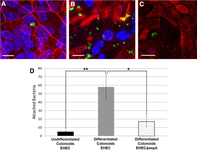
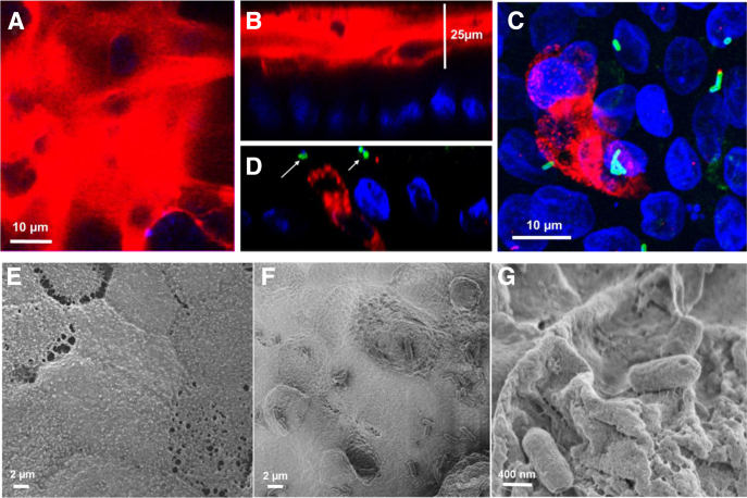
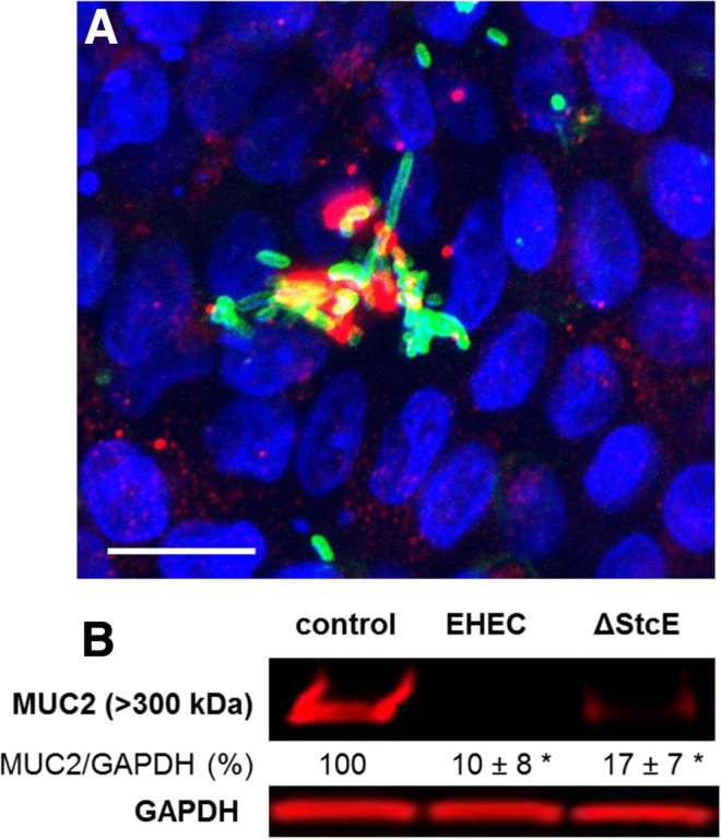
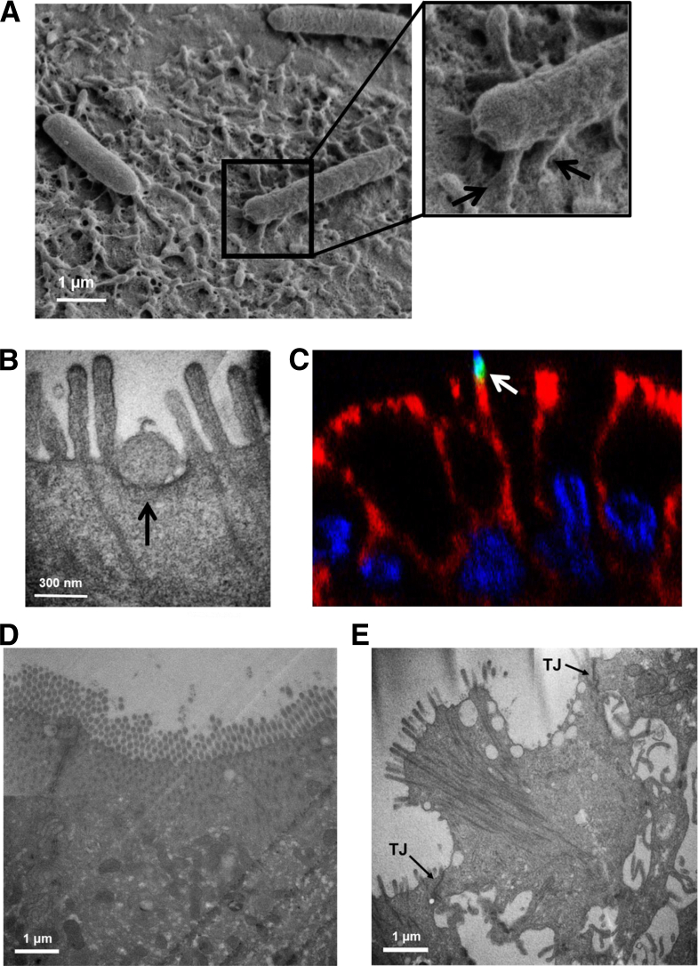
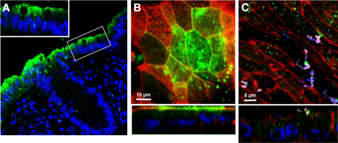
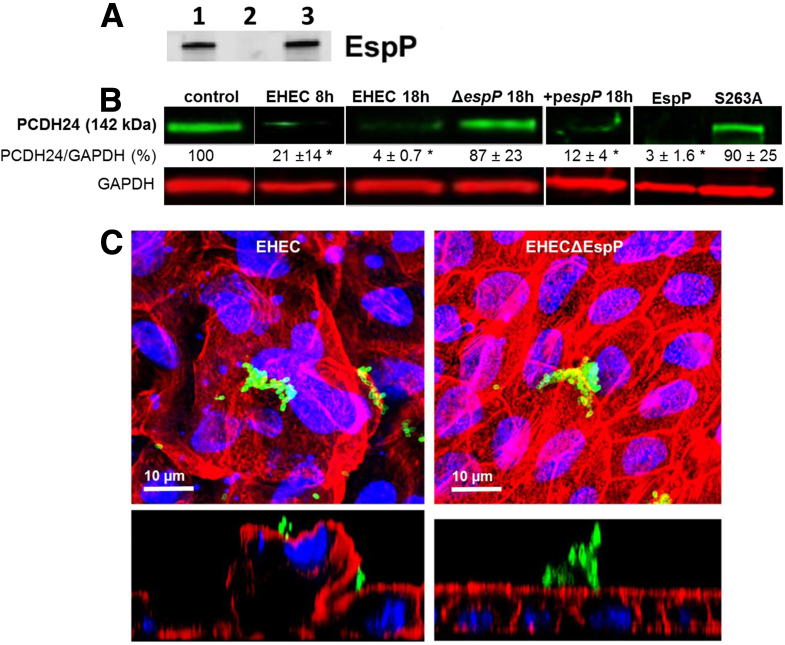
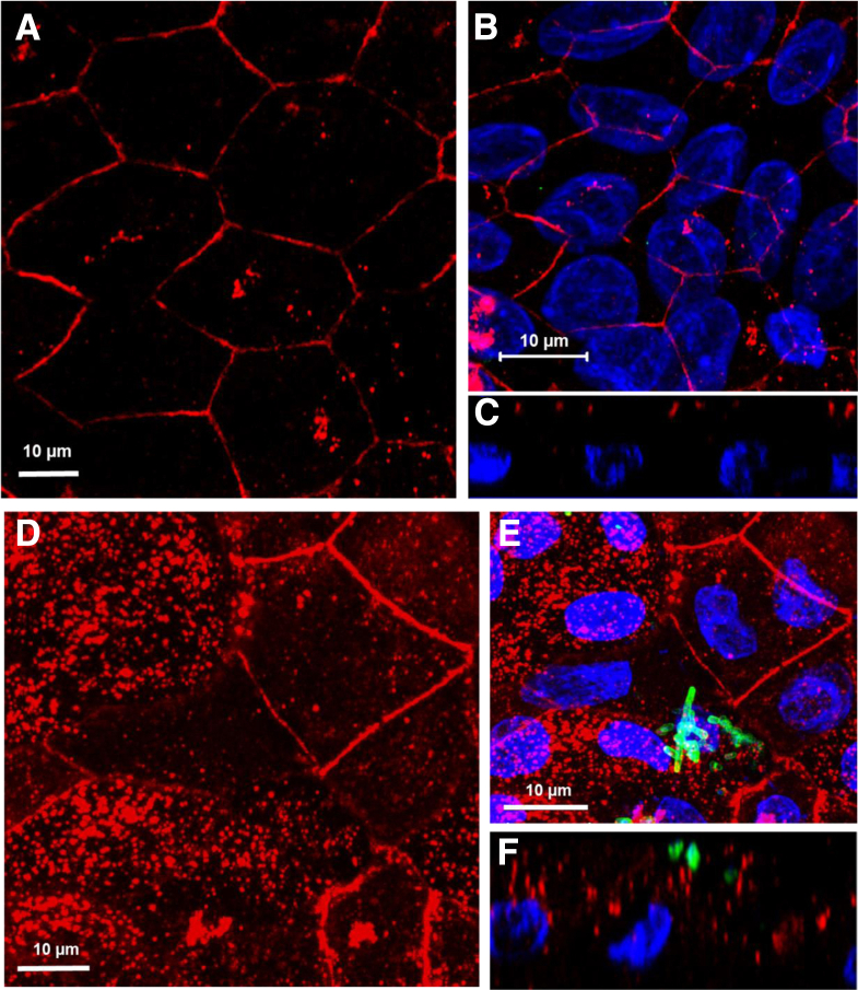
BACKGROUND AND AIMS: (EHEC) causes over 70,000 episodes of foodborne diarrhea annually in the USA. The early sequence of events which precede life-threatening hemorrhagic colitis and hemolytic uremic syndrome are not fully understood due to the initial asymptomatic phase of the disease and the lack of a suitable animal model. The aim of this study was to determine the initial molecular events in the interaction between EHEC and human colonic epithelium. METHODS: Human colonoids derived from adult proximal colonic stem cells were developed into monolayers to study EHEC-epithelial interactions. Monolayer confluency and differentiation were monitored by transepithelial electrical resistance (TER) measurements. The monolayers were apically infected with EHEC and the progression of epithelial damage over time was assessed using biochemical and imaging approaches. RESULTS: Human colonoid cultures recapitulate the differential protein expression patterns characteristic of the crypt and surface colonocytes. Mucus-producing differentiated colonoid monolayers are preferentially colonized by EHEC. Upon colonization, EHEC forms characteristic attaching and effacing lesions on the apical surface of colonoid monolayers. Mucin 2, a main component of colonic mucus, and protocadherin 24 (PCDH24), a microvillar resident protein, are targeted by EHEC at early stages of infection. The EHEC secreted serine protease, EspP, initiates brush border damage through PCDH24 reduction. CONCLUSIONS: Human colonoid monolayers are a relevant pathophysiological model which allows the study of early molecular events during enteric infections. Colonoid monolayers provide access to both apical and basolateral surfaces, thus providing an advantage over 3D cultures to study host-pathogen interactions in a controllable and tractable manner. EHEC reduces colonic mucus and affects the brush border cytoskeleton in the absence of commensal bacteria.
Overview
The claims and statistics of this paper haven’t been checked yet.
Run the check — free, no account needed. It reads the open-access text and checks the sources it cites.
Integrity signals
Registry
Cites
Statistics
Figures
Verification
Claims the paper makes
The claims and statistics of this paper haven’t been checked yet.
Run the check — free, no account needed. It reads the open-access text and checks the sources it cites.
Claims other papers make about this one
Each is checked against this paper’s own text: does it say what the citing paper says it does?
The cited source says what the claim attributes to it.
In the citing paper
“Furthermore, the recent development of organoid-derived 2D monolayer culture models has been instrumental for studies in which access to the apical surface is important [10,11,44].”
From Regulation of Enterocyte Brush Border Membrane Primary Na-Absorptive Transporters in Human Intestinal Organoid-Derived Monolayers (2024) · cites this paper as [44]
Self-check
Percentages, sums and reported differences re-computed from the paper’s own numbers.
The self-check needs the paper’s full text.
Citations
Cites
One-step inactivation of chromosomal genes in Escherichia coli K-12 using PCR products
Datsenko, K. A., & Wanner, B. L. (2000) · Proceedings of the National Academy of Sciences · doi:10.1073/pnas.120163297 · Cited by 14,881
Kaper, J. B., Nataro, J. P., & Mobley, H. L. T. (2004) · Nature Reviews Microbiology · doi:10.1038/nrmicro818 · Cited by 5,583
Self-citationSato, T., Stange, D. E., Ferrante, M., Vries, R. G. J., van Es, J. H., van den Brink, S., van Houdt, W. J., Pronk, A., van Gorp, J., Siersema, P. D., et al. (2011) · Gastroenterology · doi:10.1053/j.gastro.2011.07.050 · Cited by 3,753
Organoid Models and Applications in Biomedical Research
Xinaris, C., Brizi, V., & Remuzzi, G. (2015) · The Nephron journals/Nephron journals · doi:10.1159/000433566 · Cited by 3,519
The gastrointestinal mucus system in health and disease
Johansson, M. E. V., Sjövall, H., & Hansson, G. C. (2013) · Nature Reviews Gastroenterology & Hepatology · doi:10.1038/nrgastro.2013.35 · Cited by 1,438
Molecular mechanisms of Escherichia coli pathogenicity
Croxen, M. A., & Finlay, B. B. (2009) · Nature Reviews Microbiology · doi:10.1038/nrmicro2265 · Cited by 1,222
Origins of the E. coli Strain Causing an Outbreak of Hemolytic–Uremic Syndrome in Germany
Rasko, D. A., Webster, D. R., Sahl, J. W., Bashir, A., Boisen, N., Scheutz, F., Paxinos, E. E., Sebra, R., Chin, C.-S., Iliopoulos, D., et al. (2011) · New England Journal of Medicine · doi:10.1056/nejmoa1106920 · Cited by 835
Isolation and in vitro expansion of human colonic stem cells
Jung, P., Sato, T., Merlos-Suárez, A., Barriga, F. M., Iglesias, M., Rossell, D., Auer, H., Gallardo, M., Blasco, M. A., Sancho, E., et al. (2011) · Nature Medicine · doi:10.1038/nm.2470 · Cited by 709
Kosinski, C., Li, V. S. W., Chan, A. S. Y., Zhang, J., Ho, C., Tsui, W. Y., Chan, T. L., Mifflin, R. C., Powell, D. W., Yuen, S. T., et al. (2007) · Proceedings of the National Academy of Sciences · doi:10.1073/pnas.0707210104 · Cited by 596
Verocytotoxin-producing Escherichia coli (VTEC)
Karmali, M. A., Gannon, V. P. J., & Sargeant, J. M. (2009) · Veterinary Microbiology · doi:10.1016/j.vetmic.2009.04.011 · Cited by 543
Cited by
Sorted by citations
Items 101–200 of 238
Stem Cell Impairment at the Host-Microbiota Interface in Colorectal Cancer
Marzano, M., Fosso, B., Piancone, E., Defazio, G., Pesole, G., & Robertis, M. D. (2021) · Cancers · doi:10.3390/cancers13050996 · Cited by 40
Modeling mucus physiology and pathophysiology in human organs-on-chips
Izadifar, Z., Sontheimer-Phelps, A., Lubamba, B., Bai, H., Fadel, C. W., Stejskalová, A., Özkan, A., Dasgupta, Q., Bein, A., Junaid, A., et al. (2022) · Advanced Drug Delivery Reviews · doi:10.1016/j.addr.2022.114542 · Cited by 39
Floor, E., Su, J., Chatterjee, M., Kuipers, E. S., Ijssennagger, N., Heidari, F., Giordano, L., Wubbolts, R., Mihaila, S. M., Stapels, D. A. C., et al. (2024) · Gut Microbes · doi:10.1080/19490976.2024.2434685 · Cited by 38
Organoids, organs-on-chips and other systems, and microbiota
May, S., Evans, S., & Parry, L. (2017) · Emerging Topics in Life Sciences · doi:10.1042/etls20170047 · Cited by 36
Hasan, N. M., Johnson, K. F., Yin, J., Baetz, N. W., Fayad, L., Sherman, V., Blutt, S. E., Estes, M. K., Kumbhari, V., Zachos, N. C., et al. (2020) · Molecular Metabolism · doi:10.1016/j.molmet.2020.101129 · Cited by 36
Self-citationGastrointestinal organoids: a next-generation tool for modeling human development
Singh, A., Poling, H. M., Spence, J. R., Wells, J. M., & Helmrath, M. A. (2020) · American Journal of Physiology-Gastrointestinal and Liver Physiology · doi:10.1152/ajpgi.00199.2020 · Cited by 33
Murine Endometrial Organoids to Model Chlamydia Infection
Bishop, R. C., Boretto, M., Rutkowski, M. R., Vankelecom, H. E. J., & Derré, I. (2020) · Frontiers in Cellular and Infection Microbiology · doi:10.3389/fcimb.2020.00416 · Cited by 34
Patterson, L., Allen, J., Posey, I. R., Shaw, J. J. P., Costa‐Pinheiro, P., Walker, S. J., Gademsey, A. N., Wu, X., Wu, S., Zachos, N. C., et al. (2020) · The FASEB Journal · doi:10.1096/fj.202001669r · Cited by 32
Self-citationCurrent Perspectives on Gastrointestinal Models to Assess Probiotic-Pathogen Interactions
Anjum, M., Laitila, A., Ouwehand, A. C., & Forssten, S. D. (2022) · Frontiers in Microbiology · doi:10.3389/fmicb.2022.831455 · Cited by 35
Lemme‐Dumit, J. M., Doucet, M., Zachos, N. C., & Pasetti, M. F. (2022) · mBio · doi:10.1128/mbio.00944-22 · Cited by 31
Self-citationMelia, J., Lin, R., Xavier, R. J., Thompson, R. B., Fu, D., Wan, F., Sears, C. L., & Donowitz, M. (2019) · Biochemical and Biophysical Research Communications · doi:10.1016/j.bbrc.2019.05.137 · Cited by 30
Self-citationTwo- and Three-Dimensional Bioengineered Human Intestinal Tissue Models for Cryptosporidium
Cardenas, D., Bhalchandra, S., Lamisere, H., Chen, Y., Zeng, X., Ramani, S., Karandikar, U. C., Kaplan, D. L., Estes, M. K., & Ward, H. (2019) · Methods in molecular biology · doi:10.1007/978-1-4939-9748-0_21 · Cited by 29
Self-citationHellman, S. (2021) · Veterinary Research · doi:10.1186/s13567-021-00976-0 · Cited by 30
Hypoxia-tolerant apical-out intestinal organoids to model host-microbiome interactions
Kakni, P., Jutten, B., Carvalho, D. J., Penders, J., Truckenmüller, R. K., Habibović, P., & Giselbrecht, S. (2023) · Journal of Tissue Engineering · doi:10.1177/20417314221149208 · Cited by 34
In vitro models to evaluate ingestible devices: Present status and current trends
O’Farrell, C., Stamatopoulos, K., Simmons, M. J. H., & Batchelor, H. K. (2021) · Advanced Drug Delivery Reviews · doi:10.1016/j.addr.2021.113924 · Cited by 31
Challenges to, and prospects for, reverse engineering the gastrointestinal tract using organoids
Kakni, P., Truckenmüller, R. K., Habibović, P., & Giselbrecht, S. (2022) · Trends in biotechnology · doi:10.1016/j.tibtech.2022.01.006 · Cited by 28
In, J., Foulke-Abel, J. D., Clarke, E., & Kovbasnjuk, O. N. (2019) · Journal of Visualized Experiments · doi:10.3791/59357-v · Cited by 28
Self-citationUse of organoids to study regenerative responses to intestinal damage
Blutt, S. E., Klein, O. D., Donowitz, M., Shroyer, N. F., Guha, C., & Estes, M. K. (2019) · American Journal of Physiology-Gastrointestinal and Liver Physiology · doi:10.1152/ajpgi.00346.2018 · Cited by 26
Self-citationCasaburi, G., Wei, J., Kazi, S., Liu, J., Wang, K., Tao, G., Lin, P., Dunn, J., Henrick, B. M., Frese, S. A., et al. (2022) · Frontiers in Pediatrics · doi:10.3389/fped.2022.893059 · Cited by 27
Gonyar, L. A., Smith, R. M., Girón, J. A., Zachos, N. C., Ruı́z-Pérez, F., & Nataro, J. P. (2020) · Infection and Immunity · doi:10.1128/iai.00176-20 · Cited by 26
Self-citationGhorbaninejad, M., Asadzadeh‐Aghdaei, H., Baharvand, H., & Meyfour, A. (2023) · Life Sciences · doi:10.1016/j.lfs.2023.121506 · Cited by 27
Organoid Medicine for Inflammatory Bowel Disease
Wakisaka, Y., Sugimoto, S., & Sato, T. (2022) · Stem Cells · doi:10.1093/stmcls/sxab020 · Cited by 27
Noel, G., Doucet, M., Nataro, J. P., Kaper, J. B., Zachos, N. C., & Pasetti, M. F. (2017) · Gut Microbes · doi:10.1080/19490976.2017.1398871 · Cited by 28
Self-citationTse, C.-M., In, J. G., Yin, J.-Y., Donowitz, M., Doucet, M., Foulke-Abel, J. D., Ruiz-Pérez, F., Nataro, J., Zachos, N., Kaper, J., et al. (2018) · Toxins · doi:10.3390/toxins10090351 · Cited by 20
Self-citationHuman intestinal models to study interactions between intestine and microbes
Aguilar‐Rojas, A., Olivo‐Marín, J., & Guillén, N. (2020) · Open Biology · doi:10.1098/rsob.200199 · Cited by 26
Epithelial Organization: The Gut and Beyond
Shashikanth, N., Yeruva, S., Ong, M. L. D. M., Odenwald, M. A., Pavlyuk, R., & Turner, J. R. (2017) · Comprehensive physiology · doi:10.1002/cphy.c170003 · Cited by 34
Effect of air–liquid interface on cultured human intestinal epithelial cells
Sabapaty, A., Lin, P., & Dunn, J. (2023) · FASEB BioAdvances · doi:10.1096/fba.2023-00132 · Cited by 20
Intestinal Organoids as a Novel Complementary Model to Dissect Inflammatory Bowel Disease
Schulte, L.-A., Hohwieler, M., Müller, M., & Klaus, J. (2019) · Stem Cells International · doi:10.1155/2019/8010645 · Cited by 22
Foulke-Abel, J. D., Yu, H. M., Sunuwar, L., Lin, R., Fleckenstein, J. M., Kaper, J. B., & Donowitz, M. (2020) · Gut Microbes · doi:10.1080/19490976.2020.1752125 · Cited by 22
Self-citationIntestinal organoid coculture systems: current approaches, challenges, and future directions
Al-Qadami, G. H., Raposo, A. E., Chien, C.-C., Ma, C., Priebe, I. K., Hor, M., & Fung, K. Y. C. (2024) · American Journal of Physiology-Gastrointestinal and Liver Physiology · doi:10.1152/ajpgi.00203.2024 · Cited by 24
van Rijn, J. M., Eriksson, J., Grüttner, J., Sundbom, M., Webb, D., Hellström, P. M., Svärd, S. G., & Sellin, M. E. (2022) · mBio · doi:10.1128/mbio.00022-22 · Cited by 19
Enteropathogenic Infections: Organoids Go Bacterial
Hentschel, V., Arnold, F., Seufferlein, T. T. W., Azoitei, N., Kleger, A., & Müller, M. (2021) · Stem Cells International · doi:10.1155/2021/8847804 · Cited by 21
Martins, F. H., Rajan, A., Carter, H. E., Baniasadi, H. R., Maresso, A. W., & Sperandio, V. (2022) · mBio · doi:10.1128/mbio.01321-22 · Cited by 22
Wang, K., Tao, G., Salimi‐Jazi, F., Lin, P., Sun, Z., Liu, B., Sinclair, T. J., MOSTAGHIMI, M., Dunn, J., & Sylvester, K. G. (2022) · Pediatric Research · doi:10.1038/s41390-022-02333-z · Cited by 20
Krsek, D., Yara, D. A., Hrbáčková, H., Daniel, O., Mančíková, A., Schüller, S., & Bielaszewska, M. (2023) · Frontiers in Microbiology · doi:10.3389/fmicb.2023.1198945 · Cited by 22
Applications and research trends in organoid based infectious disease models
Ryu, J.-O., Seong, Y.-J., Lee, E., Lee, S., & Lee, D. W. (2025) · Scientific Reports · doi:10.1038/s41598-025-07816-7 · Cited by 19
Human intestinal organoids as models to study enteric bacteria and viruses
Adeniyi-Ipadeola, G. O., Nwanosike, H., & Ramani, S. (2023) · Current Opinion in Microbiology · doi:10.1016/j.mib.2023.102362 · Cited by 19
Llorente, C. (2024) · Cells · doi:10.3390/cells13100820 · Cited by 22
Oguro-Igashira, E., Murakami, M., Mori, R., Kuwahara, R., Kihara, T., Kohara, M., Fujiwara, M., Motooka, D., Okuzaki, D., Arase, M., et al. (2024) · Proceedings of the National Academy of Sciences · doi:10.1073/pnas.2318767121 · Cited by 17
Gray, M. E., Johnson, Z., Modak, D., Tamilselvan, E., Tyska, M. J., & Sotomayor, M. M. (2021) · PLoS Biology · doi:10.1371/journal.pbio.3001463 · Cited by 16
Bioinspired in vitro intestinal mucus model for 3D-dynamic culture of bacteria
Sardelli, L., Vangosa, F. B., Merli, M., Ziccarelli, A., Visentin, S., Visai, L., & Petrini, P. (2022) · Biomaterials Advances · doi:10.1016/j.bioadv.2022.213022 · Cited by 16
Adhesion-based capture stabilizes nascent microvilli at epithelial cell junctions
Cencer, C. S., Silverman, J. B., Meenderink, L. M., Krystofiak, E., Millis, B. A., & Tyska, M. J. (2023) · Developmental Cell · doi:10.1016/j.devcel.2023.09.001 · Cited by 16
Hares, M. F., Tiffney, E., Johnston, L. J., Luu, L., Stewart, C. J., Flynn, R. J., & Coombes, J. L. (2020) · Parasite Immunology · doi:10.1111/pim.12765 · Cited by 16
Duan, T., Çil, O., Tse, C., Sarker, R. I., Lin, R., Donowitz, M., & Verkman, A. S. (2019) · The FASEB Journal · doi:10.1096/fj.201901166r · Cited by 18
Self-citationSalari, A., Zhou, K., Nikolovska, K., Seidler, U. E., & Amiri, M. (2023) · International Journal of Molecular Sciences · doi:10.3390/ijms24054266 · Cited by 15
Iribarren, C., Nordlander, S., Sundin, J., Isaksson, S., Savolainen, O. I., Törnblom, H., Magnusson, M. K., Simrén, M., & Öhman, L. (2022) · Neurogastroenterology & Motility · doi:10.1111/nmo.14390 · Cited by 13
Dooley, S. A., Engevik, K. A., Digrazia, J. R., Stubler, R., Kaji, I., Krystofiak, E., & Engevik, A. C. (2022) · American Journal of Physiology-Gastrointestinal and Liver Physiology · doi:10.1152/ajpgi.00212.2022 · Cited by 13
A Role for Salivary Peptides in the Innate Defense Against Enterotoxigenic Escherichia coli
Brown, J. W., Badahdah, A., Iticovici, M. S., Vickers, T. J., Alvarado, D. M., Helmerhorst, E. J., Oppenheim, F. G., Mills, J. C., Ciorba, M. A., Fleckenstein, J. M., et al. (2018) · The Journal of Infectious Diseases · doi:10.1093/infdis/jiy032 · Cited by 14
Wang, Y., Sims, C. E., & Allbritton, N. L. (2020) · Analytical Chemistry · doi:10.1021/acs.analchem.0c02016 · Cited by 13
Myosins and membrane trafficking in intestinal brush border assembly
Engevik, M. A., & Engevik, A. C. (2022) · Current Opinion in Cell Biology · doi:10.1016/j.ceb.2022.102117 · Cited by 13
Ahn, J., Kang, M., Seo, Y., & Kim, H. (2022) · BMB Reports · doi:10.5483/bmbrep.2022-0182 · Cited by 16
Using 3D Organoid Cultures to Model Intestinal Physiology and Colorectal Cancer
Short, S. P., Costacurta, P. W., & Williams, C. S. (2017) · Current Colorectal Cancer Reports · doi:10.1007/s11888-017-0363-8 · Cited by 14
New Age Strategies To Reconstruct Mucosal Tissue Colonization and Growth in Cell Culture Systems
Fasciano, A. C., Mecsas, J. C., & Isberg, R. R. (2019) · Microbiology Spectrum · doi:10.1128/microbiolspec.bai-0013-2019 · Cited by 12
Intestinal Organoids: New Tools to Comprehend the Virulence of Bacterial Foodborne Pathogens
Garcia, M. A., Hillion, K., Cappelier, J.-M., Neunlist, M., Mahé, M. M., & Haddad, N. (2022) · Foods · doi:10.3390/foods11010108 · Cited by 13
Hosic, S., Lake, W., Stas, E., Koppes, R. A., Breault, D. T., Murthy, S. K., & Koppes, A. N. (2020) · Cellular and Molecular Bioengineering · doi:10.1007/s12195-020-00633-0 · Cited by 12
Zhang, M., Yang, K.-L., Cui, Y., Zhou, Y., Zhang, H., Wang, Q., Ye, Y., Wang, S., & Jiang, K. (2021) · Frontiers in Cell and Developmental Biology · doi:10.3389/fcell.2021.609452 · Cited by 12
Intestinal Enteroid Monolayers Model the Human Intestinal Environment for Escherichia coli Infection
Small, J. T., & Weiss, A. A. (2022) · Journal of Bacteriology · doi:10.1128/jb.00620-21 · Cited by 11
Evaluation of a Live Attenuated S. sonnei Vaccine Strain in the Human Enteroid Model
Pilla, G., Wu, T., Grassel, C. L., Moon, J., Foulke-Abel, J. D., Tang, C. M., & Barry, E. M. (2021) · Pathogens · doi:10.3390/pathogens10091079 · Cited by 12
Self-citationThree-Dimensional Scaffolds for Intestinal Cell Culture: Fabrication, Utilization, and Prospects
Liu, T., Gu, J. L., Fu, C., & Su, L. (2023) · Tissue Engineering Part B Reviews · doi:10.1089/ten.teb.2023.0124 · Cited by 12
Yin, Y., Feng, W., Chen, X., Chen, J., Wang, G., Xia, L., & Wu, K. (2024) · The Innovation Medicine · doi:10.59717/j.xinn-med.2024.100092 · Cited by 11
Haynes, J., Palaniappan, B., Tsopmegha, E., & Sundaram, U. (2022) · Translational Research · doi:10.1016/j.trsl.2022.04.008 · Cited by 11
Kulkarni, G., Apostolou, A. P., Ewart, L. C., Lucchesi, C., & Kasendra, M. J. (2022) · Journal of Visualized Experiments · doi:10.3791/63724 · Cited by 12
Zhang, D., Zhou, X., Zhou, W., Cui, S. W., & Nie, S. (2023) · Food Research International · doi:10.1016/j.foodres.2023.113109 · Cited by 11
xMD-miRNA-seq to generate near in vivo miRNA expression estimates in colon epithelial cells
Rosenberg, A. Z., Wright, C., Fox-Talbot, K., Rajpurohit, A., Williams, C. A., Porter, C. M., Kovbasnjuk, O. N., McCall, M. N., Shin, J. H., & Halushka, M. K. (2018) · Scientific Reports · doi:10.1038/s41598-018-28198-z · Cited by 9
Self-citationDaoud, A., & Múnera, J. O. (2019) · Frontiers in Medicine · doi:10.3389/fmed.2019.00297 · Cited by 11
Calvigioni, M., Mazzantini, D., Celandroni, F., Vozzi, G., & Ghelardi, E. (2024) · Microbial Biotechnology · doi:10.1111/1751-7915.70036 · Cited by 10
Mucus-on-a-chip: investigating the barrier properties of mucus with organic bioelectronics
McCoy, R. S. A., Wang, K., Treiber, J., Fu, Y., Malliaras, G. G., Salleo, A., & Owens, R. M. (2024) · Journal of Materials Chemistry B · doi:10.1039/d4tb01351d · Cited by 12
Park, N.-Y., & Koh, A. (2022) · International Journal of Stem Cells · doi:10.15283/ijsc21243 · Cited by 10
Controlled Complexity: Optimized Systems to Study the Role of the Gut Microbiome in Host Physiology
Glowacki, R. W. P., Engelhart, M. J., & Ahern, P. P. (2021) · Frontiers in Microbiology · doi:10.3389/fmicb.2021.735562 · Cited by 10
Arvans, D. L., Chang, C., Alshaikh, A., Tesar, C., Babnigg, G., Wolfgeher, D., Kron, S. J., Antonopoulos, D. A., Bashir, M. E. H., Cham, C. M., et al. (2023) · American Journal of Physiology-Cell Physiology · doi:10.1152/ajpcell.00466.2021 · Cited by 9
Enns, C. B., Harding, J. C. S., & Loewen, M. E. (2019) · American Journal of Physiology-Gastrointestinal and Liver Physiology · doi:10.1152/ajpgi.00348.2018 · Cited by 10
Organotypic human ex vivo models for coronavirus disease 2019 research and drug development
Youhanna, S. C., Wright, S. C., & Lauschke, V. M. (2021) · Current Opinion in Pharmacology · doi:10.1016/j.coph.2021.04.006 · Cited by 10
A Micro-engineered Human Colon Intestine-Chip Platform to Study Leaky BarrierPreprint
Apostolou, A. P., Panchakshari, R. A., Banerjee, A., Manatakis, D. V., Paraskevopoulou, M. D., Luc, R., Abu-Ali, G., Dimitriou, A., Lucchesi, C., Kulkarni, G., et al. (2020) · bioRxiv (Cold Spring Harbor Laboratory) · doi:10.1101/2020.08.28.271759 · Cited by 9
Tse, C., Zhang, Z., Lin, R., Sarker, R. I., Donowitz, M., & Singh, V. (2023) · International Journal of Molecular Sciences · doi:10.3390/ijms24098273 · Cited by 8
Self-citationPotential Application of Intestinal Organoids in Intestinal Diseases
Liu, W., Wang, Q., Bai, Y., Xiao, H., Li, Z., Wang, Q., Yang, J., Yang, J., & Sun, H. (2023) · Stem Cell Reviews and Reports · doi:10.1007/s12015-023-10651-w · Cited by 8
Martins, F. H., Rosay, T., Rajan, A., Carter, H. E., Turocy, T., Mejia, A. F., Crawford, J. M., Maresso, A. W., & Sperandio, V. (2024) · Nature Microbiology · doi:10.1038/s41564-024-01747-1 · Cited by 11
Nagao, I., Kawasaki, M., Goyama, T., Kim, H. J., Call, D. R., & Ambrosini, Y. M. (2024) · Microbiology Spectrum · doi:10.1128/spectrum.00961-24 · Cited by 8
Johnson, K. F., Yin, J., In, J., Kulkarni, S., Pasricha, P. J., Tse, C., & Donowitz, M. (2020) · American Journal of Physiology-Cell Physiology · doi:10.1152/ajpcell.00179.2020 · Cited by 8
Self-citationIgG Binds Escherichia coli Serine Protease EspP and Protects Mice From E. coli O157:H7 Infection
Tontanahal, A., Sperandio, V., Kovbasnjuk, O. N., Loos, S., Kristoffersson, A., Karpman, D., & Arvidsson, I. (2022) · Frontiers in Immunology · doi:10.3389/fimmu.2022.807959 · Cited by 8
Self-citationHuman iPSC colon organoid function is improved by exposure to fecal fermentates
Pearce, S. E., Weber, G. J., Doherty, L. A., & Soares, J. W. (2022) · FASEB BioAdvances · doi:10.1096/fba.2021-00166 · Cited by 7
Intestinal organoid technology and applications in probiotics
Tan, H., Chen, X., Wang, C., Song, J., Xu, J., Zhang, Y., & Suo, H. (2023) · Critical Reviews in Food Science and Nutrition · doi:10.1080/10408398.2023.2288887 · Cited by 9
Maeda, K., Zachos, N., Orzalli, M., Schmieder, S. S., Chang, D., Gwilt, K. B., Doucet, M., Baetz, N., Lee, S., Crawford, S., et al. (2022) · Cell Host and Microbe · doi:10.1016/j.chom.2021.12.011 · Cited by 8
Self-citationInsights into Modeling Inflammatory Bowel Disease from Stem Cell Derived Intestinal Organoids
Zhao, Q., Shao, M., Ma, L., & Zhou, R. (2025) · Stem Cell Reviews and Reports · doi:10.1007/s12015-025-10887-8 · Cited by 10
Matrix-free human 2D organoids recapitulate duodenal barrier and transport properties
Masete, K. V., Günzel, D., Schulzke, J., Epple, H., & Hering, N. A. (2025) · BMC Biology · doi:10.1186/s12915-024-02105-7 · Cited by 7
Dokładny, K., In, J., Kaper, J. B., & Kovbasnjuk, O. N. (2021) · Methods in molecular biology · doi:10.1007/978-1-0716-1339-9_13 · Cited by 7
Self-citationThe microvillar protocadherin CDHR5 associates with EBP50 to promote brush border assembly
Matoo, S., Graves, M. J., Choi, M. S., Idris, R. A. E. S., Acharya, P., Thapa, G., Nguyen, T., Atallah, S. Y., Tipirneni, A. K., Stevenson, P. J., et al. (2024) · Molecular Biology of the Cell · doi:10.1091/mbc.e23-02-0065 · Cited by 5
Li, L., Long, Q., Deng, N., & Tan, Z. (2025) · Frontiers in Microbiology · doi:10.3389/fmicb.2025.1567971 · Cited by 6
Shiga Toxin (Stx) Type 1a and Stx2a Translocate through a Three-Layer Intestinal Model
Bova, R. A., Lamont, A. C., Picou, T. J., Ho, V. B., Gilchrist, K. H., & Melton‐Celsa, A. R. (2023) · Toxins · doi:10.3390/toxins15030207 · Cited by 6
Kawasaki, M., & Ambrosini, Y. M. (2024) · International Journal of Molecular Sciences · doi:10.3390/ijms25094914 · Cited by 6
Advancements in understanding bacterial enteritis pathogenesis through organoids
Wu, Z., Liu, H., & Wang, X. (2024) · Molecular Biology Reports · doi:10.1007/s11033-024-09495-5 · Cited by 5
Yoshida, S., Nakazawa, M., Kawasaki, M., & Ambrosini, Y. M. (2024) · Frontiers in Veterinary Science · doi:10.3389/fvets.2024.1483421 · Cited by 5
Yang, D., Yang, Y., Qiao, P., Jiang, F., Zhang, X., Zhao, Z., Cai, T., Li, G., & Cai, W. (2023) · mBio · doi:10.1128/mbio.03152-22 · Cited by 5
Tyska, M. J. (2015) · Cellular and Molecular Gastroenterology and Hepatology · doi:10.1016/j.jcmgh.2015.11.002 · Cited by 5
Atanga, R., Romero, A. S., Hernández, A. J., Peralta-Herrera, E., Merkley, S. D., In, J., & Castillo, E. F. (2023) · bioRxiv (Cold Spring Harbor Laboratory) · doi:10.1101/2023.06.29.547119 · Cited by 5
Self-citationEnterohemorrhagic E. coli (EHEC) and the microbiome
Perraud, Q., & Sperandio, V. (2025) · PLoS Pathogens · doi:10.1371/journal.ppat.1013224 · Cited by 6
O’Sullivan, D., Arora, T., Durif, C., Uriot, O., Brun, M., Riu, M., Foguet‐Romero, E., Samarra, I., Domingo-Almenara, X., Gahan, C. G. M., et al. (2024) · Nutrients · doi:10.3390/nu16132046 · Cited by 5
In vitro Self-organized Mouse Small Intestinal Epithelial Monolayer Protocol
Altay, G., Batlle, E., Fernández‐Majada, V., & Martínez, E. (2020) · BIO-PROTOCOL · doi:10.21769/bioprotoc.3514 · Cited by 4
Single Cell Analysis of Human Colonoids Exposed to Uranium-Bearing Dust
Atanga, R., Appell, L. L., Thompson, M. N., Lauer, F. T., BREARLEY, A. J., Campen, M. J., Castillo, E. F., & In, J. (2024) · Environmental Health Perspectives · doi:10.1289/ehp13855 · Cited by 3
Self-citationAdaptation of Enterococcus faecalis to intestinal mucus revealed by a human colonic organoid model
Mikhaleva, S., Hsiao, P.-L., Kurmashev, A., Anderson, C. M., Colomer‐Winter, C., Boos, J. A., Choo, P. Y., Willett, J. L. E., Hierlemann, A. R., Kline, K. A., et al. (2026) · mSystems · doi:10.1128/msystems.01304-25 · Cited by 3
Donowitz, M., Tse, C.-M., Sarker, R., Lin, R., Dokładny, K., Rawat, M., Horwitz, I., Ye, C., McNamara, G. T., In, J., et al. (2024) · Cellular and Molecular Gastroenterology and Hepatology · doi:10.1016/j.jcmgh.2024.101383 · Cited by 3
Self-citation
Metadata
- Authors
- In, J., Foulke-Abel, J., Zachos, N. C., Hansen, A.-M., Kaper, J. B., Bernstein, H. D., Halushka, M., Blutt, S., Estes, M. K., Donowitz, M., & Kovbasnjuk, O.
- Affiliations
- Johns Hopkins University (US)
- University of Maryland, Baltimore (US)
- National Institutes of Health (US)
- Baylor College of Medicine (US)
- Corresponding author
- Kovbasnjuk, Olga
- ORCID
- Venue
- Cellular and Molecular Gastroenterology and Hepatology
- Publisher
- Elsevier BV
- Published
- 2016-01
- Volume
- 2
- Issue
- 1
- Pages
- 48-62.e3
- ISSN
- 2352-345X (ISSN-L)
- PMC
- PMC4740923
- OpenAlex
- W2193652497
- Cited by
- 239
- References
- 42
- Citations by year
- 2026: 4 · 2025: 13 · 2024: 24 · 2023: 22 · 2022: 30 · 2021: 28 · 2020: 34 · 2019: 30 · 2018: 23 · 2017: 22 · 2016: 6
- FWCI
- 14.03
- Type
- journal article
- Language
- en
- Open access
- gold
- doi.org
- Licence
- Locations
- Cellular and Molecular Gastroenterology and Hepatology (publishedVersion, cc-by-nc-nd)
- Europe PMC (PubMed Central) (publishedVersion, cc-by-nc-nd)
- PubMed Central (publishedVersion, cc-by-nc-nd)
- PubMed (publishedVersion)
- DOAJ (DOAJ: Directory of Open Access Journals) (submittedVersion, cc-by-sa)
- Funders
- Johns Hopkins University
- National Institutes of Health (5r24dk064388-07, U18TR000552)
- National Institute of Allergy and Infectious Diseases (U19AI116497)
- National Institute of Diabetes and Digestive and Kidney Diseases (T32DK007632, P30DK089502, R24DK-064388, P01DK072084, R01DK026523, R24DK099803)
- National Center for Advancing Translational Sciences
- Topics
- Life Sciences › Immunology and Microbiology › Microbiology › Escherichia coli research studies
- Health Sciences › Medicine › Infectious Diseases › Clostridium difficile and Clostridium perfringens research
- Life Sciences › Immunology and Microbiology › Microbiology › Enterobacteriaceae and Cronobacter Research
- Keywords
- enterohemorrhagic Escherichia coli, colonoids, mucus production, MUC2, attaching and effacing lesions, host-pathogen interaction, transepithelial electrical resistance
About these checks
Registry, retraction, Cites and statistics results are computed, not judged, with one exception: a printed reference that no index lists is read once by a model before it counts as a citation error. Claims are extracted by a model and kept only when their quote appears verbatim in the text; each cited source is then checked, with a model’s help, for whether it says what the claim relies on. Those verdicts can be wrong — read the claim’s quote and the cited source before relying on one.
Statistics and arithmetic are recomputed, not judged.
Confidence bands are shown only for fields calibrated on our annotated test set. None is calibrated yet, so no band is shown.
What counts as a claim ›How we measure confidence ›How we check this ›
Are you an author, or do you see a problem with this page? Request a review
