

The DOI matches its Crossref record; 42 of the 42 references it cites were found in a registry or an index.
Enterohemorrhagic Escherichia coli Reduces Mucus and Intermicrovillar Bridges in Human Stem Cell-Derived Colonoids
Open accessAbstract
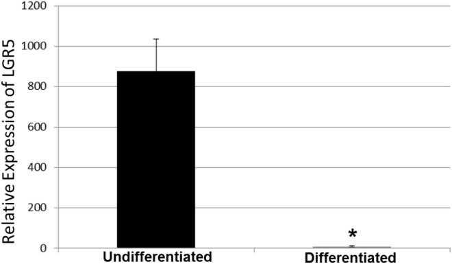
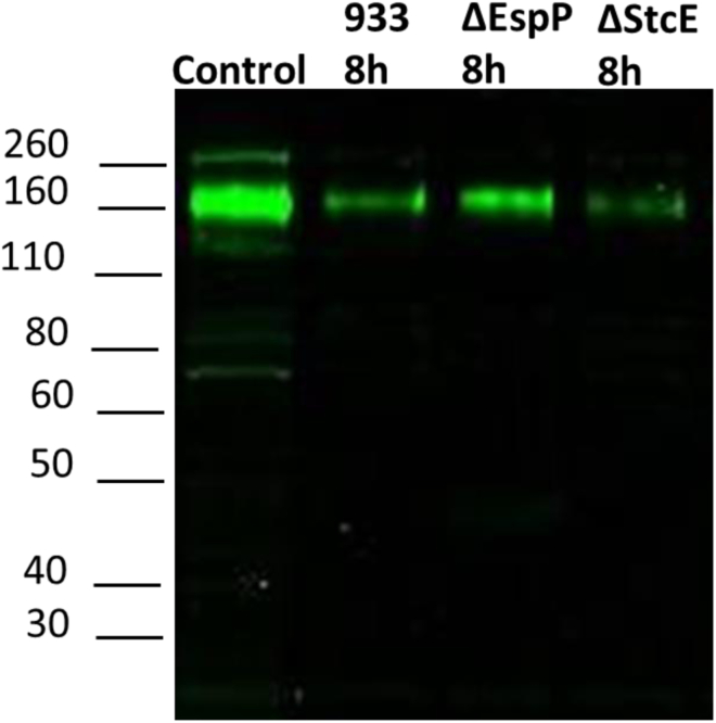
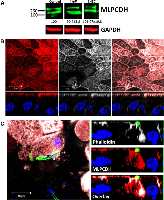
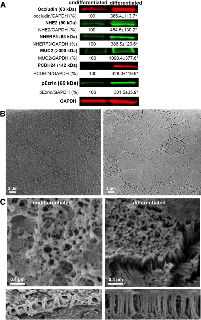
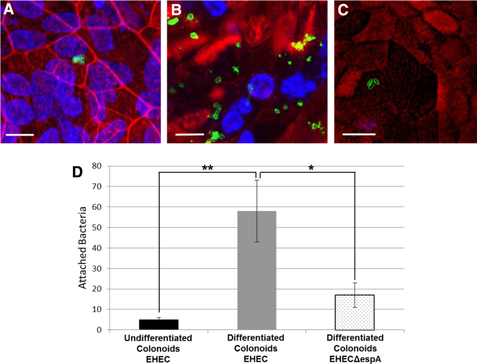
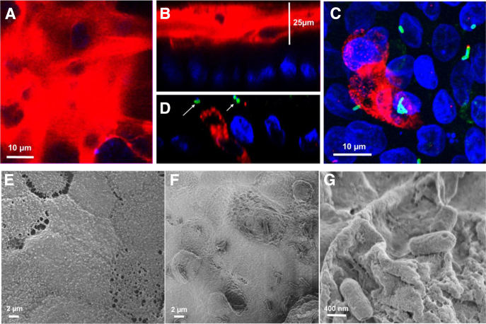
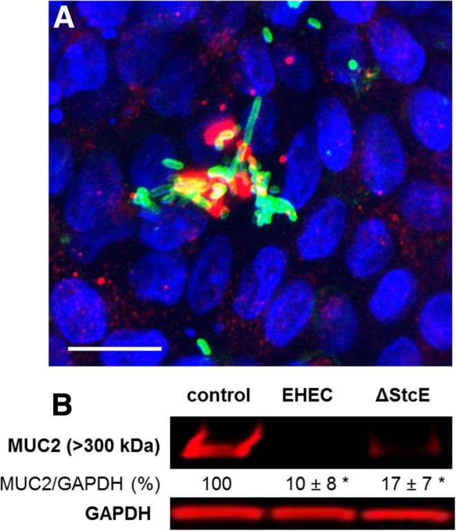
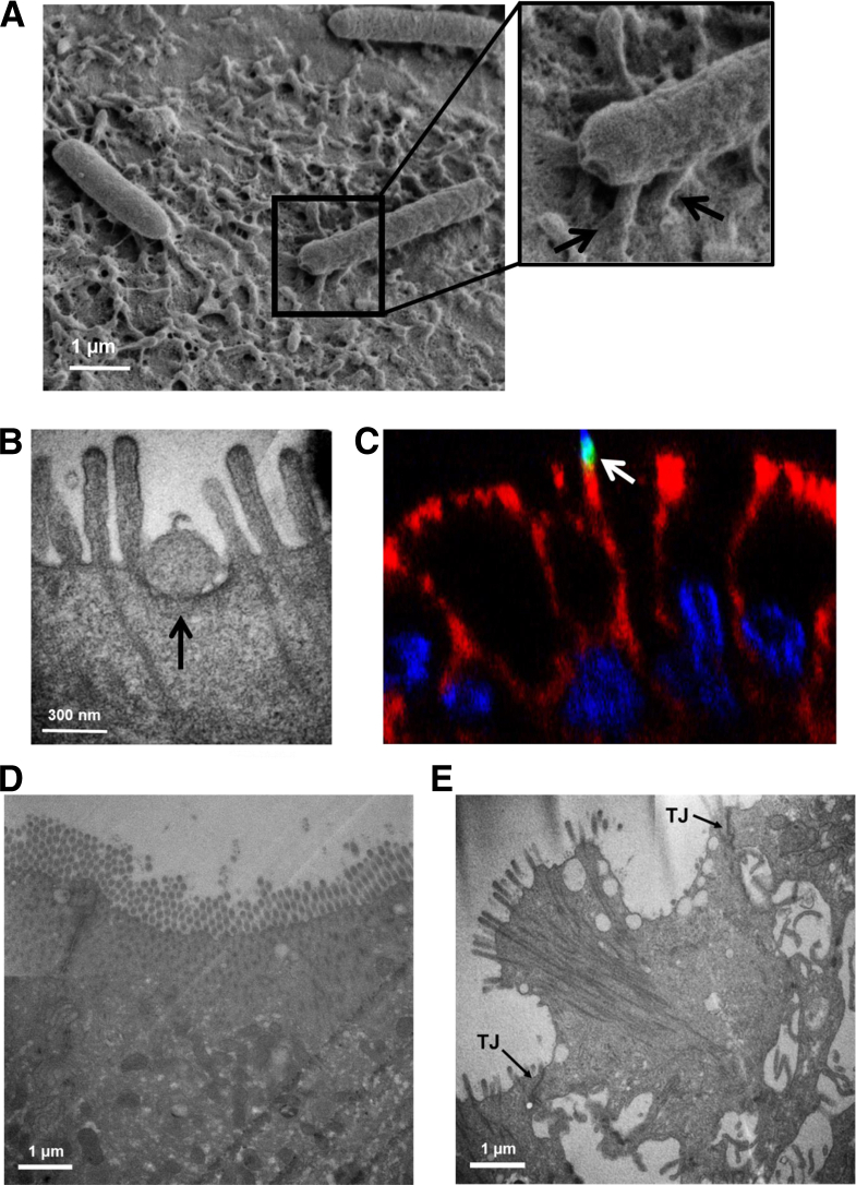
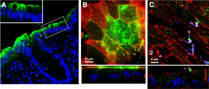
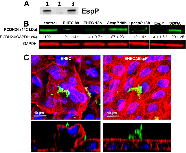
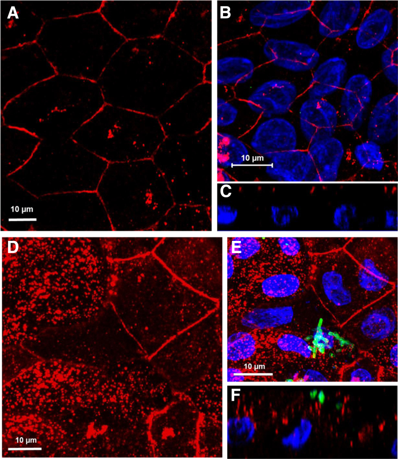
BACKGROUND AND AIMS: (EHEC) causes over 70,000 episodes of foodborne diarrhea annually in the USA. The early sequence of events which precede life-threatening hemorrhagic colitis and hemolytic uremic syndrome are not fully understood due to the initial asymptomatic phase of the disease and the lack of a suitable animal model. The aim of this study was to determine the initial molecular events in the interaction between EHEC and human colonic epithelium. METHODS: Human colonoids derived from adult proximal colonic stem cells were developed into monolayers to study EHEC-epithelial interactions. Monolayer confluency and differentiation were monitored by transepithelial electrical resistance (TER) measurements. The monolayers were apically infected with EHEC and the progression of epithelial damage over time was assessed using biochemical and imaging approaches. RESULTS: Human colonoid cultures recapitulate the differential protein expression patterns characteristic of the crypt and surface colonocytes. Mucus-producing differentiated colonoid monolayers are preferentially colonized by EHEC. Upon colonization, EHEC forms characteristic attaching and effacing lesions on the apical surface of colonoid monolayers. Mucin 2, a main component of colonic mucus, and protocadherin 24 (PCDH24), a microvillar resident protein, are targeted by EHEC at early stages of infection. The EHEC secreted serine protease, EspP, initiates brush border damage through PCDH24 reduction. CONCLUSIONS: Human colonoid monolayers are a relevant pathophysiological model which allows the study of early molecular events during enteric infections. Colonoid monolayers provide access to both apical and basolateral surfaces, thus providing an advantage over 3D cultures to study host-pathogen interactions in a controllable and tractable manner. EHEC reduces colonic mucus and affects the brush border cytoskeleton in the absence of commensal bacteria.
Overview
The claims and statistics of this paper haven’t been checked yet.
Run the check — free, no account needed. It reads the open-access text and checks the sources it cites.
Integrity signals
Registry
Cites
Statistics
Figures
Verification
Claims the paper makes
The claims and statistics of this paper haven’t been checked yet.
Run the check — free, no account needed. It reads the open-access text and checks the sources it cites.
Claims other papers make about this one
Each is checked against this paper’s own text: does it say what the citing paper says it does?
The cited source says what the claim attributes to it.
In the citing paper
“Furthermore, the recent development of organoid-derived 2D monolayer culture models has been instrumental for studies in which access to the apical surface is important [10,11,44].”
From Regulation of Enterocyte Brush Border Membrane Primary Na-Absorptive Transporters in Human Intestinal Organoid-Derived Monolayers (2024) · cites this paper as [44]
Self-check
Percentages, sums and reported differences re-computed from the paper’s own numbers.
The self-check needs the paper’s full text.
Citations
Cites
One-step inactivation of chromosomal genes in Escherichia coli K-12 using PCR products
Datsenko, K. A., & Wanner, B. L. (2000) · Proceedings of the National Academy of Sciences · doi:10.1073/pnas.120163297 · Cited by 14,881
Kaper, J. B., Nataro, J. P., & Mobley, H. L. T. (2004) · Nature Reviews Microbiology · doi:10.1038/nrmicro818 · Cited by 5,583
Self-citationSato, T., Stange, D. E., Ferrante, M., Vries, R. G. J., van Es, J. H., van den Brink, S., van Houdt, W. J., Pronk, A., van Gorp, J., Siersema, P. D., et al. (2011) · Gastroenterology · doi:10.1053/j.gastro.2011.07.050 · Cited by 3,753
Organoid Models and Applications in Biomedical Research
Xinaris, C., Brizi, V., & Remuzzi, G. (2015) · The Nephron journals/Nephron journals · doi:10.1159/000433566 · Cited by 3,519
The gastrointestinal mucus system in health and disease
Johansson, M. E. V., Sjövall, H., & Hansson, G. C. (2013) · Nature Reviews Gastroenterology & Hepatology · doi:10.1038/nrgastro.2013.35 · Cited by 1,438
Molecular mechanisms of Escherichia coli pathogenicity
Croxen, M. A., & Finlay, B. B. (2009) · Nature Reviews Microbiology · doi:10.1038/nrmicro2265 · Cited by 1,222
Origins of the E. coli Strain Causing an Outbreak of Hemolytic–Uremic Syndrome in Germany
Rasko, D. A., Webster, D. R., Sahl, J. W., Bashir, A., Boisen, N., Scheutz, F., Paxinos, E. E., Sebra, R., Chin, C.-S., Iliopoulos, D., et al. (2011) · New England Journal of Medicine · doi:10.1056/nejmoa1106920 · Cited by 835
Isolation and in vitro expansion of human colonic stem cells
Jung, P., Sato, T., Merlos-Suárez, A., Barriga, F. M., Iglesias, M., Rossell, D., Auer, H., Gallardo, M., Blasco, M. A., Sancho, E., et al. (2011) · Nature Medicine · doi:10.1038/nm.2470 · Cited by 709
Kosinski, C., Li, V. S. W., Chan, A. S. Y., Zhang, J., Ho, C., Tsui, W. Y., Chan, T. L., Mifflin, R. C., Powell, D. W., Yuen, S. T., et al. (2007) · Proceedings of the National Academy of Sciences · doi:10.1073/pnas.0707210104 · Cited by 596
Verocytotoxin-producing Escherichia coli (VTEC)
Karmali, M. A., Gannon, V. P. J., & Sargeant, J. M. (2009) · Veterinary Microbiology · doi:10.1016/j.vetmic.2009.04.011 · Cited by 543
Cited by
Sorted by citations
The intestinal epithelial barrier: a therapeutic target?
Odenwald, M. A., & Turner, J. R. (2017) · Nature Reviews Gastroenterology & Hepatology · doi:10.1038/nrgastro.2016.169 · Cited by 1,288
Martinez-Guryn, K., Hubert, N., Frazier, K., Urlass, S., Musch, M. W., Ojeda, P., Pierre, J. F., Miyoshi, J., Sontag, T. J., Cham, C. M., et al. (2018) · Cell Host & Microbe · doi:10.1016/j.chom.2018.03.011 · Cited by 663
Gut Microbiota and Colonization Resistance against Bacterial Enteric Infection
Ducarmon, Q. R., Zwittink, R. D., Hornung, B. V. H., van Schaik, W., Young, V. B., & Kuijper, E. J. (2019) · Microbiology and Molecular Biology Reviews · doi:10.1128/mmbr.00007-19 · Cited by 566
Human organoids in basic research and clinical applications
Tang, X.-Y., Wu, S., Wang, D., Chu, C., Hong, Y., Tao, M., Hu, H., Xu, M., Guo, X. L., & Liu, Y. (2022) · Signal Transduction and Targeted Therapy · doi:10.1038/s41392-022-01024-9 · Cited by 539
Nakamoto, N., Sasaki, N., Aoki, R., Miyamoto, K., Suda, W., Teratani, T., Suzuki, T., Koda, Y., Chu, P.-S., Taniki, N., et al. (2019) · Nature Microbiology · doi:10.1038/s41564-018-0333-1 · Cited by 425
Noel, G., Baetz, N. W., Staab, J. F., Donowitz, M., Kovbasnjuk, O., Pasetti, M. F., & Zachos, N. C. (2017) · Scientific Reports · doi:10.1038/srep45270 · Cited by 420
Self-citationModelling Cryptosporidium infection in human small intestinal and lung organoids
Heo, I., Dutta, D., Schaefer, D. A., Iakobachvili, N., Artegiani, B., Sachs, N., Boonekamp, K. E., Bowden, G. D., Hendrickx, A. P. A., Willems, R. J. L., et al. (2018) · Nature Microbiology · doi:10.1038/s41564-018-0177-8 · Cited by 386
Saxena, K., Blutt, S. E., Ettayebi, K., Zeng, X.-L., Broughman, J. R., Crawford, S. E., Karandikar, U. C., Sastri, N. P., Conner, M. E., Opekun, A. R., et al. (2016) · Journal of Virology · doi:10.1128/jvi.01930-15 · Cited by 375
Self-citationGut Microbiota and Intestinal Trans-Epithelial Permeability
Allam‐Ndoul, B., Castonguay-Paradis, S., & Veilleux, A. (2020) · International Journal of Molecular Sciences · doi:10.3390/ijms21176402 · Cited by 371
Rawat, M., Nighot, M., Al–Sadi, R., Gupta, Y. A., Viszwapriya, D., Yochum, G. S., Koltun, W. A., & Ma, T. Y. (2020) · Gastroenterology · doi:10.1053/j.gastro.2020.06.038 · Cited by 268
The mucosal barrier at a glance
France, M. M., & Turner, J. R. (2017) · Journal of Cell Science · doi:10.1242/jcs.193482 · Cited by 274
Williamson, I., Arnold, J. W., Samsa, L. A., Gaynor, L. T., DiSalvo, M., Cocchiaro, J. L., Carroll, I. M., Azcárate-Peril, M. A., Rawls, J. F., Allbritton, N. L., et al. (2018) · Cellular and Molecular Gastroenterology and Hepatology · doi:10.1016/j.jcmgh.2018.05.004 · Cited by 259
Sontheimer-Phelps, A., Chou, D. B., Tovaglieri, A., Ferrante, T. C., Duckworth, T., Fadel, C., Frismantas, V., Sutherland, A. D., Jalili-Firoozinezhad, S., Kasendra, M., et al. (2020) · Cellular and Molecular Gastroenterology and Hepatology · doi:10.1016/j.jcmgh.2019.11.008 · Cited by 239
Mucus models to evaluate the diffusion of drugs and particles
Lock, J. Y., Carlson, T. L., & Carrier, R. L. (2017) · Advanced Drug Delivery Reviews · doi:10.1016/j.addr.2017.11.001 · Cited by 252
Co, J. Y., Margalef-Català, M., Monack, D. M., & Amieva, M. R. (2021) · Nature Protocols · doi:10.1038/s41596-021-00607-0 · Cited by 248
Organoids and organs-on-chips: Insights into human gut-microbe interactions
Puschhof, J., Pleguezuelos‐Manzano, C., & Clevers, H. C. (2021) · Cell Host & Microbe · doi:10.1016/j.chom.2021.04.002 · Cited by 233
Self-renewing Monolayer of Primary Colonic or Rectal Epithelial Cells
Wang, Y., DiSalvo, M., Gunasekara, D. B., Dutton, J. S., Proctor, A., Lebhar, M. S., Williamson, I., Speer, J. E., Howard, R. L., Smiddy, N. M., et al. (2017) · Cellular and Molecular Gastroenterology and Hepatology · doi:10.1016/j.jcmgh.2017.02.011 · Cited by 200
Human Enteroids as a Model of Upper Small Intestinal Ion Transport Physiology and Pathophysiology
Foulke-Abel, J. D., In, J., Yin, J., Zachos, N. C., Kovbasnjuk, O. N., Estes, M. K., de Jonge, H. R., & Donowitz, M. (2015) · Gastroenterology · doi:10.1053/j.gastro.2015.11.047 · Cited by 194
Self-citationDevelopment and Characterization of a Human and Mouse Intestinal Epithelial Cell Monolayer Platform
Kozuka, K., He, Y., Koo-McCoy, S., Kumaraswamy, P., Nie, B., Shaw, K., Chan, P., Leadbetter, M., He, L., Lewis, J. G., et al. (2017) · Stem Cell Reports · doi:10.1016/j.stemcr.2017.10.013 · Cited by 179
Experimental models to study intestinal microbes–mucus interactions in health and disease
Etienne‐Mesmin, L., Chassaing, B., Desvaux, M., Paepe, K. D., Gresse, R., Sauvaitre, T., Forano, É., de Wiele, T. V., Schüller, S., Juge, N., et al. (2019) · FEMS Microbiology Reviews · doi:10.1093/femsre/fuz013 · Cited by 193
Somatic cell-derived organoids as prototypes of human epithelial tissues and diseases
Fujii, M., & Sato, T. (2020) · Nature Materials · doi:10.1038/s41563-020-0754-0 · Cited by 176
Intestinal in vitro and ex vivo Models to Study Host-Microbiome Interactions and Acute Stressors
Pearce, S., Coia, H. G., Karl, J. P., Pantoja-Feliciano, I. G., Zachos, N. C., & Racicot, K. (2018) · Frontiers in Physiology · doi:10.3389/fphys.2018.01584 · Cited by 167
Self-citationKarve, S. S., Pradhan, S. K., Ward, D. V., & Weiss, A. A. (2017) · PLoS ONE · doi:10.1371/journal.pone.0178966 · Cited by 157
Barrila, J., Crabbé, A., Yang, J., Franco, K., Nydam, S. D., Forsyth, R. J., Davis, R. R., Gangaraju, S., Ott, C. M., Coyne, C. B., et al. (2018) · Infection and Immunity · doi:10.1128/iai.00282-18 · Cited by 163
Gut organoids: mini-tissues in culture to study intestinal physiology and disease
Almeqdadi, M., Mana, M., Roper, J., & Yilmaz, Ö. H. (2019) · American Journal of Physiology-Cell Physiology · doi:10.1152/ajpcell.00300.2017 · Cited by 160
van der Hee, B., Loonen, L. M. P., Taverne, N., Taverne-Thiele, J. J., Smidt, H., & Wells, J. M. (2018) · Stem Cell Research · doi:10.1016/j.scr.2018.02.013 · Cited by 144
Gastrointestinal tract modeling using organoids engineered with cellular and microbiota niches
Min, S., Kim, S., & Cho, S. (2020) · Experimental & Molecular Medicine · doi:10.1038/s12276-020-0386-0 · Cited by 151
Translational applications of adult stem cell-derived organoids
Drost, J., & Clevers, H. C. (2017) · Development · doi:10.1242/dev.140566 · Cited by 144
Primary Cell-Derived Intestinal Models: Recapitulating Physiology
Dutton, J. S., Hinman, S. S., Kim, R., Wang, Y., & Allbritton, N. L. (2018) · Trends in biotechnology · doi:10.1016/j.tibtech.2018.12.001 · Cited by 141
Mechanisms of Intestinal Epithelial Barrier Dysfunction by Adherent-Invasive Escherichia coli
Shawki, A., & McCole, D. F. (2016) · Cellular and Molecular Gastroenterology and Hepatology · doi:10.1016/j.jcmgh.2016.10.004 · Cited by 142
Altay, G., Larrañaga, E., Tosi, S., Barriga, F. M., Batlle, E., Fernández‐Majada, V., & Martínez, E. (2019) · Scientific Reports · doi:10.1038/s41598-019-46497-x · Cited by 141
Human mini-guts: New insights into intestinal physiology and host–pathogen interactions
In, J. G., Foulke-Abel, J., Estes, M. K., Zachos, N. C., Kovbasnjuk, O., & Donowitz, M. (2016) · Nature Reviews Gastroenterology & Hepatology · doi:10.1038/nrgastro.2016.142 · Cited by 125
Self-citationSaxena, K., Simon, L. M., Zeng, X., Blutt, S. E., Crawford, S. E., Sastri, N. P., Karandikar, U. C., Ajami, N. J., Zachos, N. C., Kovbasnjuk, O. N., et al. (2017) · Proceedings of the National Academy of Sciences · doi:10.1073/pnas.1615422114 · Cited by 120
Self-citationAdvancing Intestinal Organoid Technology Toward Regenerative Medicine
Nakamura, T., & Sato, T. (2017) · Cellular and Molecular Gastroenterology and Hepatology · doi:10.1016/j.jcmgh.2017.10.006 · Cited by 124
Evaluating Shigella flexneri Pathogenesis in the Human Enteroid Model
Ranganathan, S., Doucet, M., Grassel, C. L., Delaine-Elias, B., Zachos, N. C., & Barry, E. M. (2019) · Infection and Immunity · doi:10.1128/iai.00740-18 · Cited by 105
Self-citationIntestinal organoids: A new paradigm for engineering intestinal epithelium in vitro
Rahmani, S., Breyner, N. M., Su, H.-M., Verdu, E. F., & Didar, T. F. (2019) · Biomaterials · doi:10.1016/j.biomaterials.2018.12.006 · Cited by 98
The Past, Present and Future of Intestinal In Vitro Cell Systems for Drug Absorption Studies
Youhanna, S. C., & Lauschke, V. M. (2020) · Journal of Pharmaceutical Sciences · doi:10.1016/j.xphs.2020.07.001 · Cited by 103
The Use of Stem Cell-Derived Organoids in Disease Modeling: An Update
Azar, J. H., Bahmad, H. F., Daher, D., Moubarak, M. M., Hadadeh, O., Monzer, A., Bitar, S. A., Jamal, M., Al‐Sayegh, M., & Abou-Kheir, W. G. (2021) · International Journal of Molecular Sciences · doi:10.3390/ijms22147667 · Cited by 106
Biologically inspired approaches to enhance human organoid complexity
Holloway, E. M., Capeling, M. M., & Spence, J. R. (2019) · Development · doi:10.1242/dev.166173 · Cited by 99
Silberger, D. J., Zindl, C. L., & Weaver, C. T. (2017) · Mucosal Immunology · doi:10.1038/mi.2017.47 · Cited by 93
Copper Transport and Disease: What Can We Learn from Organoids?
Pierson, H. E., Yang, H., & Lutsenko, S. (2019) · Annual Review of Nutrition · doi:10.1146/annurev-nutr-082018-124242 · Cited by 94
Wang, B. X., Wheeler, K. M., Cady, K. C., Lehoux, S. D., Cummings, R. D., Laub, M. T., & Ribbeck, K. (2020) · Current Biology · doi:10.1016/j.cub.2020.09.088 · Cited by 92
Gastrointestinal Organoids: Understanding the Molecular Basis of the Host–Microbe Interface
Hill, D. R., & Spence, J. R. (2016) · Cellular and Molecular Gastroenterology and Hepatology · doi:10.1016/j.jcmgh.2016.11.007 · Cited by 90
Organoid-based Models to Study the Role of Host-microbiota Interactions in IBD
Poletti, M., Arnauts, K., Ferrante, M., & Korcsmáros, T. (2020) · Journal of Crohn s and Colitis · doi:10.1093/ecco-jcc/jjaa257 · Cited by 93
Chronic Inflammation in Ulcerative Colitis Causes Long-Term Changes in Goblet Cell Function
Singh, V., Johnson, K. F., Yin, J., Lee, S., Lin, R., Yu, H. M., In, J., Foulke-Abel, J. D., Zachos, N. C., Donowitz, M., et al. (2021) · Cellular and Molecular Gastroenterology and Hepatology · doi:10.1016/j.jcmgh.2021.08.010 · Cited by 88
Self-citationEngineered Human Gastrointestinal Cultures to Study the Microbiome and Infectious Diseases
Blutt, S. E., Crawford, S. E., Ramani, S., Zou, W. Y., & Estes, M. K. (2017) · Cellular and Molecular Gastroenterology and Hepatology · doi:10.1016/j.jcmgh.2017.12.001 · Cited by 86
Self-citationBein, A., Fadel, C. W., Swenor, B., Cao, W., Powers, R. K., Camacho, D. M., Naziripour, A., Parsons, A., LoGrande, N., Sanjay, S. T., et al. (2022) · Nature Biomedical Engineering · doi:10.1038/s41551-022-00899-x · Cited by 81
Rajan, A., Vela, L., Zeng, X., Yu, X., Shroyer, N. F., Blutt, S. E., Poole, N. M., Carlin, L. G., Nataro, J. P., Estes, M. K., et al. (2018) · mBio · doi:10.1128/mbio.02419-17 · Cited by 81
Self-citationOrganoid Models for Infectious Disease
Blutt, S. E., & Estes, M. K. (2021) · Annual Review of Medicine · doi:10.1146/annurev-med-042320-023055 · Cited by 81
Self-citation3D bioengineered tissue model of the large intestine to study inflammatory bowel disease
Roh, T. T., Chen, Y., Paul, H. T., Guo, C., & Kaplan, D. L. (2019) · Biomaterials · doi:10.1016/j.biomaterials.2019.119517 · Cited by 79
Applications of human organoids in the personalized treatment for digestive diseases
Wang, Q., Guo, F., Jin, Y., & Ma, Y. (2022) · Signal Transduction and Targeted Therapy · doi:10.1038/s41392-022-01194-6 · Cited by 87
Hews, C. L., Tran, S.-L., Wegmann, U., Brett, B., Walsham, A. D. S., Kavanaugh, D., Ward, N. J., Juge, N., & Schüller, S. (2017) · Cellular Microbiology · doi:10.1111/cmi.12717 · Cited by 77
Allaire, J. M., Morampudi, V., Crowley, S. M., Stahl, M., Yu, H., Bhullar, K., Knodler, L. A., Bressler, B. L., Jacobson, K., & Vallance, B. A. (2018) · American Journal of Physiology-Gastrointestinal and Liver Physiology · doi:10.1152/ajpgi.00181.2017 · Cited by 79
Organoid and Enteroid Modeling of Salmonella Infection
Yin, Y., & Zhou, D. (2018) · Frontiers in Cellular and Infection Microbiology · doi:10.3389/fcimb.2018.00102 · Cited by 74
Bovine colon organoids: From 3D bioprinting to cryopreserved multi-well screening platforms
Töpfer, E., Pasotti, A., Telopoulou, A., Italiani, P., Boraschi, D., Ewart, M., & Wilde, C. J. (2019) · Toxicology in Vitro · doi:10.1016/j.tiv.2019.104606 · Cited by 75
Coculture of primary human colon monolayer with human gut bacteria
Zhang, J., Hernandez‐Gordillo, V., Trapečar, M., Wright, C. W., Taketani, M., Schneider, K., Chen, W. L. K., Stas, E., Breault, D. T., Carrier, R. L., et al. (2021) · Nature Protocols · doi:10.1038/s41596-021-00562-w · Cited by 72
Intestinal enteroids/organoids: A novel platform for drug discovery in inflammatory bowel diseases
Yoo, J. H., & Donowitz, M. (2019) · World Journal of Gastroenterology · doi:10.3748/wjg.v25.i30.4125 · Cited by 77
Self-citationBioengineered Systems and Designer Matrices That Recapitulate the Intestinal Stem Cell Niche
Wang, Y., Kim, R., Hinman, S. S., Zwarycz, B., Magness, S. T., & Allbritton, N. L. (2018) · Cellular and Molecular Gastroenterology and Hepatology · doi:10.1016/j.jcmgh.2018.01.008 · Cited by 73
Ambrosini, Y. M., Park, Y., Jergens, A. E., Shin, W., Min, S., Atherly, T. A., Borcherding, D. C., Jang, J., Allenspach, K., Mochel, J. P., et al. (2020) · PLoS ONE · doi:10.1371/journal.pone.0231423 · Cited by 68
Hinman, S. S., Wang, Y., Kim, R., & Allbritton, N. L. (2020) · Nature Protocols · doi:10.1038/s41596-020-00419-8 · Cited by 65
Co‐Culture System of Human Enteroids/Colonoids with Innate Immune Cells
Staab, J. F., Lemme‐Dumit, J. M., Latanich, R., Pasetti, M. F., & Zachos, N. C. (2020) · Current Protocols in Immunology · doi:10.1002/cpim.113 · Cited by 71
Self-citationApostolou, A. P., Panchakshari, R. A., Banerjee, A., Manatakis, D. V., Paraskevopoulou, M. D., Luc, R., Abu-Ali, G., Dimitriou, A., Lucchesi, C., Kulkarni, G., et al. (2021) · Cellular and Molecular Gastroenterology and Hepatology · doi:10.1016/j.jcmgh.2021.07.004 · Cited by 66
Bacterial detection by NAIP/NLRC4 elicits prompt contractions of intestinal epithelial cell layers
Ventayol, P. S., Geiser, P., Martino, M. L. D., Florbrant, A., Fattinger, S. A., Walder, N., Sima, E., Shao, F., Gekara, N. O., Sundbom, M., et al. (2021) · Proceedings of the National Academy of Sciences · doi:10.1073/pnas.2013963118 · Cited by 65
Epigenetic Changes Induced by Bacteroides fragilis Toxin
Allen, J., Hao, S., Sears, C. L., & Timp, W. (2019) · Infection and Immunity · doi:10.1128/iai.00447-18 · Cited by 68
Creating a More Perfect Union: Modeling Intestinal Bacteria-Epithelial Interactions Using Organoids
Han, X., Mslati, M., Davies, E., Chen, Y., Allaire, J. M., & Vallance, B. A. (2021) · Cellular and Molecular Gastroenterology and Hepatology · doi:10.1016/j.jcmgh.2021.04.010 · Cited by 67
Wang, Y., Kim, R., Sims, C. E., & Allbritton, N. L. (2019) · Cellular and Molecular Gastroenterology and Hepatology · doi:10.1016/j.jcmgh.2019.07.009 · Cited by 62
Advances in modelling the human microbiome–gut–brain axis in vitro
Moysidou, C., & Owens, R. M. (2021) · Biochemical Society Transactions · doi:10.1042/bst20200338 · Cited by 60
In vitro models to study human gut-microbiota interactions: Applications, advances, and limitations
Qi, Y., Yu, L., Tian, F., Zhao, J., & Zhai, Q. (2023) · Microbiological Research · doi:10.1016/j.micres.2023.127336 · Cited by 65
Enterotoxigenic Escherichia coli–blood group A interactions intensify diarrheal severity
Kumar, P., Kuhlmann, F., Chakraborty, S., Bourgeois, A. L., Foulke-Abel, J. D., Tumala, B., Vickers, T. J., Sack, D. A., DeNearing, B., Harro, C. D., et al. (2018) · Journal of Clinical Investigation · doi:10.1172/jci97659 · Cited by 61
Self-citationPoole, N. M., Green, S. I., Rajan, A., Vela, L. E., Zeng, X., Estes, M. K., & Maresso, A. W. (2017) · Infection and Immunity · doi:10.1128/iai.00581-17 · Cited by 58
Self-citationYin, J., Tse, C.-M., Avula, L. R., Singh, V., Foulke-Abel, J., de Jonge, H. R., & Donowitz, M. (2018) · Cellular and Molecular Gastroenterology and Hepatology · doi:10.1016/j.jcmgh.2018.02.002 · Cited by 58
Self-citationCarvalho, I., Silva, N. A., Carrola, J. S., SILVA, V. Y. N. E. D., Currie, C., Igrejas, G., & Poeta, P. A. C. Q. D. (2019) · doi:10.1002/9781119282549.ch11 · Cited by 58
Cassotta, M., Forbes-Hernández, T. Y., Iglesias, R. C., Ruíz, R., Elexpuru-Zabaleta, M., Giampieri, F., & Battino, M. A. (2020) · Nutrients · doi:10.3390/nu12061827 · Cited by 59
Mucus layer modeling of human colonoids during infection with enteroaggragative E. coli
Liu, L., Saitz, W., Smith, R., Gonyar, L. A., In, J., Kovbasnjuk, O. N., Zachos, N. C., Donowitz, M., Nataro, J. P., & Ruı́z-Pérez, F. (2020) · Scientific Reports · doi:10.1038/s41598-020-67104-4 · Cited by 58
Self-citationYouhanna, S. C., Kemas, A. M., Preiss, L. C., Zhou, Y., Shen, J. X., Cakal, S. D., Paqualini, F. S., Goparaju, S. K., Shafagh, R. Z., Lind, J. U., et al. (2022) · Pharmacological Reviews · doi:10.1124/pharmrev.120.000238 · Cited by 61
Rapid Prototyping of Multilayer Microphysiological Systems
Hosic, S., Bindas, A. J., Puzan, M. L., Lake, W., Soucy, J. R., Zhou, F., Koppes, R. A., Breault, D. T., Murthy, S. K., & Koppes, A. N. (2020) · ACS Biomaterials Science & Engineering · doi:10.1021/acsbiomaterials.0c00190 · Cited by 54
Fernando, E. H., Dicay, M. S., Stahl, M., Gordon, M., Vegso, A. J., Baggio, C. H., Alston, L. A., Lopes, F., Baker, K., Hirota, S. A., et al. (2017) · American Journal of Physiology-Gastrointestinal and Liver Physiology · doi:10.1152/ajpgi.00152.2017 · Cited by 54
Jubelin, G., Desvaux, M., Schüller, S., Etienne‐Mesmin, L., Muniesa, M., & Blanquet‐Diot, S. (2018) · Microorganisms · doi:10.3390/microorganisms6040115 · Cited by 55
Microbe–Mucus Interface in the Pathogenesis of Colorectal Cancer
Coleman, O. I., & Haller, D. (2021) · Cancers · doi:10.3390/cancers13040616 · Cited by 54
Tse, C.-M., Yin, J., Singh, V., Sarker, R., Lin, R., Verkman, A. S., Turner, J. R., & Donowitz, M. (2019) · Cellular and Molecular Gastroenterology and Hepatology · doi:10.1016/j.jcmgh.2019.01.002 · Cited by 53
Self-citationGut-on-a-Chip Models: Current and Future Perspectives for Host–Microbial Interactions Research
Morelli, M., Kurek, D., Ng, C. P., & Queiroz, K. C. S. (2023) · Biomedicines · doi:10.3390/biomedicines11020619 · Cited by 58
Alpha-defensin-dependent enhancement of enteric viral infection
Wilson, S. S., Bromme, B. A., Holly, M. K., Wiens, M. E., Gounder, A. P., Sul, Y., & Smith, J. G. (2017) · PLoS Pathogens · doi:10.1371/journal.ppat.1006446 · Cited by 52
Impact of dietary fibers in infant formulas on gut microbiota and the intestinal immune barrier
Kong, C., Faas, M. M., de Vos, P., & Akkerman, R. (2020) · Food & Function · doi:10.1039/d0fo01700k · Cited by 51
Border Control: The Role of the Microbiome in Regulating Epithelial Barrier Function
Schreiber, F., Balas, I., Robinson, M. J., & Bakdash, G. (2024) · Cells · doi:10.3390/cells13060477 · Cited by 55
Physiologically relevant human tissue models for infectious diseases
Mills, M., & Estes, M. K. (2016) · Drug Discovery Today · doi:10.1016/j.drudis.2016.06.020 · Cited by 50
Self-citationThe Contributions of Human Mini-Intestines to the Study of Intestinal Physiology and Pathophysiology
Yu, H. M., Hasan, N. M., In, J., Estes, M. K., Kovbasnjuk, O. N., Zachos, N. C., & Donowitz, M. (2017) · Annual Review of Physiology · doi:10.1146/annurev-physiol-021115-105211 · Cited by 51
Self-citationMini-gut: a promising model for drug development
Yin, Y., de Jonge, H. R., Wu, X., & Yin, Y. (2019) · Drug Discovery Today · doi:10.1016/j.drudis.2019.06.006 · Cited by 49
Potential Use of Human Stem Cell-Derived Intestinal Organoids to Study Inflammatory Bowel Diseases.
Dotti, I., & Salas, A. (2018) · Europe PMC (PubMed Central) · doi:10.1093/ibd/izy275 · Cited by 49
Bioengineered Co-culture of organoids to recapitulate host-microbe interactions
Kim, M. B., Hwangbo, S., Jang, S., & Jo, Y. K. (2022) · Materials Today Bio · doi:10.1016/j.mtbio.2022.100345 · Cited by 49
Fluid shear stress enhances differentiation of jejunal human enteroids in Intestine-Chip
Yin, J., Sunuwar, L., Kasendra, M. J., Yu, H. M., Tse, C., Talbot, C. C., Boronina, T. N., Cole, R. N., Karalis, K., & Donowitz, M. (2020) · American Journal of Physiology-Gastrointestinal and Liver Physiology · doi:10.1152/ajpgi.00282.2020 · Cited by 45
Self-citationEverything You Always Wanted to Know About Organoid-Based Models (and Never Dared to Ask)
Hautefort, I., Poletti, M., Papp, D., & Korcsmáros, T. (2022) · Cellular and Molecular Gastroenterology and Hepatology · doi:10.1016/j.jcmgh.2022.04.012 · Cited by 49
Modeling Intestinal Stem Cell Function with Organoids
Takahashi, T., Fujishima, K., & Kengaku, M. (2021) · International Journal of Molecular Sciences · doi:10.3390/ijms222010912 · Cited by 45
In, J., Foulke-Abel, J. D., Clarke, E., & Kovbasnjuk, O. N. (2019) · Journal of Visualized Experiments · doi:10.3791/59357 · Cited by 44
Self-citationWang, Y., Kim, R., Hwang, S.-H. J., Dutton, J. S., Sims, C. E., & Allbritton, N. L. (2018) · Analytical Chemistry · doi:10.1021/acs.analchem.8b02835 · Cited by 40
Brush border protocadherin CDHR2 promotes the elongation and maximized packing of microvilli in vivo
Pinette, J. A., Mao, S., Millis, B. A., Krystofiak, E., Faust, J. J., & Tyska, M. J. (2018) · Molecular Biology of the Cell · doi:10.1091/mbc.e18-09-0558 · Cited by 40
Roles of the Cell Surface Architecture of Bacteroides and Bifidobacterium in the Gut Colonization
Nishiyama, K., Yokoi, T., Sugiyama, M., Osawa, R., Mukai, T., & Okada, N. (2021) · Frontiers in Microbiology · doi:10.3389/fmicb.2021.754819 · Cited by 41
Ranganathan, S., Smith, E. M., Foulke-Abel, J. D., & Barry, E. M. (2020) · Gut Microbes · doi:10.1080/19490976.2020.1795389 · Cited by 39
Self-citationGastrointestinal microphysiological systems
Blutt, S. E., Broughman, J. R., Zou, W. Y., Zeng, X., Karandikar, U. C., In, J., Zachos, N. C., Kovbasnjuk, O. N., Donowitz, M., & Estes, M. K. (2017) · Experimental Biology and Medicine · doi:10.1177/1535370217710638 · Cited by 41
Self-citationRecent progress in organoid culture to model intestinal epithelial barrier functions
Nakamura, T. (2018) · International Immunology · doi:10.1093/intimm/dxy065 · Cited by 40
Rajan, A., Robertson, M. J., Carter, H. E., Poole, N. M., Clark, J. R., Green, S. I., Criss, Z. K., Zhao, B., Karandikar, U. C., Xing, Y., et al. (2020) · PLoS Pathogens · doi:10.1371/journal.ppat.1008851 · Cited by 38
Self-citation
Metadata
- Authors
- In, J., Foulke-Abel, J., Zachos, N. C., Hansen, A.-M., Kaper, J. B., Bernstein, H. D., Halushka, M., Blutt, S., Estes, M. K., Donowitz, M., & Kovbasnjuk, O.
- Affiliations
- Johns Hopkins University (US)
- University of Maryland, Baltimore (US)
- National Institutes of Health (US)
- Baylor College of Medicine (US)
- Corresponding author
- Kovbasnjuk, Olga
- ORCID
- Venue
- Cellular and Molecular Gastroenterology and Hepatology
- Publisher
- Elsevier BV
- Published
- 2016-01
- Volume
- 2
- Issue
- 1
- Pages
- 48-62.e3
- ISSN
- 2352-345X (ISSN-L)
- PMC
- PMC4740923
- OpenAlex
- W2193652497
- Cited by
- 239
- References
- 42
- Citations by year
- 2026: 4 · 2025: 13 · 2024: 24 · 2023: 22 · 2022: 30 · 2021: 28 · 2020: 34 · 2019: 30 · 2018: 23 · 2017: 22 · 2016: 6
- FWCI
- 14.03
- Type
- journal article
- Language
- en
- Open access
- gold
- doi.org
- Licence
- Locations
- Cellular and Molecular Gastroenterology and Hepatology (publishedVersion, cc-by-nc-nd)
- Europe PMC (PubMed Central) (publishedVersion, cc-by-nc-nd)
- PubMed Central (publishedVersion, cc-by-nc-nd)
- PubMed (publishedVersion)
- DOAJ (DOAJ: Directory of Open Access Journals) (submittedVersion, cc-by-sa)
- Funders
- Johns Hopkins University
- National Institutes of Health (5r24dk064388-07, U18TR000552)
- National Institute of Allergy and Infectious Diseases (U19AI116497)
- National Institute of Diabetes and Digestive and Kidney Diseases (T32DK007632, P30DK089502, R24DK-064388, P01DK072084, R01DK026523, R24DK099803)
- National Center for Advancing Translational Sciences
- Topics
- Life Sciences › Immunology and Microbiology › Microbiology › Escherichia coli research studies
- Health Sciences › Medicine › Infectious Diseases › Clostridium difficile and Clostridium perfringens research
- Life Sciences › Immunology and Microbiology › Microbiology › Enterobacteriaceae and Cronobacter Research
- Keywords
- enterohemorrhagic Escherichia coli, colonoids, mucus production, MUC2, attaching and effacing lesions, host-pathogen interaction, transepithelial electrical resistance
About these checks
Registry, retraction, Cites and statistics results are computed, not judged, with one exception: a printed reference that no index lists is read once by a model before it counts as a citation error. Claims are extracted by a model and kept only when their quote appears verbatim in the text; each cited source is then checked, with a model’s help, for whether it says what the claim relies on. Those verdicts can be wrong — read the claim’s quote and the cited source before relying on one.
Statistics and arithmetic are recomputed, not judged.
Confidence bands are shown only for fields calibrated on our annotated test set. None is calibrated yet, so no band is shown.
What counts as a claim ›How we measure confidence ›How we check this ›
Are you an author, or do you see a problem with this page? Request a review
