

The DOI matches its Crossref record; 37 of the 37 references it cites were found in a registry or an index.
Abnormal blood–brain barrier permeability in normal appearing white matter in multiple sclerosis investigated by MRI
Open accessAbstract
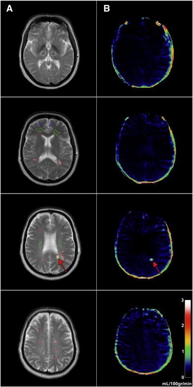
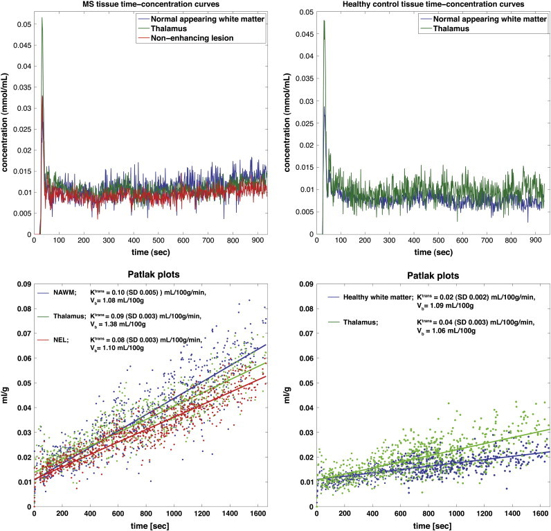
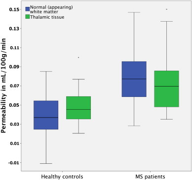
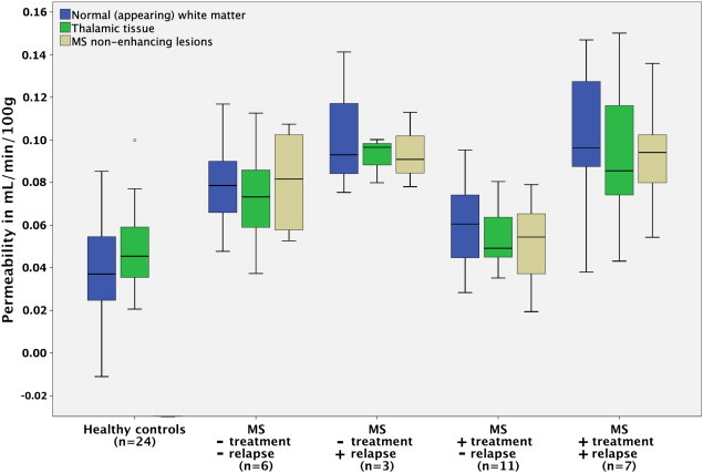
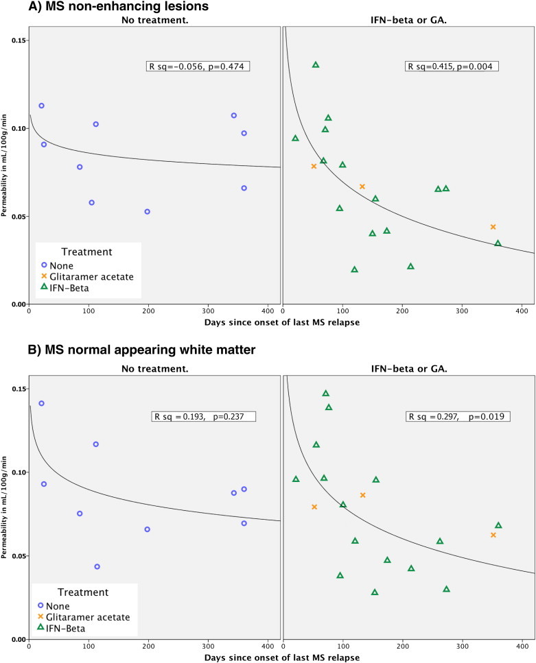
OBJECTIVES: To investigate whether blood-brain barrier (BBB) permeability is disrupted in normal appearing white matter in MS patients, when compared to healthy controls and whether it is correlated with MS clinical characteristics. METHODS: Dynamic contrast-enhanced MRI was used to measure BBB permeability in 27 patients with MS and compared to 24 matched healthy controls. RESULTS: Permeability measured as K(trans) was significantly higher in periventricular normal appearing white matter (NAWM) and thalamic gray matter in MS patients when compared to healthy controls, with periventricular NAWM showing the most pronounced difference. Recent relapse coincided with significantly higher permeability in periventricular NAWM, thalamic gray matter, and MS lesions. Immunomodulatory treatment and recent relapse were significant predictors of permeability in MS lesions and periventricular NAWM. Our results suggest that after an MS relapse permeability gradually decreases, possibly an effect of immunomodulatory treatment. CONCLUSIONS: Our results emphasize the importance of BBB pathology in MS, which we find to be most prominent in the periventricular NAWM, an area prone to development of MS lesions. Both the facts that recent relapse appears to cause widespread BBB disruption and that immunomodulatory treatment seems to attenuate this effect indicate that BBB permeability is intricately linked to the presence of MS relapse activity. This may reveal further insights into the pathophysiology of MS.
Overview
The claims and statistics of this paper haven’t been checked yet.
Run the check — free, no account needed. It reads the open-access text and checks the sources it cites.
Integrity signals
Registry
Cites
Statistics
Figures
Verification
Claims the paper makes
The claims and statistics of this paper haven’t been checked yet.
Run the check — free, no account needed. It reads the open-access text and checks the sources it cites.
Claims other papers make about this one
Each is checked against this paper’s own text: does it say what the citing paper says it does?
The source backs part of the claim, not all of it — or backs it for a different population, condition or comparison.
In the citing paper
“More recently, an increase in BBB permeability has been shown in MS patients by DCE-MRI in the cortex and adjacent white matter (392), corpus callosum and internal capsule (121-123, 421, 574), as well as thalamic gray matter (123).”
From Blood-Brain Barrier: From Physiology to Disease and Back (2019) · cites this paper as [123]
Experimental — accuracy not yet measured
Self-check
Percentages, sums and reported differences re-computed from the paper’s own numbers.
The self-check needs the paper’s full text.
Citations
Cites
Schumacher, G. A., Beebe, G., Kibler, R. F., Kurland, L. T., Kurtzke, J. F., McDowell, F., Nagler, B., Sibley, W. A., Tourtellotte, W. W., & Willmon, T. L. (1965) · Annals of the New York Academy of Sciences · doi:10.1111/j.1749-6632.1965.tb20235.x · Cited by 1,438 · cited 1×
“Relapsewasdefined as new or worsening symptoms that lasted 24 h and occurred in the absence of fever or infection (Schumacher et al., 1965).”
BackgroundProgressive multiple sclerosis: pathology and pathogenesis
Lassmann, H., van Horssen, J., & Mahad, D. (2012) · Nature Reviews Neurology · doi:10.1038/nrneurol.2012.168 · Cited by 1,033 · cited 5×
“…did not include secondary progressive MS patients, where the relationship between inflammation and damage to the BBB is less obvious than in RRMS (Lassmann et al., 2012), secondly, our cohorts were all evaluated for treatment intensification, thus presumably having a high relative disease…”
BackgroundInfluentialBlood-brain barrier disruption in multiple sclerosis
Minagar, A., & Alexander, J. S. (2003) · Multiple Sclerosis · doi:10.1191/1352458503ms965oa · Cited by 685 · cited 2×
“However, the MS disease process itself seems capable of modifying the permeability of the BBB (Larochelle et al., 2011; Minagar and Alexander, 2003) but we observed no correlation between permeability and age, EDSS or years sinceMS onset indicating that BBB abnormalities do not seem toworsen with disease progression.”
BackgroundKERMODE, A. G., THOMPSON, A. J., TOFTS, P., MACMANUS, D. G., KENDALL, B. E., KINGSLEY, D. P. E., MOSELEY, I. F., RUDGE, P., & MCDONALD, W. I. (1990) · Brain · doi:10.1093/brain/113.5.1477 · Cited by 578 · cited 1×
“All rights reser inactive MS lesions as well as normal appearing white matter (NAWM) (Kirk et al., 2003; McQuaid et al., 2009; Plumb et al., 2002; Vos et al., 2005), and serial MRI studies have indicated that structural changes (Filippi et al., 1998; Werring, 2000) and BBB dysfunction (Goodkin et…”
BackgroundHow do immune cells overcome the blood-brain barrier in multiple sclerosis?
Larochelle, C., Alvarez, J. I., & Prat, A. (2011) · FEBS Letters · doi:10.1016/j.febslet.2011.04.066 · Cited by 405 · cited 3×
“On the other hand, activated leukocytes themselves are capable of altering BBB permeability through expression and secretion of inflammatory cytokines (Larochelle et al., 2011).”
BackgroundFilippi, M., Rocca, M. A., Martino, G., Horsfield, M. A., & Comi, G. (1998) · Annals of Neurology · doi:10.1002/ana.410430616 · Cited by 395 · cited 2×
“…white matter (NAWM) (Kirk et al., 2003; McQuaid et al., 2009; Plumb et al., 2002; Vos et al., 2005), and serial MRI studies have indicated that structural changes (Filippi et al., 1998; Werring, 2000) and BBB dysfunction (Goodkin et al., 1998) may precedemyelin damage and leukocyte infiltration.”
BackgroundKirk, J., Plumb, J., Mirakhur, M., & McQuaid, S. (2003) · The Journal of Pathology · doi:10.1002/path.1434 · Cited by 375 · cited 2×
“All rights reser inactive MS lesions as well as normal appearing white matter (NAWM) (Kirk et al., 2003; McQuaid et al., 2009; Plumb et al., 2002; Vos et al., 2005), and serial MRI studies have indicated that structural changes (Filippi et al., 1998; Werring, 2000) and BBB dysfunction (Goodkin et…”
BackgroundHarris, J. O., Frank, J., Patronas, N., Mcfarlin, D. E., & McFarland, H. (1991) · Annals of Neurology · doi:10.1002/ana.410290515 · Cited by 334
Werring, D. J. (2000) · Brain · doi:10.1093/brain/123.8.1667 · Cited by 323 · cited 3×
“…white matter (NAWM) (Kirk et al., 2003; McQuaid et al., 2009; Plumb et al., 2002; Vos et al., 2005), and serial MRI studies have indicated that structural changes (Filippi et al., 1998; Werring, 2000) and BBB dysfunction (Goodkin et al., 1998) may precedemyelin damage and leukocyte infiltration.”
BackgroundEpstein-Barr virus infection is not a characteristic feature of multiple sclerosis brain.
Willis, S., Stadelmann, C., Rodig, S., Caron, T. J., Gattenloehner, S., Mallozzi, S. S., Roughan, J. E., Almendinger, S. E., Blewett, M. M., Brück, W., et al. (2009) · Brain : a journal of neurology · doi:10.1093/brain/awp200 · Cited by 304 · cited 1×
“Only a minority of cells need be infected and express viral genes, which would serve to explain the inconsistent detection of EBV in MS brains (Willis et al., 2009) and CSF (Mancuso et al., 2010).”
Background
Cited by
Sorted by citations
Items 101–200 of 206
Neuroimaging Studies Illustrate the Commonalities Between Ageing and Brain Diseases
Cole, J. H. (2018) · BioEssays · doi:10.1002/bies.201700221 · Cited by 18
Impaired Cerebral Perfusion in Multiple Sclerosis: Relevance of Endothelial Factors
Monti, L., Morbidelli, L., & Rossi, A. (2018) · Biomarker Insights · doi:10.1177/1177271918774800 · Cited by 20
Dynamic Contrast-Enhanced MRI and Its Applications in Various Central Nervous System Diseases
Kim, K. J., Park, M., Joo, B., Ahn, S. J., & Suh, S. H. (2022) · Investigative Magnetic Resonance Imaging · doi:10.13104/imri.2022.26.4.256 · Cited by 24
Ritson, M., Wheeler-Jones, C. P. D., & Stolp, H. B. (2024) · Journal of Neuroimmunology · doi:10.1016/j.jneuroim.2024.578363 · Cited by 23
Leone, P., Arenas, Y. M., Balzano, T., Mincheva, G., Martínez-García, M., Montoliu, C., Llansola, M., & Felipo, V. (2023) · European Journal of Neurology · doi:10.1111/ene.15935 · Cited by 19
“[36] analyzed the BBB permeability and indicated that it is linked to the presence of multiple sclerosis relapse activity.”
BackgroundAkaishi, T., Takahashi, T., & Nakashima, I. (2018) · Journal of the Neurological Sciences · doi:10.1016/j.jns.2018.02.020 · Cited by 20
Nwafor, D. C., Brichacek, A. L., Ali, A., & Brown, C. M. (2021) · International Journal of Molecular Sciences · doi:10.3390/ijms22105257 · Cited by 22
Genomic, proteomic, and systems biology approaches in biomarker discovery for multiple sclerosis
Huizar, C. C., Raphael, I., & Forsthuber, T. G. (2020) · Cellular Immunology · doi:10.1016/j.cellimm.2020.104219 · Cited by 26
Cramer, S. P., Simonsen, H. J., Varatharaj, A., Galea, I., Frederiksen, J. L., & Larsson, H. B. W. (2018) · Annals of Neurology · doi:10.1002/ana.25219 · Cited by 21
“453 8mm(3)], field of view 5 2303 119mm(2)) with same orientation and slice thickness (8mm) as our DCE-MRI sequence, to manually draw ROIs in the periventricular NAWM, and in the normal-appearing thalamic gray matter in both hemispheres, avoiding inclusion of, or proximity to, any MS lesions or diffusely abnormal whit…”
BackgroundMethodSelf-citationRole of Blood-Brain barrier in bacterial translocation
Aborode, A. T., Adesola, R. O., Scott, G. Y., Adepoju, V. A., Akan, O. D., Ogunyemi, A. D., Adetunji, A. P.-G., Arogundade, F. Q., Kwaku, S. D., Emmanuel, A. O., et al. (2025) · Neuroscience · doi:10.1016/j.neuroscience.2025.06.037 · Cited by 18
Cerebrovascular Reactivity and Neurovascular Coupling in Multiple Sclerosis—A Systematic Review
Vestergaard, M. B., Frederiksen, J. L., Larsson, H. B. W., & Cramer, S. P. (2022) · Frontiers in Neurology · doi:10.3389/fneur.2022.912828 · Cited by 23
Self-citationDisease-directed engineering for physiology-driven treatment interventions in neurological disorders
Wood, T. R., & Nance, E. A. (2019) · APL Bioengineering · doi:10.1063/1.5117299 · Cited by 21
Nonconventional MRI biomarkers for in vivo monitoring of pathogenesis in multiple sclerosis
Londoño, A. C., & Mora, C. A. (2014) · Neurology Neuroimmunology & Neuroinflammation · doi:10.1212/nxi.0000000000000045 · Cited by 16
“Also, it has been shown that immunomodulatory therapy has the ability to repair the dysfunction of the BBB in MS.(16) To our knowledge, evidence of BBB dysfunction in NABT other than the presence of contrastenhancing lesions has not been reported in CIS or RIS.”
BackgroundInfluentialEvidence of emerging BBB changes in mid‐age apolipoprotein E epsilon‐4 carriers
Alruwais, N. M., Rusted, J. M., Tabet, N., & Dowell, N. G. (2022) · Brain and Behavior · doi:10.1002/brb3.2806 · Cited by 17
“The sample size was computed using nQuery power calculator (https://www.statsols.com/nquery) and was motivated by prior research (Cramer et al., 2014; Dowell et al., 2016; Montagne et al., 2016).”
MethodWaller, R., Narramore, R., Simpson, J. E., Heath, P. R., Verma, N., Tinsley, M., Barnes, J. R., Haris, H. T., Henderson, F. E., Matthews, F. E., et al. (2020) · Brain Pathology · doi:10.1111/bpa.12928 · Cited by 17
“This has been previously identified in the ageing white matter as evidenced by the accumulation of serum protein in CD astrocytes (21), but BBB disruption has also been identified in many other disorders including multiple sclerosis (55), amyotrophic lateral sclerosis (56), chronic traumatic encephalopathy (57), Parki…”
BackgroundDziedzic, A., Morel, A., Miller, E. D., Bijak, M., Śliwiński, T., Synowiec, E., Ceremuga, M., & Saluk, J. (2020) · Oxidative Medicine and Cellular Longevity · doi:10.1155/2020/2868014 · Cited by 16
“MS is an inflammatory demyelinating disease of CNS, nonetheless, tightly related to the injury of blood vessels, mainly as a result of augmented permeability of the blood-brain barrier (BBB) [6, 7].”
BackgroundNeuroprotective natural products against experimental autoimmune encephalomyelitis: A review
Mohtashami, L., Shakeri, A., & Javadi, B. (2019) · Neurochemistry International · doi:10.1016/j.neuint.2019.104516 · Cited by 17
Metabolic encephalopathy secondary to diabetic ketoacidosis: a case report
Tomkins, M., McCormack, R. A., O'Connell, K., Agha, A., & Merwick, Á. (2019) · BMC Endocrine Disorders · doi:10.1186/s12902-019-0398-8 · Cited by 15
Fawaz, C. N., Makki, I., Kazan, J. M., Gebara, N. Y., Andary, F. S., Itani, M. M., El-Sayyed, M., Zeidan, A., Quartarone, A., Darwish, H., et al. (2015) · Expert Review of Proteomics · doi:10.1586/14789450.2015.1099435 · Cited by 13
Cramer, S. P., Larsson, H. B. W., Knudsen, M. H., Simonsen, H. J., Vestergaard, M. B., & Lindberg, U. (2022) · Journal of Magnetic Resonance Imaging · doi:10.1002/jmri.28380 · Cited by 13
“For each tissue type, the mean value was used to reduce noise (eg four regions of WM were drawn, of which the mean was used to represent WM), as previously described.(2,5) See Fig.”
MethodSelf-citationHobson, N., Polster, S., Cao, Y., Flemming, K. D., Shu, Y., Huston, J., Gerrard, C., Selwyn, R., Mabray, M., Zafar, A., et al. (2019) · Journal of Magnetic Resonance Imaging · doi:10.1002/jmri.26927 · Cited by 14
Intracerebral Hemorrhage in Multiple Sclerosis: A Retrospective Cohort Study.
Zulfiqar, M., Qeadan, F., Ikram, A., Farooqui, M., Richardson, S., Calder, C. S., Quadri, S. A., Mathur, P., Ford, C., Gutierrez, S. O., et al. (2019) · Journal of Stroke & Cerebrovascular Diseases · doi:10.1016/j.jstrokecerebrovasdis.2018.09.050 · Cited by 14
Bell, L., Koeniger, T., Tacke, S., & Küerten, S. (2019) · Histochemistry and Cell Biology · doi:10.1007/s00418-019-01768-6 · Cited by 17
Koch, T., Flemisch, B., Helmig, R., Wiest, R., & Obrist, D. (2018) · bioRxiv · doi:10.1002/cnm.3298 · Cited by 14
“That this effect may indeed occur is suggested in several clinical studies.(8,73) However, this effect cannot be seen for the numerical experiment where σ is assumed 10 times higher (Figure 10, right).”
BackgroundModeling compartmentalized chronic immune-mediated demyelinating CNS disease in the Biozzi ABH mouse
Nishri, Y., Fainstein, N., Goldfarb, S., Hampton, D., Macrini, C., Meinl, E., Chandran, S., & Ben-Hur, T. (2021) · Journal of Neuroimmunology · doi:10.1016/j.jneuroim.2021.577582 · Cited by 14
Oghabian, M. A., Fatemidokht, A., & Harirchian, M. H. (2022) · Basic and Clinical Neuroscience Journal · doi:10.32598/bcn.2022.575.1 · Cited by 17
“They showed an increase in measured parameters, which had significant differences between NAWM and WM lesions in MS brains (Cramer et al., 2014; Taheri et al., 2013).”
BackgroundMethodInfluential7T dynamic contrast‐enhanced MRI for the detection of subtle blood–brain barrier leakage
Canjels, L., Jansen, J., van den Kerkhof, M., Alers, R., Poser, B., Wiggins, C., Schiffer, V., van de Ven, V., Rouhl, R., Palm, W. M., et al. (2021) · Journal of Neuroimaging · doi:10.1111/jon.12894 · Cited by 13
Choi, S., Spini, M., Hua, J., & Harrison, D. M. (2021) · PLoS ONE · doi:10.1371/journal.pone.0249973 · Cited by 11
“In fact, prior DCE MRI studies suggest increased permeability in non-enhancing WMLs and NAWM in patients with MS [8].”
BackgroundRestoring brain barriers: an innovative approach for treating neurological disorders
Lemarchant, S., Engelhardt, B., Cicchetti, F., Bix, G. J., Janus, A., Godfrin, Y., Blasco, H., Campbell, M., & Jacquet, A. D. R. (2025) · Fluids and Barriers of the CNS · doi:10.1186/s12987-025-00688-z · Cited by 16
Knudsen, M. H., Lindberg, U., Frederiksen, J. L., Vestergaard, M. B., Simonsen, H. J., Varatharaj, A., Galea, I., Blinkenberg, M., Sellebjerg, F. T., Larsson, H. B. W., et al. (2022) · Multiple Sclerosis and Related Disorders · doi:10.1016/j.msard.2022.103891 · Cited by 13
Self-citationGarcía, F., & Heiman, M. (2025) · Molecular Neurodegeneration · doi:10.1186/s13024-025-00799-z · Cited by 14
“Similar findings have been found within the substantia nigra of PD patients [137], caudate nucleus of HD patients [138], and white matter regions of MS patients [139].”
Lu, Q., Ma, J., Zhao, Y., Ding, G., Wang, Y., Qiao, X., & Cheng, X. (2024) · Biomedicine & Pharmacotherapy · doi:10.1016/j.biopha.2024.117521 · Cited by 11
Shalom, E. S., Kim, H., van der Heijden, R. A., Ahmed, Z., Patel, R., Hormuth, D. A., DiCarlo, J. C., Yankeelov, T. E., Sisco, N. J., Dortch, R. D., et al. (2023) · Magnetic Resonance in Medicine · doi:10.1002/mrm.29909 · Cited by 14
“The K trans values in NAWM have been shown to be small but non-zero in several studies at high field strengths, 44–46 with a distinguishable difference in values also reported between some patient groups with healthy controls.”
Bateman, G., Bateman, A. R., & Lechner-Scott, J. (2023) · Multiple Sclerosis and Related Disorders · doi:10.1016/j.msard.2023.104843 · Cited by 11
Li, K.-L., Zhu, X.-P., Zhao, S., & Jackson, A. (2017) · Journal of Magnetic Resonance Imaging · doi:10.1002/jmri.25573 · Cited by 11
A peptide targeting inflammatory CNS lesions in the EAE rat model of multiple sclerosis
Boiziau, C., Nikolski, M., Mordelet, E., Aussudre, J., Vargas-Sanchez, K., & Petry, K. G. (2018) · Inflammation · doi:10.1007/s10753-018-0748-0 · Cited by 10
Li, K.-L., Lewis, D., Coope, D., Roncaroli, F., Agushi, E., Pathmanaban, O., King, A., Zhao, S., Jackson, A., Cootes, T., et al. (2021) · Magnetic Resonance in Medicine · doi:10.1002/mrm.28842 · Cited by 10
The development and impact of cladribine on lymphoid and myeloid cells in multiple sclerosis
Voo, V. T. F., Butzkueven, H., Stankovich, J. M., O'Brien, T. J., & Monif, M. (2021) · Multiple Sclerosis and Related Disorders · doi:10.1016/j.msard.2021.102962 · Cited by 11
Lezhnyova, V., Martynova, E. V., Khaiboullin, T. I., Urbanowicz, R. A., Khaiboullina, S. F., & Rizvanov, A. A. (2020) · Biology · doi:10.3390/biology9120464 · Cited by 13
Xiong, H., Yin, P., Li, X., Yang, C., Zhang, D., Huang, X., & Tang, Z. (2019) · Therapeutics and Clinical Risk Management · doi:10.2147/tcrm.s189598 · Cited by 10
“NE lesions and NAWM regions, which is consistent with previous study.(23) Cramer et al(4) found a slightly higher K”
ResultChaos theory for clinical manifestations in multiple sclerosis
Akaishi, T., Takahashi, T., & Nakashima, I. (2018) · Medical Hypotheses · doi:10.1016/j.mehy.2018.04.004 · Cited by 14
Assessing Treatment Response Through Generalized Pharmacokinetic Modeling of DCE-MRI Data
Kontopodis, E., Kanli, G., Manikis, G. C., van Cauter, S., & Marias, K. (2015) · Cancer Informatics · doi:10.4137/cin.s19342 · Cited by 10
“This way, the tissue concentration is given by Equation (4):”
MethodBlood–brain barrier and brain structural changes in lung cancer patients with non-brain metastases
Zhang, D.-F., Ma, H., Yang, G.-J., Zhang, Z.-P., He, Y., Feng, M., Shan, B.-C., Xu, X.-F., Ding, Y.-Y., & Cheng, Y.-Q. (2022) · Frontiers in Oncology · doi:10.3389/fonc.2022.1015011 · Cited by 9
“This parameter has been confirmed to be increased in patients with BMs (44), multiple sclerosis (22), stroke (23), traumatic brain injury (20), and dementia (24).”
BackgroundArtzi, M., Liberman, G., Blumenthal, D., Bokstein, F., Aizenstein, O., & Bashat, D. B. (2018) · Journal of Neuro-Oncology · doi:10.1007/s11060-018-03006-w · Cited by 9
Cen, Q., Cui, Y., Jin, J., Feng, J., Xin, Y., Zhang, Z., Li, J., Wang, J., & Zhang, A. (2025) · Gut Microbes · doi:10.1080/19490976.2025.2576657 · Cited by 8
Amin, N. S., & Tayebi, H. M. E. (2022) · Multiple Sclerosis and Related Disorders · doi:10.1016/j.msard.2022.104401 · Cited by 9
Gadolinium leakage in ocular structures in optic neuritis.
Förster, A., Böhme, J., Groden, C., & Wenz, H. (2019) · Journal of clinical neuroscience · doi:10.1016/j.jocn.2019.05.050 · Cited by 8
Samiee, F., & Samiee, K. (2016) · Electromagnetic Biology and Medicine · doi:10.3109/15368378.2016.1144064 · Cited by 7
Mokarizadeh, A., Esmaeili, P., Soraya, H., Hassanzadeh, K., Jalili, A., Abdi, M., & Faryabi, M. R. (2015) · Journal of Medical Hypotheses and Ideas · doi:10.1016/j.jmhi.2015.02.002 · Cited by 6
Blood-brain barrier at the interface of air pollution-associated neurotoxicity and neuroinflammation
Young, T. L., Zychowski, K. E., Denson, J. L., & Campen, M. J. (2018) · Advances in neurotoxicology · doi:10.1016/bs.ant.2018.10.010 · Cited by 11
Chen, X., Luo, J., Song, M., Pan, L., Qu, Z., Huang, B., Yu, S., & Shu, H. (2024) · Frontiers in Aging Neuroscience · doi:10.3389/fnagi.2024.1342366 · Cited by 5
Hirata, T., Itokazu, T., Sasaki, A., Sugihara, F., & Yamashita, T. (2022) · Frontiers in Immunology · doi:10.3389/fimmu.2022.870126 · Cited by 9
Mortazavi, M., Hizarci, Ö., Gerdes, L. A., Havla, J. B., Kümpfel, T., Hohlfeld, R., Stoecklein, S., Keeser, D., & Ertl-Wagner, B. B. (2021) · NeuroImage Clinical · doi:10.1016/j.nicl.2021.102734 · Cited by 10
Alatrash, R., Golubenko, M., Martynova, E. V., Garanina, E. E., Mukhamedshina, Y. O., Khaiboullina, S. F., Rizvanov, A. A., Salafutdinov, I. I., & Arkhipova, S. S. (2023) · International Journal of Molecular Sciences · doi:10.3390/ijms24098332 · Cited by 4
“This is also accompanied by the loss of terminal processes around small blood vessels and increased blood–brain barrier (BBB) permeability, leading to CNS inflammation and perivascular edema [61,62].”
BackgroundTemporal Huber Regularization for DCE-MRI
Hanhela, M., Kettunen, M., Gröhn, O., Vauhkonen, M., & Kolehmainen, V. (2020) · Journal of Mathematical Imaging and Vision · doi:10.1007/s10851-020-00985-2 · Cited by 5
Koch, T. (2020) · doi:10.18419/opus-10975 · Cited by 5
“That this effect may indeed occur, is suggested in several clinical studies (Ingrisch et al., 2012; Cramer et al., 2014).”
BackgroundCramer, S., Hamrouni, N., Simonsen, H. J., Vestergaard, M., Varatharaj, A., Galea, I., Lindberg, U., Frederiksen, J., & Larsson, H. (2025) · Frontiers in Neuroscience · doi:10.3389/fnins.2025.1546236 · Cited by 4
Self-citationLindland, E. S., Solheim, A. M., Andreassen, S., Bugge, R., Eikeland, R., Reiso, H., Lorentzen, Å. R., Harbo, H. F., Beyer, M. K., & Bjørnerud, A. (2023) · European Radiology Experimental · doi:10.1186/s41747-023-00365-6 · Cited by 7
“Among reported findings are increased leakage in hippocampus associated with ageing and mild cognitive impairment [9], and in white matter and thalamus of patients with multiple sclerosis which was predicted by disease activity [7].”
BackgroundResultMassacesi, L., Mariottini, A., & Nicoletti, F. (2022) · Journal of Clinical Medicine · doi:10.3390/jcm11154288 · Cited by 4
“It may be tempting to speculate that such molecules can achieve their target within the leptomeningeal follicle-like structures where the BEE is dysfunctional, or that they might migrate to CNS parenchyma through the BBB in areas involved by smouldering inflammation, even in the absence of gadolinium leakage; indeed,…”
BackgroundEmblem, K., Larsson, C., Groote, I., & Bjørnerud, A. (2020) · Springer eBooks · doi:10.1007/978-3-030-48419-4_11 · Cited by 4
Hills, C., Hernandez, C. A., Gabbay, V., & Berman, J. W. (2026) · Cells · doi:10.3390/cells15050399 · Cited by 3
El-Taibany, A. A., Heydarian, P., Porada, D. A., Seeds, M. C., & Atala, A. (2025) · Frontiers in Immunology · doi:10.3389/fimmu.2025.1595276 · Cited by 11
Treating acute ischemic stroke in a patient with multiple sclerosis: A challenging issue.
de Falco, A., Simone, M. D., D'Onofrio, F., Spitaleri, D., & de Falco, F. D. (2020) · Multiple Sclerosis and Related Disorders · doi:10.1016/j.msard.2020.101962 · Cited by 3
Wang, S., Liu, M., Shi, X., Xiong, T., Li, R., Zheng, W., Huang, Q., Nan, Y., & Ai, K. (2026) · 中国科学通报:英文版 · doi:10.1016/j.scib.2026.05.020 · Cited by 2
Spatial memory in Alzheimer’s disease 5XFAD mice is enhanced by XPO1 inhibitor KPT-330
Wong, S., Ouellette, A., Harrison, L., McNamara, A. D., Tam, R. A., Alexandrov, A., Nawrocik-Madrid, A., Sanchez, J. J., Ginsburg, B. C., Andrade, A. A., et al. (2026) · GeroScience · doi:10.1007/s11357-026-02122-0 · Cited by 2
“Some neurological pathologies such as schizophrenia and multiple sclerosis, as well as traumatic brain injury, have been shown to weaken the blood–brain barrier at the thalamus [30–32].”
Age-related decline in cerebral oxygen consumption in multiple sclerosis
Knudsen, M. H., Vestergaard, M., Lindberg, U., Simonsen, H. J., Frederiksen, J. L., Cramer, S., & Larsson, H. B. W. (2024) · Journal of Cerebral Blood Flow and Metabolism · doi:10.1177/0271678x231224502 · Cited by 2
“67 Pericytes govern the integrity of the BBB and regulate perfusion at the capillary level.”
BackgroundSelf-citationKetabforoush, A., Tajik, A., Habibi, M. A., & Khoshsirat, N. (2024) · Current Therapeutic Research · doi:10.1016/j.curtheres.2024.100732 · Cited by 2
Li, K.-L., Lewis, D., Zhu, X.-P., Coope, D., Djoukhadar, I., King, A. T., Cootes, T., & Jackson, A. (2023) · Pharmaceuticals · doi:10.3390/ph16091282 · Cited by 2
“In keeping with previous literature demonstrating higher blood volume, higher vascular surface area, and higher transfer constant estimates in grey compared to white matter [15,55,56], our study data demonstrated higher Ktrans estimates within NAGM compared to NAWM.”
ResultSparse precontrast T1 mapping for high‐resolution whole‐brain DCE‐MRI
Zhu, Z.-B., Lebel, R., Bliesener, Y., Acharya, J., Frayne, R., & Nayak, K. (2021) · Magnetic Resonance in Medicine · doi:10.1002/mrm.28849 · Cited by 2
do Amaral, L. L. F., Fragoso, D. C., & da Rocha, A. J. (2019) · Arquivos de Neuro-Psiquiatria · doi:10.1590/0004-282x20190082 · Cited by 2
Cerebral blood flow as a mechanistic framework across neurological disease
Patel, S., Nischal, S. A., & Patel, N. K. (2026) · Discover Neuroscience · doi:10.1186/s13064-026-00269-2 · Cited by 1
“Evidence suggests that hypoxic ischaemia, a key consequence of impaired CBF, can activate inflammatory cascades and promote generation of reactive oxygen species (ROS), which damages the BBB and increases its permeability [110, 111].”
Petitclerc, L., Durrant, H., Hirschler, L., Václavů, L., & van Osch, M. V. (2026) · Journal of Cerebral Blood Flow and Metabolism · doi:10.1177/0271678x261429042 · Cited by 1
Bloodstream infections and Blood-Brain barrier Permeability: An observational cohort study
Underwood, J., Davies, K., Loveless, S., Cercignani, M., Dowell, N. G., Periche-Tomas, E., Ronen, I., Evans, J., McLaren, J. E., & Harrison, N. A. (2026) · Brain Behavior and Immunity · doi:10.1016/j.bbi.2026.106518 · Cited by 1
“BBB dysfunction is found early in many neurodegenerative conditions such as AD and multiple sclerosis and can be assessed in vivo using dynamic contrast-enhanced magnetic resonance imaging, DCE-MRI (Cramer et al., 2014; Varatharaj and Galea, 2017).”
InfluentialPassali, M., Knudsen, M. H., Josefsen, K. E., Antvorskov, J. C., Hindsholm, A. M., Lindberg, U., Frederiksen, J. L., Larsson, H. B. W., & Cramer, S. P. (2025) · Multiple Sclerosis Journal - Experimental Translational and Clinical · doi:10.1177/20552173251346979
Self-citationMaciak, K., Dziedzic, A., Szymański, J., Studzian, M., Redlicka, J., Miller, E. D., Michlewska, S., Jóźwiak, P., & Saluk, J. (2024) · Journal of Molecular Biology · doi:10.1016/j.jmb.2024.168885 · Cited by 3
Bressler, I., Bashat, D. B., Buchsweiler, Y., Aizenstein, O., Limon, D., Bokestein, F., Blumenthal, T., Nevo, U., & Artzi, M. (2022) · Magnetic Resonance Materials in Physics, Biology and Medicine · doi:10.1007/s10334-022-01045-z · Cited by 1
Shin, D. J., Choi, S.-H., Yoo, R.-E., Kang, K. M., Yun, T., Kim, J.-H., Sohn, C., Jo, S., & Lee, E. J. (2021) · Korean Journal of Radiology · doi:10.3348/kjr.2020.1201 · Cited by 1
“Some studies revealed that the permeability changes measured by DCE MRI in brain lesions without visible contrast enhancement, such as small vessel disease [32-34].”
BackgroundEmara, D. M., Abd, A. E. E., & Fatatry, A. M. E. (2018) · doi:10.1016/j.ejrnm.2017.12.006 · Cited by 1
Enhancing our understanding of white matter changes in early multiple sclerosis.
Naismith, R., & Cross, A. (2015) · Brain : a journal of neurology · doi:10.1093/brain/awv196 · Cited by 1
“Previously, this same group has demonstrated by using DCE that patients with established relapsingremitting multiple sclerosis have higher BBB permeability than controls (Cramer et al., 2014).”
Background[Contrast agents in MRI-diagnosis of multiple sclerosis].
Abakumova, T., Nukolova, N., Gusev, E., & Chekhonin, V. (2015) · Zhurnal Nevrologii i Psikhiatrii imeni S.S. Korsakova · doi:10.17116/jnevro20151151158-65 · Cited by 1
“Кроме этого, при РС наблюдается повышенная проницаемость гематоэнцефалического барьера (ГЭБ), что также позволяет улучшить визуализацию на Т1-взвешенных томограммах с помощью не только низкомолекулярных, но и макромолекулярных КВ [60, 61].”
Khalid, S., Abid, M., Bilal, H. M., Alharbi, S. S., & Aldawsari, M. (2026) · Computer Science Review · doi:10.1016/j.cosrev.2026.100982
Cooney, L. A., Lim, N., Harris, K. M., Smilek, D. E., Bathala, P., Stengelin, M. K., Sigal, G. B., Wohlstadter, J. N., Cencioni, M. T., Fernández-Velasco, J. I., et al. (2026) · Neurology Neuroimmunology & Neuroinflammation · doi:10.1212/nxi.0000000000200653
Tireli, E. D., Cramer, S., Lindberg, U., Tireli, D., Vestergaard, M., & Larsson, H. B. W. (2026) · Magnetic Resonance in Medicine · doi:10.1002/mrm.70554
Self-citationDuque, C., Sargento‐Freitas, J., Balabanov, R. D., Cohen, B., & Sorond, F. A. (2026) · Therapeutic Advances in Neurological Disorders · doi:10.1177/17562864261481753
Diffusion MRI reveals CNS microstructural alterations in DLBCL patients at risk of CNS relapse.
Nørskov, M. P., Cramer, E., Lindberg, U., Tireli, D., Hansen, A. E., Carlsen, J. F., Brown, P. D. N., Rinker, D. A., Bjørnerud, A., Larsson, H. B. W., et al. (2026) · Magnetic Resonance Imaging · doi:10.1016/j.mri.2026.110783
Self-citationNørskov, M. P., Cramer, E. P., Lindberg, U., Hansen, A. E., Carlsen, J. F., Brown, P. D. N., Larsson, H. B. W., Hutchings, M., & Cramer, S. P. (2026) · Tissue Barriers · doi:10.1080/21688370.2026.2696764
Self-citationZhang, D.-F., Ma, H., Zhang, Z.-P., Ai, J., cao, W., & Li, Z.-H. (2026) · Annals medicus · doi:10.1080/07853890.2026.2662776
“Cramer and Larsson’s study [22] pointed out that at low leakage rates, i.e. leakage rates below 0.3 ml/100g/min (approximately equal to 0.003 min −1 ), the Patlak model is recommended for analysis.”
InfluentialLarsson, H. B. W., Cramer, S., Tireli, E. D., Larsson, T. S. W., Lindberg, U., & Vestergaard, M. (2026) · EJNMMI Physics · doi:10.1186/s40658-026-00886-0
“The extraction fraction is estimated to around 0.04% and the corresponding K 1 to 0.02 ml/100 ml/ min, which is within the expected range, as seen in previous DCE studies [13, 14].”
InfluentialSelf-citationSemnic, I., Bonifačić, D., & Vuletić, V. (2026) · Brazilian Journal of Case Reports · doi:10.52600/2763-583x.bjcr.2026.6.1.bjcr170
“MS itself is associated with chronic neuroinflammation, BBB disruption, and endothelial dysfunction, all of which may predispose patients to cerebrovascular pathology [10,11].”
Senthil, S., Tagge, I., Fetco, D., Hsieh, C. H., Assemlal, H.-E., Karimaghaloo, Z., Fetco, E., Moore, G. R. W., Arnold, D. L., Rudko, D. A., et al. (2026) · Journal of Neuroimaging · doi:10.1111/jon.70132 · Cited by 4
Tireli, E. D., Cramer, S., Lindberg, U., Tireli, D., Vestergaard, M., & Larsson, H. (2026) · bioRxiv · doi:10.64898/2026.02.15.705995
“Accurate quantification of BBB permeability can be used to study neurological diseases such as multiple sclerosis, brain tumors, and neurodegenerative disorders (Cramer et al., 2014; Okuchi et al., 2019; Heye et al., 2014; Chagnot et al., 2021; Raja et al., 2018).”
Self-citationSundvall, L., Mikkelsen, I. K., Hansen, B., Eskildsen, S. F., Hjørringgaard, M. M., Nygaard, M. K. E., Rasmussen, P. V., Petersen, T., & Østergaard, L. (2025) · Neuroimage Reports · doi:10.1016/j.ynirp.2025.100309 · Cited by 1
“…damage (Witte et al., 2014; Chandler et al., 2023), oxidative stress (Trapp and Stys, 2009; Mahad et al., 2015; Haider et al., 2011; Lu et al., 2000), and endothelial dysfunction (Cashion et al., 2023; Cramer et al., 2014; Iadecola, 2017), contribute to both MS neurodegeneration and aging.”
The Effect of Gadolinium on Synthetic Magnetic Resonance Quantitative Imaging
Novakova, L., Rosenstein, I., Axelsson, M., & Ouellette, R. (2025) · Journal of Neuroimaging · doi:10.1111/jon.70104
Li, J.-F., Xie, L.-J., Xiao, L., Lu, Y.-X., Qin, L.-P., Lin, H., Zha, Y.-M., Zhang, B.-J., Yang, J., Qiu, W., et al. (2025) · BMC Neurology · doi:10.1186/s12883-025-04454-4
“Previous studies have indicated that the assessment of BBB permeability dysfunction serves as a hallmark of active lesions in multiple sclerosis, a classic autoimmune disease of the nervous system [22, 23].”
Erol, S., & Cebeci, H. (2025) · Genel Tıp Dergisi · doi:10.54005/geneltip.1599021
Multiple sclerosis: what have we learned and can we still learn from electron microscopy
Oost, W., Meilof, J. F., & Baron, W. (2025) · Cellular and Molecular Life Sciences · doi:10.1007/s00018-025-05690-0 · Cited by 7
“Several MRI studies revealed abnormalities in NAGM and NAWM, with more extensive changes in NAWM [51, 52], including disruption of blood–brain barrier (BBB) permeability [53, 54].”
Senthil, S., Tagge, I., Fetco, D., Hsieh, C. H., Assemlal, H.-E., Karimaghaloo, Z., Fetco, E., Moore, G. R. W., Arnold, D. L., Rudko, D. A., et al. (2025) · medRxiv · doi:10.1101/2025.03.26.25324694 · Cited by 2
“While immune cell trafficking into the central nervous system (CNS) through disruptions in the blood-brain barrier (BBB) is a well-established feature of MS (1– 4), recent studies have implicated the choroid plexus (ChP) in MS pathogenesis (5–7).”
Wang, F., Che, X., Yang, Q., Wang, R., Zeng, J., Chen, Y., Su, X., & Miao, J. (2022) · Neurology India · doi:10.4103/0028-3886.344667
Extended perfusion protocol for MS lesion quantification
Kontopodis, E., Kostas Marias, A., Manikis, G. C., Nikiforaki, K., Venianaki, M., Maris, T. G., Mastorodemos, V. C., Papadakis, G. Z., & Papadaki, E. Z. (2020) · Open Medicine · doi:10.1515/med-2020-0100
Glial cell interactions in neonatal hypoxic-ischemic injury
Ramljak, M., Andrade, M. F., Lacoste, B., Aguilar‐Valles, A., & Salmaso, N. (2025) · Glial health research. · doi:10.1016/j.ghres.2025.100006
Metadata
- Authors
- Cramer, S. P., Simonsen, H., Frederiksen, J. L., Rostrup, E., & Larsson, H. B. W.
- Affiliations
- University of Copenhagen (DK)
- Corresponding author
- Cramer, S.P.
- Venue
- NeuroImage: Clinical
- Publisher
- Elsevier BV
- Published
- 2014
- Volume
- 4
- Pages
- 182-189
- ISSN
- 2213-1582 (ISSN-L)
- PMC
- PMC3872721
- OpenAlex
- W2039561850
- Cited by
- 232
- References
- 37
- Citations by year
- 2026: 16 · 2025: 20 · 2024: 16 · 2023: 20 · 2022: 24 · 2021: 16 · 2020: 14 · 2019: 20 · 2018: 26 · 2017: 13 · 2016: 16 · 2015: 21 · 2014: 9
- FWCI
- 11.49
- Type
- journal article
- Language
- en
- Open access
- gold
- doi.org
- Licence
- Locations
- NeuroImage Clinical (publishedVersion, cc-by-nc-sa)
- Europe PMC (PubMed Central) (publishedVersion, cc-by-nc-sa)
- PubMed Central (publishedVersion, cc-by-nc-sa)
- PubMed (publishedVersion)
- BIBSYS Brage (BIBSYS (Norway)) (submittedVersion)
- DOAJ (DOAJ: Directory of Open Access Journals) (submittedVersion, cc-by-sa)
- NORA - Norwegian Open Research Archives (submittedVersion, cc-by-nc-sa)
- Research at the University of Copenhagen (University of Copenhagen) (submittedVersion)
- Funders
- Biogen
- Scleroseforeningen (14588)
- Multiple Sclerosis Society (14588)
- Region Hovedstaden
- Topics
- Health Sciences › Medicine › Clinical Neurology › Multiple Sclerosis Research Studies
- Life Sciences › Neuroscience › Neurology › Barrier Structure and Function Studies
- Health Sciences › Medicine › Radiology, Nuclear Medicine and Imaging › Advanced MRI Techniques and Applications
- Keywords
- multiple sclerosis, blood-brain barrier permeability, normal-appearing white matter, periventricular white matter, dynamic contrast-enhanced MRI, MS relapse, immunomodulatory therapy, MS lesions, blood-brain barrier disruption
- MeSH terms
- Adult, Blood-Brain Barrier, Capillary Permeability, Contrast Media, Female, Humans, Magnetic Resonance Imaging, Male, Middle Aged, Multiple Sclerosis, Severity of Illness Index, Imaging, Three-Dimensional, Perfusion Imaging, White Matter
About these checks
Registry, retraction, Cites and statistics results are computed, not judged, with one exception: a printed reference that no index lists is read once by a model before it counts as a citation error. Claims are extracted by a model and kept only when their quote appears verbatim in the text; each cited source is then checked, with a model’s help, for whether it says what the claim relies on. Those verdicts can be wrong — read the claim’s quote and the cited source before relying on one.
Statistics and arithmetic are recomputed, not judged.
Confidence bands are shown only for fields calibrated on our annotated test set. None is calibrated yet, so no band is shown.
What counts as a claim ›How we measure confidence ›How we check this ›
Are you an author, or do you see a problem with this page? Request a review
