

The DOI matches its Crossref record; 37 of the 37 references it cites were found in a registry or an index.
Abnormal blood–brain barrier permeability in normal appearing white matter in multiple sclerosis investigated by MRI
Open accessAbstract
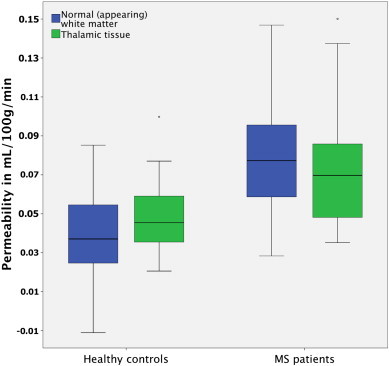
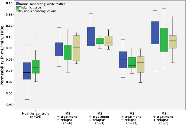
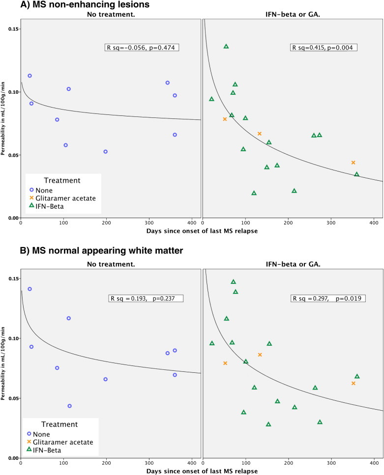
OBJECTIVES: To investigate whether blood-brain barrier (BBB) permeability is disrupted in normal appearing white matter in MS patients, when compared to healthy controls and whether it is correlated with MS clinical characteristics. METHODS: Dynamic contrast-enhanced MRI was used to measure BBB permeability in 27 patients with MS and compared to 24 matched healthy controls. RESULTS: Permeability measured as K(trans) was significantly higher in periventricular normal appearing white matter (NAWM) and thalamic gray matter in MS patients when compared to healthy controls, with periventricular NAWM showing the most pronounced difference. Recent relapse coincided with significantly higher permeability in periventricular NAWM, thalamic gray matter, and MS lesions. Immunomodulatory treatment and recent relapse were significant predictors of permeability in MS lesions and periventricular NAWM. Our results suggest that after an MS relapse permeability gradually decreases, possibly an effect of immunomodulatory treatment. CONCLUSIONS: Our results emphasize the importance of BBB pathology in MS, which we find to be most prominent in the periventricular NAWM, an area prone to development of MS lesions. Both the facts that recent relapse appears to cause widespread BBB disruption and that immunomodulatory treatment seems to attenuate this effect indicate that BBB permeability is intricately linked to the presence of MS relapse activity. This may reveal further insights into the pathophysiology of MS.
Overview
The claims and statistics of this paper haven’t been checked yet.
Run the check — free, no account needed. It reads the open-access text and checks the sources it cites.
Integrity signals
Registry
Cites
Statistics
Figures
Verification
Claims the paper makes
The claims and statistics of this paper haven’t been checked yet.
Run the check — free, no account needed. It reads the open-access text and checks the sources it cites.
Claims other papers make about this one
Each is checked against this paper’s own text: does it say what the citing paper says it does?
The source backs part of the claim, not all of it — or backs it for a different population, condition or comparison.
In the citing paper
“More recently, an increase in BBB permeability has been shown in MS patients by DCE-MRI in the cortex and adjacent white matter (392), corpus callosum and internal capsule (121-123, 421, 574), as well as thalamic gray matter (123).”
From Blood-Brain Barrier: From Physiology to Disease and Back (2019) · cites this paper as [123]
Experimental — accuracy not yet measured
Self-check
Percentages, sums and reported differences re-computed from the paper’s own numbers.
The self-check needs the paper’s full text.
Citations
Cites
Schumacher, G. A., Beebe, G., Kibler, R. F., Kurland, L. T., Kurtzke, J. F., McDowell, F., Nagler, B., Sibley, W. A., Tourtellotte, W. W., & Willmon, T. L. (1965) · Annals of the New York Academy of Sciences · doi:10.1111/j.1749-6632.1965.tb20235.x · Cited by 1,438 · cited 1×
“Relapsewasdefined as new or worsening symptoms that lasted 24 h and occurred in the absence of fever or infection (Schumacher et al., 1965).”
BackgroundProgressive multiple sclerosis: pathology and pathogenesis
Lassmann, H., van Horssen, J., & Mahad, D. (2012) · Nature Reviews Neurology · doi:10.1038/nrneurol.2012.168 · Cited by 1,033 · cited 5×
“…did not include secondary progressive MS patients, where the relationship between inflammation and damage to the BBB is less obvious than in RRMS (Lassmann et al., 2012), secondly, our cohorts were all evaluated for treatment intensification, thus presumably having a high relative disease…”
BackgroundInfluentialBlood-brain barrier disruption in multiple sclerosis
Minagar, A., & Alexander, J. S. (2003) · Multiple Sclerosis · doi:10.1191/1352458503ms965oa · Cited by 685 · cited 2×
“However, the MS disease process itself seems capable of modifying the permeability of the BBB (Larochelle et al., 2011; Minagar and Alexander, 2003) but we observed no correlation between permeability and age, EDSS or years sinceMS onset indicating that BBB abnormalities do not seem toworsen with disease progression.”
BackgroundKERMODE, A. G., THOMPSON, A. J., TOFTS, P., MACMANUS, D. G., KENDALL, B. E., KINGSLEY, D. P. E., MOSELEY, I. F., RUDGE, P., & MCDONALD, W. I. (1990) · Brain · doi:10.1093/brain/113.5.1477 · Cited by 578 · cited 1×
“All rights reser inactive MS lesions as well as normal appearing white matter (NAWM) (Kirk et al., 2003; McQuaid et al., 2009; Plumb et al., 2002; Vos et al., 2005), and serial MRI studies have indicated that structural changes (Filippi et al., 1998; Werring, 2000) and BBB dysfunction (Goodkin et…”
BackgroundHow do immune cells overcome the blood-brain barrier in multiple sclerosis?
Larochelle, C., Alvarez, J. I., & Prat, A. (2011) · FEBS Letters · doi:10.1016/j.febslet.2011.04.066 · Cited by 405 · cited 3×
“On the other hand, activated leukocytes themselves are capable of altering BBB permeability through expression and secretion of inflammatory cytokines (Larochelle et al., 2011).”
BackgroundFilippi, M., Rocca, M. A., Martino, G., Horsfield, M. A., & Comi, G. (1998) · Annals of Neurology · doi:10.1002/ana.410430616 · Cited by 395 · cited 2×
“…white matter (NAWM) (Kirk et al., 2003; McQuaid et al., 2009; Plumb et al., 2002; Vos et al., 2005), and serial MRI studies have indicated that structural changes (Filippi et al., 1998; Werring, 2000) and BBB dysfunction (Goodkin et al., 1998) may precedemyelin damage and leukocyte infiltration.”
BackgroundKirk, J., Plumb, J., Mirakhur, M., & McQuaid, S. (2003) · The Journal of Pathology · doi:10.1002/path.1434 · Cited by 375 · cited 2×
“All rights reser inactive MS lesions as well as normal appearing white matter (NAWM) (Kirk et al., 2003; McQuaid et al., 2009; Plumb et al., 2002; Vos et al., 2005), and serial MRI studies have indicated that structural changes (Filippi et al., 1998; Werring, 2000) and BBB dysfunction (Goodkin et…”
BackgroundHarris, J. O., Frank, J., Patronas, N., Mcfarlin, D. E., & McFarland, H. (1991) · Annals of Neurology · doi:10.1002/ana.410290515 · Cited by 334
Werring, D. J. (2000) · Brain · doi:10.1093/brain/123.8.1667 · Cited by 323 · cited 3×
“…white matter (NAWM) (Kirk et al., 2003; McQuaid et al., 2009; Plumb et al., 2002; Vos et al., 2005), and serial MRI studies have indicated that structural changes (Filippi et al., 1998; Werring, 2000) and BBB dysfunction (Goodkin et al., 1998) may precedemyelin damage and leukocyte infiltration.”
BackgroundEpstein-Barr virus infection is not a characteristic feature of multiple sclerosis brain.
Willis, S., Stadelmann, C., Rodig, S., Caron, T. J., Gattenloehner, S., Mallozzi, S. S., Roughan, J. E., Almendinger, S. E., Blewett, M. M., Brück, W., et al. (2009) · Brain : a journal of neurology · doi:10.1093/brain/awp200 · Cited by 304 · cited 1×
“Only a minority of cells need be infected and express viral genes, which would serve to explain the inconsistent detection of EBV in MS brains (Willis et al., 2009) and CSF (Mancuso et al., 2010).”
Background
Cited by
Sorted by citations
Blood–brain barrier breakdown in Alzheimer disease and other neurodegenerative disorders
Sweeney, M. D., Sagare, A. P., & Zlokovic, B. V. (2018) · Nature Reviews Neurology · doi:10.1038/nrneurol.2017.188 · Cited by 3,076
Blood-Brain Barrier: From Physiology to Disease and Back
Sweeney, M. D., Zhao, Z., Montagne, A., Nelson, A. R., & Zlokovic, B. V. (2019) · Physiological Reviews · doi:10.1152/physrev.00050.2017 · Cited by 2,312
Dominy, S. S., Lynch, C., Ermini, F., Benedyk, M., Marczyk, A., Konradi, A., Nguyen, M., Haditsch, U., Raha, D., Griffin, C., et al. (2019) · Science Advances · doi:10.1126/sciadv.aau3333 · Cited by 1,966
The blood-brain barrier in systemic inflammation
Varatharaj, A., & Galea, I. (2017) · Brain, Behavior, and Immunity · doi:10.1016/j.bbi.2016.03.010 · Cited by 1,172
“Imaging studies show BBB disruption in normal-appearing white matter in MS (Cramer et al., 2014), and BBB breakdown precedes the development of new lesions (Alvarez et al., 2015).”
The role of brain vasculature in neurodegenerative disorders
Sweeney, M. D., Kisler, K., Montagne, A., Toga, A. W., & Zlokovic, B. V. (2018) · Nature Neuroscience · doi:10.1038/s41593-018-0234-x · Cited by 998
The blood-brain barrier in aging and neurodegeneration
Knox, E. G., Aburto, M. R., Clarke, G., Cryan, J. F., & O’Driscoll, C. M. (2022) · Molecular Psychiatry · doi:10.1038/s41380-022-01511-z · Cited by 738
Heye, A. K., Culling, R. D., Hernández, M. D. C. V., Thrippleton, M. J., & Wardlaw, J. M. (2014) · NeuroImage Clinical · doi:10.1016/j.nicl.2014.09.002 · Cited by 408
“…(Starr et al., 2003), vascular cognitive impairment (Taheri et al., 2011a), normal-appearing and lesion tissue in multiple sclerosis patients (Cramer et al., 2014; Taheri et al., 2011b), mild cognitive impairment (Taheri et al., 2011b), zinc de fi ciency (Noseworthy and Bray, 2000) and small…”
InfluentialRole of the Blood–Brain Barrier in Multiple Sclerosis
Ortiz, G. G., Pacheco-Moisés, F. P., Macías-Islas, M. Á., Flores-Alvarado, L. J., Mireles-Ramírez, M. A., González-Renovato, E. D., Hernández-Navarro, V. E., Sánchez-López, A. L., & Alatorre-Jiménez, M. A. (2014) · Archives of Medical Research · doi:10.1016/j.arcmed.2014.11.013 · Cited by 373
“According to one major theory proposed by Poser in 1986 (10), four elements are required in order to develop MS: a) genetic susceptibility, b) environmental and probably viral immune-mediated event, c) alteration of the BBB function d) myelinoclastic plaqueforming capability in the CNS (19).”
BackgroundRajeev, V., Fann, D. Y., Dinh, Q. N., Kim, H. A., Silva, T. M. D., Lai, M. K. P., Chen, C., Drummond, G. R., Sobey, C. G., & Arumugam, T. V. (2022) · Theranostics · doi:10.7150/thno.68304 · Cited by 320
Michinaga, S., & Koyama, Y. (2019) · International Journal of Molecular Sciences · doi:10.3390/ijms20030571 · Cited by 263
Neurovascular unit impairment in early Alzheimer's disease measured with magnetic resonance imaging
van de Haar, H. J., Jansen, J. F. A., van Osch, M. J. P., van Buchem, M. A., Muller, M., Wong, S. M., Hofman, P. A. M., Burgmans, S., Verhey, F. R. J., & Backes, W. H. (2016) · Neurobiology of Aging · doi:10.1016/j.neurobiolaging.2016.06.006 · Cited by 207
Tracer kinetic modelling for DCE-MRI quantification of subtle blood–brain barrier permeability
Heye, A. K., Thrippleton, M. J., Armitage, P. A., Valdés Hernández, M. D. C., Makin, S. D., Glatz, A., Sakka, E., & Wardlaw, J. M. (2016) · NeuroImage · doi:10.1016/j.neuroimage.2015.10.018 · Cited by 187
“…2015; Taheri et al., 2011a), healthy subjects (Cramer and Larsson, 2014), and in the normalappearing tissue of patients with multiple sclerosis (Cramer et al., 2014), primary brain tumours (Larsson et al., 2009; Sourbron et al., 2009) and acute stroke (Thornhill et al., 2010), involving a…”
BackgroundPatel, J. P., & Frey, B. N. (2015) · Neural Plasticity · doi:10.1155/2015/708306 · Cited by 186
“periventricular normal appearing white matter (NAWM) in patients with MS [63].”
BackgroundCurrent Evidence for a Role of the Kynurenine Pathway of Tryptophan Metabolism in Multiple Sclerosis
Lovelace, M. D., Varney, B., Sundaram, G., Franco, N. F., Ng, M. L., Pai, S., Lim, C. K., Guillemin, G. J., & Brew, B. J. (2016) · Frontiers in Immunology · doi:10.3389/fimmu.2016.00246 · Cited by 161
Ryan, K. J., White, C. C., Patel, K. R., Xu, J., Olah, M., Replogle, J. M., Frangieh, M., Cimpean, M., Winn, P. A., McHenry, A., et al. (2017) · Science Translational Medicine · doi:10.1126/scitranslmed.aai7635 · Cited by 159
“There is also strong evidence that in diseases such as stroke, brain trauma (17), AD (18, 19), PD (20, 21), and MS (22), peripheral leukocytes, including monocytes, infiltrate the brain and differentiate into effector cells such as macrophages that are found at the site of pathology.”
BackgroundChronic inflammation in multiple sclerosis - seeing what was always there
Matthews, P. M. (2019) · Nature Reviews Neurology · doi:10.1038/s41582-019-0240-y · Cited by 128
Dulamea, A. O. (2017) · Advances in Experimental Medicine and Biology · doi:10.1007/978-3-319-47861-6_7 · Cited by 128
Cerebrospinal Fluid Biomarkers of Neurovascular Dysfunction in Mild Dementia and Alzheimer'S Disease
Sweeney, M. D., Sagare, A. P., & Zlokovic, B. V. (2015) · Journal of Cerebral Blood Flow & Metabolism · doi:10.1038/jcbfm.2015.76 · Cited by 122
Local cholesterol metabolism orchestrates remyelination
Berghoff, S. A., Spieth, L., & Saher, G. (2022) · Trends in Neurosciences · doi:10.1016/j.tins.2022.01.001 · Cited by 145
Barnes, S. R., Ng, T. S. C., Montagne, A., Law, M., Zlokovic, B. V., & Jacobs, R. E. (2016) · Magnetic Resonance in Medicine · doi:10.1002/mrm.25793 · Cited by 115
“More recent studies acquired at higher field strengths and using more sophisticated kinetic modeling have determined that the Ktrans of healthy BBB is low but not zero and detected differences in MS patients compared to controls (19,26,28) as well as increases during normal aging and in MCI patients(20).”
BackgroundInfluentialCerebral Cavernous Malformation: From Mechanism to Therapy
Snellings, D. A., Hong, C. C., Ren, A. A., Lopez‐Ramirez, M. A., Girard, R., Srinath, A., Marchuk, D. A., Ginsberg, M. H., Awad, I. A., & Kahn, M. L. (2021) · Circulation Research · doi:10.1161/circresaha.121.318174 · Cited by 207
Cramer, S. P., & Larsson, H. B. (2014) · Journal of Cerebral Blood Flow & Metabolism · doi:10.1038/jcbfm.2014.126 · Cited by 117
Self-citationCai, Y., Zhang, Y., Leng, S., Ma, Y., Jiang, Q., Wen, Q., Ju, S., & Hu, J. (2024) · Neurobiology of Disease · doi:10.1016/j.nbd.2024.106426 · Cited by 120
“DCE-MRI studies have similarly established the presence of increased BBB permeability in white matter in MS (Montagne et al., 2015; Cramer et al., 2015; Cramer et al., 2014; Taheri et al., 2011), particularly in active MS lesions (Cramer et al., 2015; Gaitan et al., 2011).”
Blood-brain barrier imaging as a potential biomarker for bipolar disorder progression
Kamintsky, L., Cairns, K., Veksler, R., Bowen, C., Beyea, S., Friedman, A., & Calkin, C. (2019) · NeuroImage: Clinical · doi:10.1016/j.nicl.2019.102049 · Cited by 108
“However, DCE-MRI studies have been used to identify BBB dysfunction in disorders such as multiple sclerosis (Ingrisch et al., 2012; Cramer et al., 2014; Varatharaj, 2019; Haider et al., 2019), stroke (Merali et al., 2017; Kassner et al., 2009; Serlin et al., 2019), brain tumors (Bergamino et al.,…”
MethodBlood brain barrier: A review of its anatomy and physiology in health and disease
Sharif, Y., Jumah, F., Coplan, L., Krosser, A., Sharif, Κ., & Tubbs, R. S. (2018) · Clinical Anatomy · doi:10.1002/ca.23083 · Cited by 151
“These effects could be due to the disease itself or to side effects of immunomodulatory treatments (Cramer et al. 2014).”
Blood-brain barrier hyperpermeability precedes demyelination in the cuprizone model
Berghoff, S. A., Düking, T., Spieth, L., Winchenbach, J., Stumpf, S. K., Gerndt, N., Kusch, K., Ruhwedel, T., Möbius, W., & Saher, G. (2017) · Acta Neuropathologica Communications · doi:10.1186/s40478-017-0497-6 · Cited by 100
“MS is an acquired inflammatory demyelinating disease of the CNS in which BBB permeability is increased in both newly forming demyelinating lesions and even in normal appearing white matter [18, 59].”
Approaches to CNS Drug Delivery with a Focus on Transporter-Mediated Transcytosis
Razzak, R. A., Florence, G. J., & Gunn‐Moore, F. J. (2019) · International Journal of Molecular Sciences · doi:10.3390/ijms20123108 · Cited by 118
Cramer, S. P., Modvig, S., Simonsen, H. J., Frederiksen, J. L., & Larsson, H. B. W. (2015) · Brain · doi:10.1093/brain/awv203 · Cited by 94
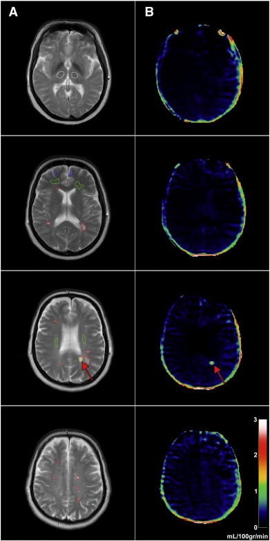
“We used an axial T2weighted MRI sequence [five slices, echo time = 100 ms, repetition time = 3000 ms, acquired voxel-size 0.57/0.76/8 mm3 (interpolated to 0.45/0.45/8 mm3), field of view = 230 119 mm2] with same orientation and slice thickness (8 mm) as our DCE-MRI sequence, in order to manually…”
BackgroundMethodInfluentialSelf-citationMiller, E. D., Dziedzic, A., Saluk, J., & Bijak, M. (2019) · Nutrients · doi:10.3390/nu11071528 · Cited by 106
“, endothelial cells, microglia, astrocytes, and pericytes) [9].”
Advances in the immunopathogenesis of multiple sclerosis
Yadav, S. K., Mindur, J. E., Ito, K., & Dhib-Jalbut, S. S. (2015) · Current Opinion in Neurology · doi:10.1097/wco.0000000000000205 · Cited by 173
Vascular Permeability in Cerebral Cavernous Malformations
Mikati, A., Khanna, O., Zhang, L.-J., Girard, R., Shenkar, R., Guo, X., Shah, A., Larsson, H., Tan, H., Li, L., et al. (2015) · Journal of Cerebral Blood Flow and Metabolism · doi:10.1038/jcbfm.2015.98 · Cited by 90
Self-citationDosSantos, M. F. H., Afonso, R. C. D. H., de Lima, R. L., DaSilva, A. F. M., & Moura‐Neto, V. (2014) · Frontiers in Cellular Neuroscience · doi:10.3389/fncel.2014.00302 · Cited by 94
“…a concept that is supported not only by experimental (Morrissey et al., 1996; Morgan et al., 2007; Kooij et al., 2009; Reijerkerk et al., 2012) but also clinical data (Plumb et al., 2002; Kirk et al., 2003; Minagar and Alexander, 2003; Leech et al., 2007; Padden et al., 2007; Cramer et al., 2014).”
BackgroundWang, Y., Wu, J., Wang, J., He, L., Lai, H., Zhang, T., Wang, X., & Li, W. (2023) · Mitochondrion · doi:10.1016/j.mito.2023.01.007 · Cited by 86
Varatharaj, A., Liljeroth, M., Darekar, A., Larsson, H. B. W., Galea, I., & Cramer, S. P. (2018) · The Journal of Physiology · doi:10.1113/jp276887 · Cited by 88
“Dynamic contrast-enhanced magnetic resonance imaging (DCE-MRI) is a newer technique which can measure all levels of BBB leakiness, including levels invisible to conventional imaging (Cramer et al. 2014).”
BackgroundResultInfluentialSelf-citationCladribine: mechanisms and mysteries in multiple sclerosis
Jacobs, B. M., Ammoscato, F., Giovannoni, G., Baker, D., & Schmierer, K. (2018) · Journal of Neurology Neurosurgery & Psychiatry · doi:10.1136/jnnp-2017-317411 · Cited by 98
Uchida, Y., Kan, H., Sakurai, K., Oishi, K., & Matsukawa, N. (2023) · Frontiers in Aging Neuroscience · doi:10.3389/fnagi.2023.1111448 · Cited by 70
“, 2013), multiple sclerosis (Gaitán et al., 2011; Jelescu et al., 2011; Shinohara et al., 2011; Ingrisch et al., 2012; Cramer et al., 2014), and ischemic strokes (Kassner et al.”
BackgroundFrom Neurodevelopmental to Neurodegenerative Disorders: The Vascular Continuum
Ouellette, J., & Lacoste, B. (2021) · Frontiers in Aging Neuroscience · doi:10.3389/fnagi.2021.749026 · Cited by 91
In vivo methods for imaging blood–brain barrier function and dysfunction
Harris, W. J., Asselin, M., Hinz, R., Parkes, L. M., Allan, S. M., Schießl, I., Boutin, H., & Dickie, B. (2022) · European Journal of Nuclear Medicine and Molecular Imaging · doi:10.1007/s00259-022-05997-1 · Cited by 91
Antipsychotic drugs counteract autophagy and mitophagy in multiple sclerosis
Patergnani, S., Bonora, M., Ingusci, S., Previati, M., Marchi, S., Zucchini, S., Perrone, M., Wieckowski, M. R., Castellazzi, M., Pugliatti, M., et al. (2021) · Proceedings of the National Academy of Sciences · doi:10.1073/pnas.2020078118 · Cited by 78
“We found that this marker was increased in MS patients in the active phase of the disease (13, 14), identified on response to Gadolinium (Gd) (28).”
BackgroundYang, H. (2025) · Pharmaceutics · doi:10.3390/pharmaceutics17081041 · Cited by 78
“Similarly, neuroinflammation in multiple sclerosis and PD increases BBB permeability through inflammatory mediator release [37,38].”
Advanced Neuroimaging of Cerebral Small Vessel Disease
Blair, G. W., Hernandez, M. V., Thrippleton, M., Doubal, F., & Wardlaw, J. (2017) · Current Treatment Options in Cardiovascular Medicine · doi:10.1007/s11936-017-0555-1 · Cited by 70
“Several groups have reported on the pitfalls and limitations of the technique, and the results of which should be interpreted with caution [78, 82 – 84].”
BackgroundKamintsky, L., Beyea, S., Fisk, J., Hashmi, J., Omisade, A., Calkin, C., Bardouille, T., Bowen, C., Quraan, M., Mitnitski, A., et al. (2020) · Annals of the Rheumatic Diseases · doi:10.1136/annrheumdis-2020-218004 · Cited by 69
Li, Y., Li, M., Zuo, L., Shi, Q., Qin, W., Yang, L., Jiang, T., & Hu, W. (2018) · Frontiers in Neurology · doi:10.3389/fneur.2018.00221 · Cited by 69
“Regions of Interest We used an axial T2 FLAIR MRI sequence with the same orientation and slice thickness as our DCE sequence to manually draw ROIs in the NAWM, WMH, CGM, and DGM in both hemispheres (Figure 1) (16).”
MethodIntact blood−brain barrier during spontaneous attacks of migraine without aura: a 3T DCE‐MRI study
Amin, F., Hougaard, A., Cramer, S., Christensen, C., Wolfram, F., Larsson, H., & Ashina, M. (2017) · European Journal of Neurology · doi:10.1111/ene.13341 · Cited by 66
“The standard deviation (30%) for the Ki in our power calculation was based on results from three previous studies using the same DCE-MRI method including a total of 125 subjects (healthy controls and patients with multiple sclerosis and optic neuritis) [11,17,18].”
BackgroundMethodInfluentialSelf-citationYu, Z., Xue, F., Liu, W., Sun, R. X., Zhou, J., Pu, Y., Zhao, M., Sun, D., Xiang, Z., Liu, P., et al. (2022) · Advanced Science · doi:10.1002/advs.202105442 · Cited by 71
Uher, T., Horáková, D., Týblová, M., Zeman, D., Krasulová, E., Mrázová, K., Seidl, Z., Vaněčková, M., Krásenský, J., Weinstock‐Guttman, B., et al. (2015) · Multiple Sclerosis Journal · doi:10.1177/1352458515601903 · Cited by 56
How does neurovascular unit dysfunction contribute to multiple sclerosis?
Cashion, J. M., Young, K. M., & Sutherland, B. A. (2023) · Neurobiology of Disease · doi:10.1016/j.nbd.2023.106028 · Cited by 58
“It is well established that the BBB becomes compromised and leaky in multiple sclerosis, resulting in immune cell infiltration into the brain parenchyma (Cramer et al., 2014; Montagne et al., 2015).”
Remyelination in multiple sclerosis: Cellular mechanisms and novel therapeutic approaches
Olsen, J. A., & Akirav, E. M. (2014) · Journal of Neuroscience Research · doi:10.1002/jnr.23493 · Cited by 51
“, 2011) as a result of increased permeabilization of the blood–brain barrier, observable by dynamic contrastenhanced magnetic resonance imaging (MRI; Cramer et al., 2013), and produce an autoimmune response to various myelin and nonmyelin antigens (Kerlero de Rosbo et al.”
BackgroundRegulatory T cell therapy for multiple sclerosis: Breaching (blood-brain) barriers
Verreycken, J., Baeten, P., & Broux, B. (2022) · Human Vaccines & Immunotherapeutics · doi:10.1080/21645515.2022.2153534 · Cited by 61
“This leads to a compromised migration of these Th1 cells in the CNS. Additionally, with the expansion of Tregs, the BBB was limitedly damaged and remained more intact, indicating a Treg-driven protective effect on the BBB. Enhancement of Tregs also decreases the migration of mast cells through the BBB via a down-regul…”
BackgroundInfluentialInsulin Resistance and Blood-Brain Barrier Dysfunction Underlie Neuroprogression in Bipolar Disorder
Calkin, C., McClelland, C., Cairns, K., Kamintsky, L., & Friedman, A. (2021) · Frontiers in Psychiatry · doi:10.3389/fpsyt.2021.636174 · Cited by 47
“Recent advances in MRI techniques have allowed direct assessment of BBB functionality in living patients, demonstrating BBBD in pathological conditions such as traumatic and ischemic injuries (86, 87), epilepsy (77), dementia (78) multiple sclerosis (79), and systemic lupus erythematosus (SLE) (75).”
BackgroundHouston, Z., Bunt, J., Chen, K.-S., Puttick, S., Howard, C. B., Fletcher, N., Fuchs, A., Cui, J.-W., Ju, Y., Cowin, G., et al. (2020) · ACS Central Science · doi:10.1021/acscentsci.9b01299 · Cited by 47
Role of proteoglycans in neuro-inflammation and central nervous system fibrosis
Heindryckx, F., & Li, J. (2018) · Matrix Biology · doi:10.1016/j.matbio.2018.01.015 · Cited by 50
Bester, M., Forkert, N., Stellmann, J., Aly, L., Drabik, A., Young, K., Heesen, C., Fiehler, J., & Siemonsen, S. (2015) · PLoS ONE · doi:10.1371/journal.pone.0119356 · Cited by 46
“Cramer et al [44] investigated the integrity of the BBB and describe a gradual decrease of permeability after an MS relapse.”
Loss of corticospinal tract integrity in early MS disease stages
Pawlitzki, M., Neumann, J. H. L., Kaufmann, J., Heidel, J., Stadler, E., Sweeney‐Reed, C. M., Sailer, M., & Schreiber, S. (2017) · Neurology Neuroimmunology & Neuroinflammation · doi:10.1212/nxi.0000000000000399 · Cited by 48
“FA andMD values could thus serve as alternative markers of inflammation, probably indicating ongoing demyelination(24) and predicting new relapses.(25) DTI measures could thus have the potential to serve as a biomarker disclosing ongoing (inflammatory) disease activity, especially in secondary or primary progressive M…”
BackgroundCGRP and Brain Functioning: Cautions for Migraine Treatment
Borkum, J. M. (2019) · Headache The Journal of Head and Face Pain · doi:10.1111/head.13591 · Cited by 46
“This might be relevant clinically for people with MS, a condition in which the prevalence of migraines is three times greater than in the general population,(129) and in which the BBB is compromised for up to several months after a flare-up.(77) As we have seen, CGRP, injected into a cerebral ventricle, reduces diseas…”
BackgroundXu, Z., Zeng, W., Sun, J., Chen, W., Zhang, R., Yang, Z., Yao, Z., Wang, L., Song, L., Chen, Y., et al. (2016) · NeuroImage · doi:10.1016/j.neuroimage.2016.07.017 · Cited by 46
Imaging blood–brain barrier disruption in neuroinflammation and Alzheimer’s disease
Lee, R., & Funk, K. E. (2023) · Frontiers in Aging Neuroscience · doi:10.3389/fnagi.2023.1144036 · Cited by 42
Direct Estimation of Tracer-Kinetic Parameter Maps from Highly Undersampled Brain DCE-MRI
Guo, Y., Lingala, S., Zhu, Y.-H., Lebel, R., & Nayak, K. (2016) · Magnetic Resonance in Medicine · doi:10.1002/mrm.26540 · Cited by 41
Hecker, M., Bühring, J., Fitzner, B., Rommer, P. S., & Zettl, U. K. (2021) · Biomolecules · doi:10.3390/biom11101510 · Cited by 49
“Key steps in the pathogenesis of MS are presumably the peripheral expansion of autoreactive immune cells, the breakdown of the blood–brain barrier [56], the infiltration of activated cells into the CNS and the subsequent generation of”
BackgroundSphingosine-1-Phosphate Receptor Modulators and Oligodendroglial Cells: Beyond Immunomodulation
Roggeri, A., Schepers, M., Tiane, A., Rombaut, B., van Veggel, L., Hellings, N., Prickaerts, J., Pittaluga, A., & Vanmierlo, T. (2020) · International Journal of Molecular Sciences · doi:10.3390/ijms21207537 · Cited by 40
“Alterations of tight junction proteins [35–37] and the consequent increased BBB permeability, ease the passage of autoreactive T-cells towards the CNS [35,38].”
BackgroundPro-Thrombotic Activity of Blood Platelets in Multiple Sclerosis
Saluk, J., Dziedzic, A., & Bijak, M. (2019) · Cells · doi:10.3390/cells8020110 · Cited by 42
Charabati, M., Zandee, S., Fournier, A. P., Tastet, O., Thai, K., Zaminpeyma, R., Lécuyer, M., Bourbonnière, L., Larouche, S., Klément, W., et al. (2022) · Brain · doi:10.1093/brain/awac389 · Cited by 48
“Its junctional integrity is disturbed and it becomes permeable to circulating macromolecules.(2,4,5) In the clinic, CNS vascular breakdown is”
BackgroundKluge, A., Lukas, M., Tóth, V., Pyka, T., Zimmer, C., & Preibisch, C. (2016) · Magnetic Resonance Imaging · doi:10.1016/j.mri.2015.12.015 · Cited by 37
“Previous studies with dynamic contrast-enhanced MRI [15,22] suggest that even in healthy tissue a small amount of leakage can occur.”
BackgroundResultCommon comorbidities and survival in MS: Risk for stroke, type 1 diabetes and infections.
Murtonen, A., Kurki, S., Hänninen, K., Soilu-Hänninen, M., & Sumelahti, M. (2018) · Multiple Sclerosis and Related Disorders · doi:10.1016/j.msard.2017.10.019 · Cited by 37
Morel, A., Miller, E. D., Bijak, M., & Saluk, J. (2016) · Molecular and Cellular Biochemistry · doi:10.1007/s11010-016-2770-6 · Cited by 40
“MS is considered as a heterogeneous neurological disease with various pathophysiological mechanisms and multiple clinical course, but closely related to the damage of intracerebral blood vessels, mainly as a result of increased permeability of blood–brain barrier (BBB) as well vessel occlusion [2].”
BackgroundBlood–Brain Barrier and Bilirubin: Clinical Aspects and Experimental Data
Brito, M. A., Palmela, I., Cardoso, F. L., Sá-Pereira, I., & Brites, D. M. T. O. (2014) · Archives of Medical Research · doi:10.1016/j.arcmed.2014.11.015 · Cited by 42
Wengler, K., Bangiyev, L., Canli, T., Duong, T., Schweitzer, M., & He, X. (2019) · NeuroImage · doi:10.1016/j.neuroimage.2019.01.035 · Cited by 34
“This could lead to a better understanding of the pathogenesis of some diseases, such as multiple sclerosis and Alzheimer's disease, where females show higher prevalence than males (Janelidze et al., 2017; Wilson et al., 2008; Cramer et al., 2014).”
Aspirin and multiple sclerosis
Tsau, S., Emerson, M. R., LYNCH, S. G., & LeVine, S. M. (2015) · BMC Medicine · doi:10.1186/s12916-015-0394-4 · Cited by 31
“However, the slight risk of increased intracerebral bleeding observed in stroke patients given ASA [79] is troubling, since MS patients have a compromised BBB [7, 8] and ASA treatment could further disrupt the BBB (Table 2).”
BackgroundInfluentialMa, X.-X., Zhang, L., Huang, D.-H., Lyu, J., Fang, M., Hu, J.-X., Zang, Y.-L., Zhang, D.-K., Shao, H., Ma, L., et al. (2018) · Journal of Magnetic Resonance Imaging · doi:10.1002/jmri.26287 · Cited by 33
“Lesions were selected if the lesion diameter was greater than 3 mm in the slice with the largest cross-section, and if the lesion was in the periventricular, juxtacortical, or infratentorial regions, or the subcortical white or deep gray matter.(28) Axial T1WI and coronal T2 FLAIR imaging were used to identify and del…”
MethodEisen, A., Greenberg, B., Bowen, J., Arnold, D., & Caggiano, A. (2017) · Multiple Sclerosis Journal - Experimental, Translational and Clinical · doi:10.1177/2055217317743097 · Cited by 33
Therapeutic perspective on vascular cognitive impairment
Toyama, K., Spin, J. M., Mogi, M., & Tsao, P. S. (2019) · Pharmacological Research · doi:10.1016/j.phrs.2019.104266 · Cited by 31
Hemostasis biomarkers in multiple sclerosis
Ziliotto, N., Bernardi, F., Jakimovski, D., Baroni, M., Marchetti, G., Bergsland, N., Ramasamy, D., Weinstock-Guttman, B., Schweser, F., Zamboni, P., et al. (2018) · European Journal of Neurology · doi:10.1111/ene.13681 · Cited by 32
“The pathogenesis of MS involves blood–brain barrier (BBB) breakdown, extravasation of immune cells, inflammation and neurodegeneration in the CNS, which results in the formation of lesions and development of brain atrophy [2].”
BackgroundInflammation-induced TRPV4 channels exacerbate blood–brain barrier dysfunction in multiple sclerosis
Hansen, C. E., Kamermans, A., Mol, K., Berve, K., Rodriguez-Mogeda, C., Fung, W. K., Hof, B. V. H., Fontijn, R. D., van der Pol, S. M. A., Michalick, L., et al. (2024) · Journal of Neuroinflammation · doi:10.1186/s12974-024-03069-9 · Cited by 33
“Hence, BBB dysfunction and inflammation are driving events in MS lesion formation, which presents a mandate for elucidating the underlying mechanisms of these processes [16, 17].”
BackgroundKim, Y. S., Kim, M., Choi, S.-H., You, S.-H., Yoo, R.-E., Kang, K. M., Yun, T., Lee, S.-T., Moon, J., & Shin, Y. (2019) · Radiology · doi:10.1148/radiol.2019182566 · Cited by 31
Kamrava, M., Sepahdari, A., Leu, K., Wang, P.-C., Roberts, K., Demanes, D., Mccannel, T., & Ellingson, B. (2015) · Neuroradiology · doi:10.1007/s00234-015-1546-0 · Cited by 31
Bäumer, P., Reimann, M., Decker, C., Radbruch, A., Bendszus, M., Heiland, S., & Pham, M. (2014) · Investigative Radiology · doi:10.1097/rli.0000000000000046 · Cited by 31
Wei, Z., Liu, H., Lin, Z., Yao, M., Li, R., Liu, C., Li, Y., Xu, J., Duan, W., & Lu, H. (2023) · NeuroImage · doi:10.1016/j.neuroimage.2023.119870 · Cited by 37
Huang, J., Han, S. H., Sun, Q., Zhao, Y., Liu, J., Yuan, X., Mao, W., Peng, B., Liu, W., Yin, J., et al. (2017) · Cell & Bioscience · doi:10.1186/s13578-017-0158-2 · Cited by 34
“Increased BBB permeability is the hallmark of acute inflammatory lesions in MS patients [12], and appears in the periventricular normal-appearing white matter of MS patients using dynamic contrast-enhanced MRI imaging [32].”
Larsson, H. B. W., Vestergaard, M. B., Lindberg, U., Iversen, H. K., & Cramer, S. P. (2016) · Journal of Magnetic Resonance Imaging · doi:10.1002/jmri.25488 · Cited by 31
“It has previously been demonstrated that subtle leakage can be measured in healthy subjects, and a higher but still small leakage can be measured in periventricular normal appearing white matter in patients with multiple sclerosis and patients with optic neuritis.(10,26) In patients with brain tumors, leakage is often…”
BackgroundSelf-citationGuo, Y., Lebel, R., Zhu, Y.-H., Lingala, S., Shiroishi, M., Law, M., & Nayak, K. (2016) · Medical Physics (Lancaster) · doi:10.1118/1.4944736 · Cited by 29
Novel MRI and PET markers of neuroinflammation in multiple sclerosis
Hagens, M. H. J., van Berckel, B. N. M., & Barkhof, F. (2016) · Current Opinion in Neurology · doi:10.1097/wco.0000000000000326 · Cited by 29
Bakhtiari, A., Vestergaard, M. B., Benedek, K., Fagerlund, B., Mortensen, E. L., Osler, M., Lauritzen, M., Larsson, H. B. W., & Lindberg, U. (2022) · GeroScience · doi:10.1007/s11357-022-00712-2 · Cited by 25
Self-citationFang, K., Wang, Z., Li, Z.-Q., Wang, B.-J., Han, G., Cheng, Z.-W., Chen, Z.-H., Lan, C., Zhang, Y., Zhao, P., et al. (2020) · Journal of Magnetic Resonance Imaging · doi:10.1002/jmri.27495 · Cited by 27
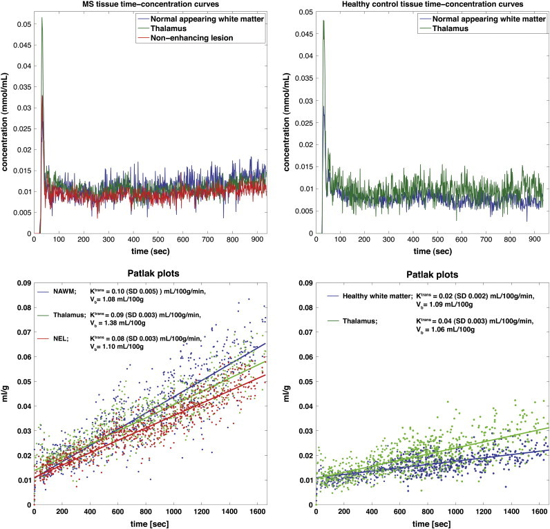
“3 As the contrast agent (CA) enters and subsequently leaves the tissue, different tissues produce unique intensity time curves.”
BackgroundMethodNatural products: Potential therapeutic agents in multiple sclerosis
Yu, S., Liu, M., & Hu, K. (2018) · International Immunopharmacology · doi:10.1016/j.intimp.2018.11.036 · Cited by 33
Popa, A., Stan, G. E., Beşleagă, C., Ion, L., Maraloiu, V., Tulyaganov, D. U., & Ferreira, J. M. F. (2016) · ACS Applied Materials and Interfaces · doi:10.1021/acsami.6b00606 · Cited by 27
Nichols, P., Urriola, J., Miller, S., Bjorkman, T., Mahady, K., Vegh, V., Nasrallah, F., & Winter, C. (2021) · NeuroImage: Clinical · doi:10.1016/j.nicl.2021.102741 · Cited by 26
“It has been shown that in normal-appearing GM and WM, the K value is low but not zero [26-28].”
Safety and efficacy of cladribine in multiple sclerosis: a systematic review and meta-analysis
Nabizadeh, F., Mohamadi, M., Rahmani, S., Rajabi, R., Afrashteh, F., Najdaghi, S., & Mirmosayyeb, O. (2023) · Neurological Sciences · doi:10.1007/s10072-023-06794-w · Cited by 28
“However, with blood–brain barrier (BBB) disruption as a common pathological finding at all MS disease stages, cladribine can penetrate better to the CNS of MS patients [55–57].”
Mokhtarzade, M., Motl, R., Negaresh, R., Zimmer, P., Khodadoost, M., Baker, J., Patel, D. I., Majdinasab, N., & Ranjbar, R. (2018) · Neuropeptides · doi:10.1016/j.npep.2018.05.010 · Cited by 25
Elevated plasma fibrinogen levels in multiple sclerosis patients during relapse
Acuña, J. A. S., de la Cruz, M. H., Ros, A. L., Prasca, S., Ginés, M. L. M., & Frutos, C. D. D. A. (2017) · Multiple Sclerosis and Related Disorders · doi:10.1016/j.msard.2017.09.033 · Cited by 23
Li, H.-P., Liu, X., Wang, R., Lu, A.-L., Ma, Z., Wu, S., Lu, H.-J., Du, Y., Deng, K., Wang, L.-X., et al. (2023) · Epilepsia · doi:10.1111/epi.17576 · Cited by 23
Developing a transwell millifluidic device for studying blood–brain barrier endothelium
Harding, I. C., O’Hare, N. R., Vigliotti, M., Caraballo, A., Lee, C. I., Millican, K., Herman, I. M., & Ebong, E. E. (2022) · Lab on a Chip · doi:10.1039/d2lc00657j · Cited by 28
Hanly, J., Legge, A., Kamintsky, L., Friedman, A., Hashmi, J., Beyea, S., Fisk, J., Omisade, A., Calkin, C., Bardouille, T., et al. (2022) · Lupus Science and Medicine · doi:10.1136/lupus-2022-000668 · Cited by 22
Choi, S., Saxena, N., Dhammu, T. S., Khan, M., Singh, A. K., Singh, I., & Won, J. (2018) · Nitric Oxide · doi:10.1016/j.niox.2018.12.007 · Cited by 21
“Brain imaging studies have shown that patients with relapsing-remitting MS (RRMS), the most common type of MS (> 80 %), have generally increase in BBB permeability [60; 61].”
BackgroundNotch signaling is impaired during inflammation in a Lunatic Fringe-dependent manner
Troletti, C. D., Pinheiro, M. A. L., Charabati, M., Gowing, E., Hof, B. V. H., van der Pol, S. M. A., Geerts, D., Prat, A., Fontijn, R. D., Unger, W. W. J., et al. (2017) · Brain Behavior and Immunity · doi:10.1016/j.bbi.2017.12.016 · Cited by 25
“In addition, reduced brain endothelial cell-cell contacts and BBB dysfunction have been suggested to result in the leakage of serum components into the CNS thereby contributing to MS pathogenesis (Cramer et al., 2014; Kirk et al., 2003; Leech et al., 2007; Plumb et al., 2002).”
Guo, Y., Lingala, S., Bliesener, Y., Lebel, R., Zhu, Y.-H., & Nayak, K. (2017) · Magnetic Resonance in Medicine · doi:10.1002/mrm.26904 · Cited by 22
Ingrisch, M., Sourbron, S., Herberich, S., Schneider, M., Kümpfel, T., Hohlfeld, R., Reiser, M., & Ertl-Wagner, B. (2016) · Investigative Radiology · doi:10.1097/rli.0000000000000320 · Cited by 22
“These results are not in line with previous reports that studied NAWMperfusion in MS. Early nuclear medicine studies1–3 have found a significantly reduced CBF in patients with MS.”
ResultInfluentialRicciardelli, A., Robledo, A., Fish, J. E., Kan, P. T. M., Harris, T. H., & Wythe, J. D. (2023) · Biomedicines · doi:10.3390/biomedicines11112876 · Cited by 24
“…hypoperfusion) can precede cognitive decline and clinical symptoms in numerous neurological pathologies [106], including aging, multiple sclerosis [107–109], stroke, Alzheimer’s [110–114], and epilepsy, with the disruption of vascular function in the CNS causing ion disturbance, edema, and…”
BackgroundPansieri, J., Hadley, G., Lockhart, A., Pisa, M., & DeLuca, G. C. (2023) · Frontiers in Neurology · doi:10.3389/fneur.2023.1199491 · Cited by 21
Kovbasyuk, Z., Tefera, E., Li, C., Baete, S. H., & Steriade, C. (2025) · Journal of Neuroinflammation · doi:10.1186/s12974-025-03598-x · Cited by 23
“Reports of higher K i in NAWM of RRMS patients compared to WM from HCs, suggests that DCE-MRI can detect subtle BBB abnormalities typically identified only histologically [2, 106].”
The blood–brain barrier as a treatment target for neurodegenerative disorders
Rust, R., Sagare, A. P., Zhang, M., Zloković, B. V., & Kisler, K. (2025) · Expert Opinion on Drug Delivery · doi:10.1080/17425247.2025.2480654 · Cited by 21
“Likewise, changes in BBB permeability and incidence of cerebral microbleeds (brain vascular microhemorrhages) have been reported in patients with HD [51,52], PD [53–55], and MS [56–58].”
Metadata
- Authors
- Cramer, S. P., Simonsen, H., Frederiksen, J. L., Rostrup, E., & Larsson, H. B. W.
- Affiliations
- University of Copenhagen (DK)
- Corresponding author
- Cramer, S.P.
- Venue
- NeuroImage: Clinical
- Publisher
- Elsevier BV
- Published
- 2014
- Volume
- 4
- Pages
- 182-189
- ISSN
- 2213-1582 (ISSN-L)
- PMC
- PMC3872721
- OpenAlex
- W2039561850
- Cited by
- 232
- References
- 37
- Citations by year
- 2026: 16 · 2025: 20 · 2024: 16 · 2023: 20 · 2022: 24 · 2021: 16 · 2020: 14 · 2019: 20 · 2018: 26 · 2017: 13 · 2016: 16 · 2015: 21 · 2014: 9
- FWCI
- 11.49
- Type
- journal article
- Language
- en
- Open access
- gold
- doi.org
- Licence
- Locations
- NeuroImage Clinical (publishedVersion, cc-by-nc-sa)
- Europe PMC (PubMed Central) (publishedVersion, cc-by-nc-sa)
- PubMed Central (publishedVersion, cc-by-nc-sa)
- PubMed (publishedVersion)
- BIBSYS Brage (BIBSYS (Norway)) (submittedVersion)
- DOAJ (DOAJ: Directory of Open Access Journals) (submittedVersion, cc-by-sa)
- NORA - Norwegian Open Research Archives (submittedVersion, cc-by-nc-sa)
- Research at the University of Copenhagen (University of Copenhagen) (submittedVersion)
- Funders
- Biogen
- Scleroseforeningen (14588)
- Multiple Sclerosis Society (14588)
- Region Hovedstaden
- Topics
- Health Sciences › Medicine › Clinical Neurology › Multiple Sclerosis Research Studies
- Life Sciences › Neuroscience › Neurology › Barrier Structure and Function Studies
- Health Sciences › Medicine › Radiology, Nuclear Medicine and Imaging › Advanced MRI Techniques and Applications
- Keywords
- multiple sclerosis, blood-brain barrier permeability, normal-appearing white matter, periventricular white matter, dynamic contrast-enhanced MRI, MS relapse, immunomodulatory therapy, MS lesions, blood-brain barrier disruption
- MeSH terms
- Adult, Blood-Brain Barrier, Capillary Permeability, Contrast Media, Female, Humans, Magnetic Resonance Imaging, Male, Middle Aged, Multiple Sclerosis, Severity of Illness Index, Imaging, Three-Dimensional, Perfusion Imaging, White Matter
About these checks
Registry, retraction, Cites and statistics results are computed, not judged, with one exception: a printed reference that no index lists is read once by a model before it counts as a citation error. Claims are extracted by a model and kept only when their quote appears verbatim in the text; each cited source is then checked, with a model’s help, for whether it says what the claim relies on. Those verdicts can be wrong — read the claim’s quote and the cited source before relying on one.
Statistics and arithmetic are recomputed, not judged.
Confidence bands are shown only for fields calibrated on our annotated test set. None is calibrated yet, so no band is shown.
What counts as a claim ›How we measure confidence ›How we check this ›
Are you an author, or do you see a problem with this page? Request a review
