

The DOI matches its Crossref record; 152 of the 152 references it cites were found in a registry or an index.
Roles of Supplementary Motor Areas in Auditory Processing and Auditory Imagery
Open accessAbstract
Although the supplementary and pre-supplementary motor areas have been intensely investigated in relation to their motor functions, they are also consistently reported in studies of auditory processing and auditory imagery. This involvement is commonly overlooked, in contrast to lateral premotor and inferior prefrontal areas. We argue here for the engagement of supplementary motor areas across a variety of sound categories, including speech, vocalizations, and music, and we discuss how our understanding of auditory processes in these regions relate to findings and hypotheses from the motor literature. We suggest that supplementary and pre-supplementary motor areas play a role in facilitating spontaneous motor responses to sound, and in supporting a flexible engagement of sensorimotor processes to enable imagery and to guide auditory perception.
Overview
The claims and statistics of this paper haven’t been checked yet.
Run the check — free, no account needed. It reads the open-access text and checks the sources it cites.
Integrity signals
Registry
Cites
Statistics
Figures
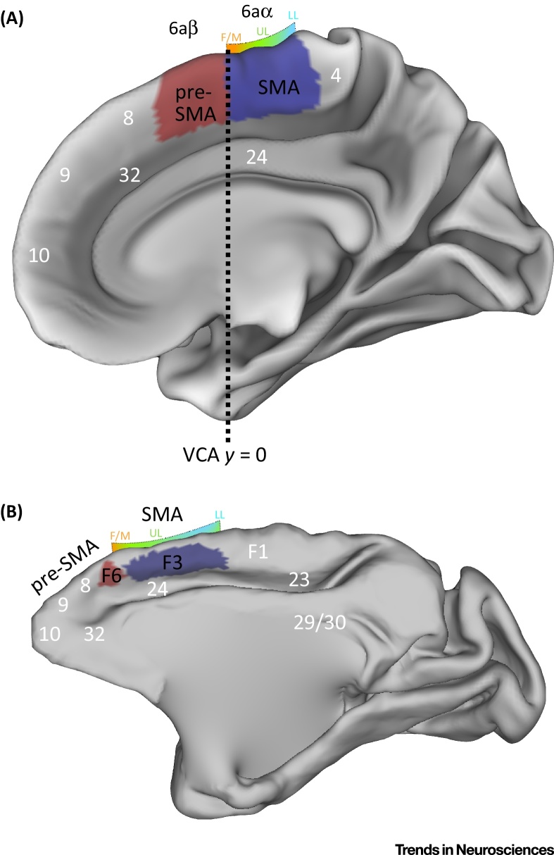
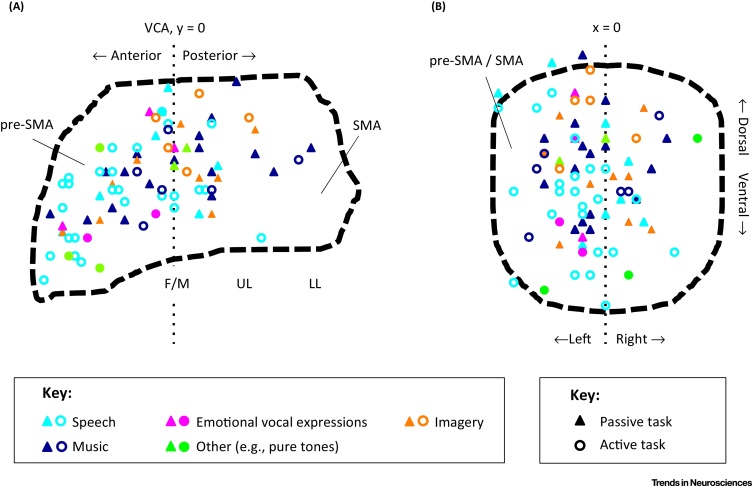
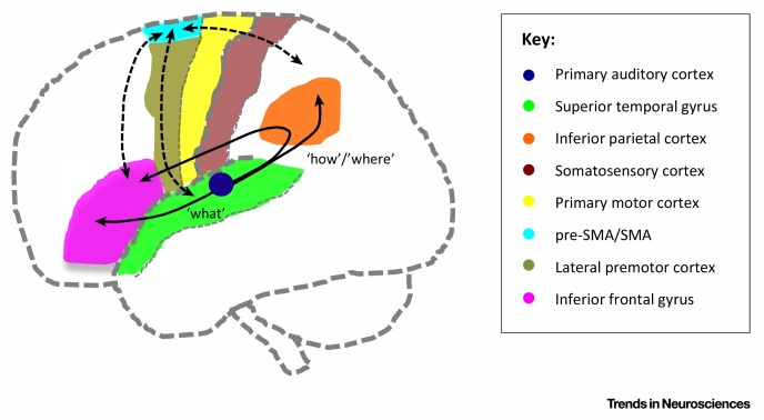
Verification
Claims the paper makes
The claims and statistics of this paper haven’t been checked yet.
Run the check — free, no account needed. It reads the open-access text and checks the sources it cites.
Claims other papers make about this one
Each is checked against this paper’s own text: does it say what the citing paper says it does?
In the citing paper
“The experience of NCVH likely also involves regions associated with the generation and monitoring of speech-motor imagery, as well as sensorimotor processes, such as the supplementary and pre-supplementary motor areas (SMA/pre-SMA; Linden et al., 2011;Lima et al., 2016).”
From Distinct processing of ambiguous speech in people with non-clinical auditory verbal hallucinations (2017) · cites this paper as Lima et al., 2016
Self-check
Percentages, sums and reported differences re-computed from the paper’s own numbers.
The self-check needs the paper’s full text.
Citations
Cites
The role of the medial frontal cortex in cognitive control
Ridderinkhof, K. R., Ullsperger, M., Crone, E. A., & Nieuwenhuis, S. (2004) · Science · doi:10.1126/science.1100301 · Cited by 2,974
Where do you know what you know? The representation of semantic knowledge in the human brain
Patterson, K., Nestor, P. J., & Rogers, T. T. (2007) · Nature Reviews Neuroscience · doi:10.1038/nrn2277 · Cited by 2,706
Functional role of the supplementary and pre-supplementary motor areas
Nachev, P., Kennard, C., & Husain, M. (2008) · Nature Reviews Neuroscience · doi:10.1038/nrn2478 · Cited by 1,928
When the brain plays music: auditory–motor interactions in music perception and production
Zatorre, R. J., Chen, J. L., & Penhune, V. B. (2007) · Nature reviews. Neuroscience · doi:10.1038/nrn2152 · Cited by 1,896
Maps and streams in the auditory cortex: nonhuman primates illuminate human speech processing
Rauschecker, J. P., & Scott, S. K. (2009) · Nature Neuroscience · doi:10.1038/nn.2331 · Cited by 1,766
Motor Areas of the Medial Wall: A Review of Their Location and Functional Activation
Picard, N., & Strick, P. L. (1996) · Cerebral Cortex · doi:10.1093/cercor/6.3.342 · Cited by 1,712
Brain Structures Differ between Musicians and Non-Musicians
Christian Gaser, B., & Gottfried Schlaug, A. (2003) · Journal of Neuroscience · doi:10.1523/jneurosci.23-27-09240.2003 · Cited by 1,626
The origin of corticospinal projections from the premotor areas in the frontal lobe
Dum, R., & Strick, P. (1991) · Journal of Neuroscience · doi:10.1523/jneurosci.11-03-00667.1991 · Cited by 1,447
Neuroplasticity Subserving Motor Skill Learning
Dayan, E., & Cohen, L. G. (2011) · Neuron · doi:10.1016/j.neuron.2011.10.008 · Cited by 1,408
The emulation theory of representation: Motor control, imagery, and perception
Grush, R. (2004) · Behavioral and Brain Sciences · doi:10.1017/s0140525x04000093 · Cited by 1,387 · cited 1×
“These ideas resonate with the ‘emulation’ framework discussed in relation to action control, and generalized to motor imagery, perception, and prediction [112,113].”
Research works printed but not linked: 1
- Vogt, 1919, Allgemeine ergebnisse unserer hirnforschung, Journal für Psychologie und Neurologie, 25, 277
Cited by
Sorted by citations
Large-scale cortical networks for hierarchical prediction and prediction error in the primate brain
Chao, Z. C., Takaura, K., Wang, L., Fujii, N., & Dehaene, S. (2018) · Neuron · doi:10.1016/j.neuron.2018.10.004 · Cited by 238
How Beat Perception Co-opts Motor Neurophysiology
Cannon, J. J., & Patel, A. D. (2020) · Trends in Cognitive Sciences · doi:10.1016/j.tics.2020.11.002 · Cited by 232
Haslbeck, F. B., András, J., Held, U., Bassler, D., Bucher, H.-U., & Hagmann, C. F. (2020) · NeuroImage Clinical · doi:10.1016/j.nicl.2020.102171 · Cited by 146
Distinct processing of ambiguous speech in people with non-clinical auditory verbal hallucinations
Alderson-Day, B., Lima, C. F., Evans, S., Krishnan, S., Shanmugalingam, P., Fernyhough, C., & Scott, S. K. (2017) · Brain · doi:10.1093/brain/awx206 · Cited by 108
Towards a neural model of infant cry perception
Witteman, J., Van IJzendoorn, M. H., Rilling, J. K., Bos, P. A., Schiller, N. O., & Bakermans-Kranenburg, M. J. (2019) · Neuroscience & Biobehavioral Reviews · doi:10.1016/j.neubiorev.2019.01.026 · Cited by 94
Metadata
- Authors
- Lima, C. F., Krishnan, S., & Scott, S. K.
- Affiliations
- University College London (GB)
- University of Oxford (GB)
- Corresponding author
- Scott, Sophie K.
- Venue
- Trends in Neurosciences
- Publisher
- Elsevier BV
- Published
- 2016-08
- Volume
- 39
- Issue
- 8
- Pages
- 527-542
- ISSN
- 0166-2236 (ISSN-L)
- PMC
- PMC5441995
- OpenAlex
- W2473628615
- Cited by
- 227
- References
- 152
- Citations by year
- 2026: 17 · 2025: 20 · 2024: 29 · 2023: 21 · 2022: 21 · 2021: 38 · 2020: 25 · 2019: 31 · 2018: 17 · 2017: 8
- FWCI
- 1.97
- Type
- journal article
- Language
- en
- Open access
- hybrid
- doi.org
- Locations
- Trends in Neurosciences (publishedVersion, cc-by)
- Europe PMC (PubMed Central) (publishedVersion, cc-by)
- PubMed Central (publishedVersion, cc-by)
- PubMed (publishedVersion)
- UCL Discovery (University College London) (submittedVersion)
- Portuguese National Funding Agency for Science, Research and Technology (RCAAP Project by FCT) (submittedVersion)
- Funders
- Wellcome Trust (WT090961MA, 090961)
- Topics
- Life Sciences › Neuroscience › Cognitive Neuroscience › Neuroscience and Music Perception
- Life Sciences › Neuroscience › Cognitive Neuroscience › Neurobiology of Language and Bilingualism
- Social Sciences › Psychology › Experimental and Cognitive Psychology › Multisensory perception and integration
- Keywords
- supplementary motor area, pre-supplementary motor area, auditory processing, auditory imagery, sensorimotor integration, speech perception, vocalizations, music perception, auditory perception
- MeSH terms
- Animals, Auditory Perception, Humans, Imagination, Motor Activity, Motor Cortex
About these checks
Registry, retraction, Cites and statistics results are computed, not judged, with one exception: a printed reference that no index lists is read once by a model before it counts as a citation error. Claims are extracted by a model and kept only when their quote appears verbatim in the text; each cited source is then checked, with a model’s help, for whether it says what the claim relies on. Those verdicts can be wrong — read the claim’s quote and the cited source before relying on one.
Statistics and arithmetic are recomputed, not judged.
Confidence bands are shown only for fields calibrated on our annotated test set. None is calibrated yet, so no band is shown.
What counts as a claim ›How we measure confidence ›How we check this ›
Are you an author, or do you see a problem with this page? Request a review
