

The DOI matches its Crossref record; 83 of the 83 references it cites were found in a registry or an index.
Dysfunctional autophagy in RPE, a contributing factor in age-related macular degeneration
Open accessAbstract
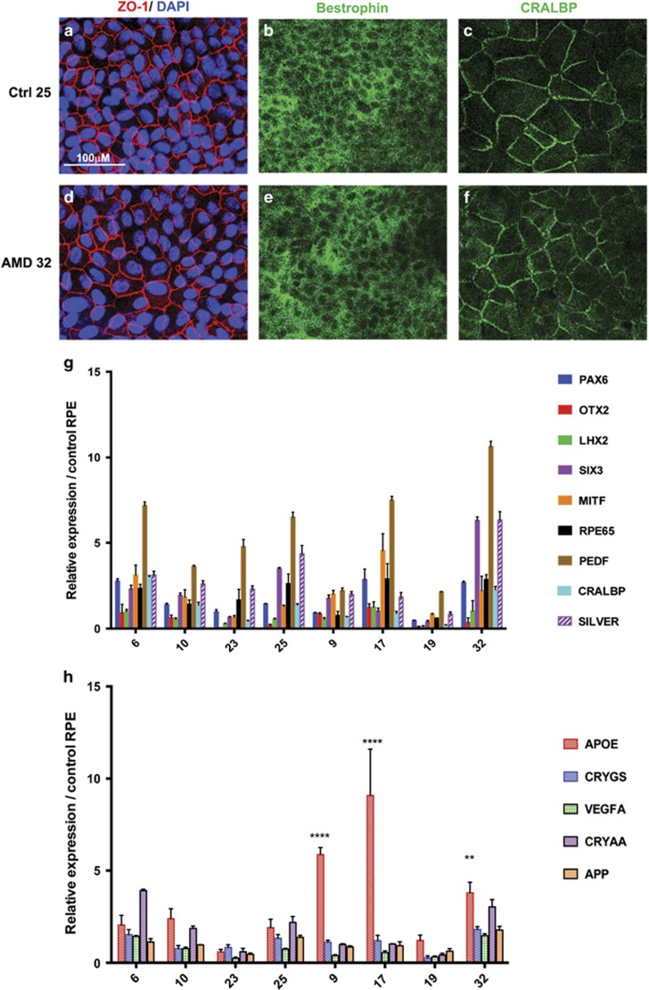
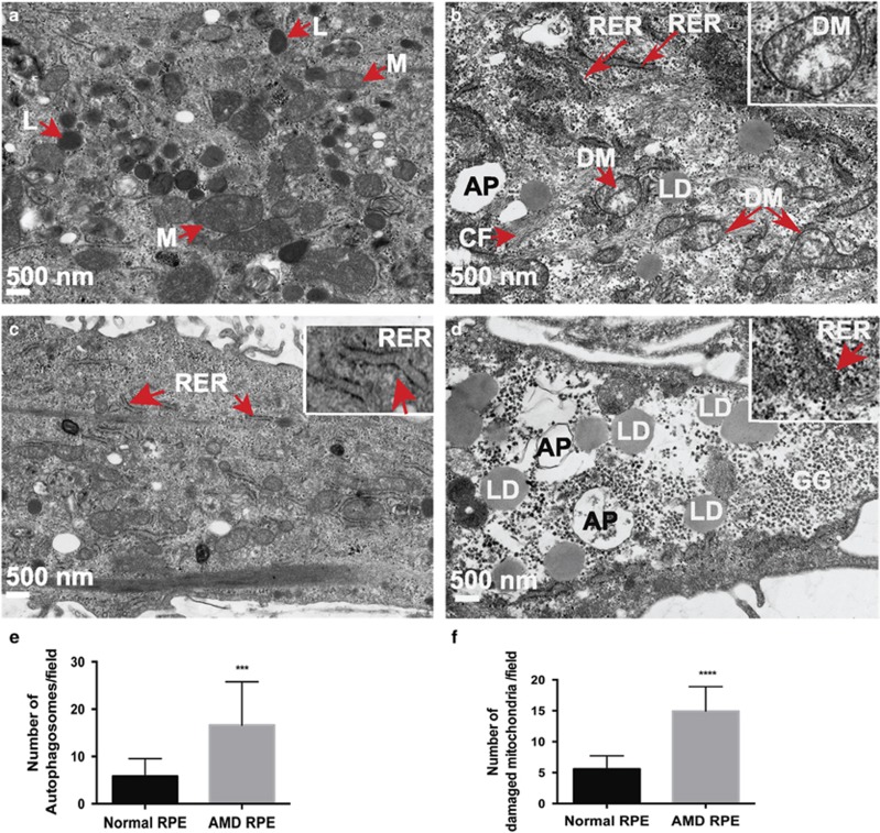
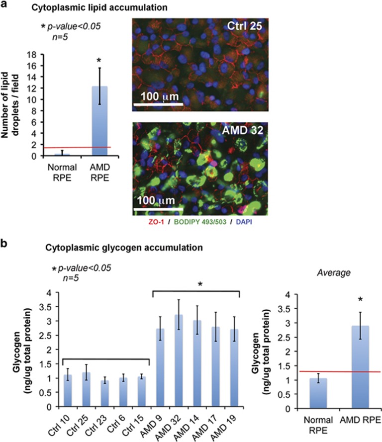
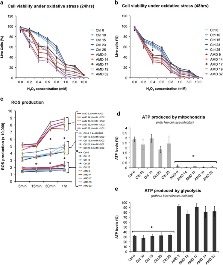
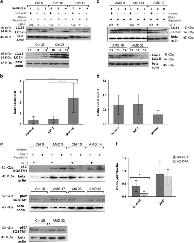
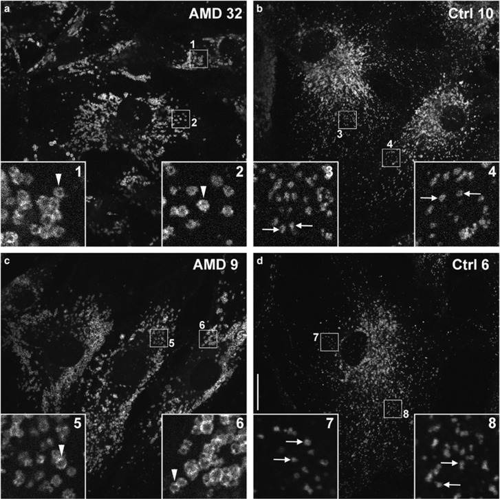
Age-related macular degeneration (AMD) is a devastating neurodegenerative disease and a major cause of blindness in the developed world. Owing to its complexity and the lack of an adequate human model that recapitulates key aspects of the disease, the molecular mechanisms of AMD pathogenesis remain poorly understood. Here we show that cultured human retinal pigment epithelium (RPE) from AMD donors (AMD RPE) are functionally impaired and exhibit distinct phenotypes compared with RPE cultured from normal donors (normal RPE). Accumulation of lipid droplets and glycogen granules, disintegration of mitochondria, and an increase in autophagosomes were observed in AMD RPE cultures. Compared with normal RPE, AMD RPE exhibit increased susceptibility to oxidative stress, produce higher levels of reactive oxygen species (ROS) under stress conditions, and showed reduced mitochondrial activity. Measurement of the ratio of LC3-II/ LC3-I, revealed impaired autophagy in AMD RPE as compared with normal RPE. Autophagic flux was also reduced in AMD RPE as compared with normal RPE, as shown by inability of AMD RPE to downregulate p62 levels during starvation. Impaired autophagic pathways were further shown by analyzing late autophagic vesicles; immunostaining with lysosome-associated membrane protein 1 (LAMP-1) antibody revealed enlarged and annular LAMP-1-positive organelles in AMD RPE as opposed to smaller discrete puncta observed in normal RPE. Our study provides insights into AMD cellular and molecular mechanisms, proposes dysfunctional autophagy as an underlying mechanism contributing to the pathophysiology of the disease, and opens up new avenues for development of novel treatment strategies.
Overview
The claims and statistics of this paper haven’t been checked yet.
Run the check — free, no account needed. It reads the open-access text and checks the sources it cites.
Integrity signals
Registry
Cites
Statistics
Figures
Verification
Claims the paper makes
The claims and statistics of this paper haven’t been checked yet.
Run the check — free, no account needed. It reads the open-access text and checks the sources it cites.
Claims other papers make about this one
Each is checked against this paper’s own text: does it say what the citing paper says it does?
In the citing paper
“RPE cultures established from AMD donors exhibited several features of disrupted autophagy, including decreased LC3B-II expression, swollen autolysosomes, and increased p62 (Golestaneh et al., 2017).”
From The cell biology of the retinal pigment epithelium (2020) · cites this paper as Golestaneh et al., 2017
In the citing paper
“AMD donor RPE cultures also accumulated lipid droplets, glycogen granules, and fragmented mitochondria, indicating a global breakdown of cellular clearance pathways.”
From The cell biology of the retinal pigment epithelium (2020) · cites this paper as Golestaneh et al., 2017
In the citing paper
“Lysosomes in cultured human RPE from AMD donors were found to be enlarged and annular; however, whether this abnormal lysosome morphology is associated with decreased hydrolytic function is not yet known (Golestaneh et al., 2017).”
From The cell biology of the retinal pigment epithelium (2020) · cites this paper as Golestaneh et al., 2017
In the citing paper
“Lipid oxidation is a critical factor in the development of AMD (Balaratnasingam et al., 2017; Golestaneh et al., 2017; Handa et al., 2017; Xu et al., 2017).”
From Microtubule-associated protein 1 light chain 3B, (LC3B) is necessary to maintain lipid-mediated homeostasis in the retinal pigment epithelium (2018) · cites this paper as Golestaneh et al., 2017
In the citing paper
“LC3II/LC3I ratios were lower in RPE from AMD patients with decreased autophagic flux and mitochondrial accumulation (suggestive of altered mitophagy) (Golestaneh et al., 2017)”
From Microtubule-associated protein 1 light chain 3B, (LC3B) is necessary to maintain lipid-mediated homeostasis in the retinal pigment epithelium (2018) · cites this paper as Golestaneh et al., 2017
In the citing paper
“leading these and other investigators doing in vitro work to conclude that dysregulated or dysfunctional autophagy contributes to AMD.”
From Microtubule-associated protein 1 light chain 3B, (LC3B) is necessary to maintain lipid-mediated homeostasis in the retinal pigment epithelium (2018) · cites this paper as Golestaneh et al., 2017
In the citing paper
“RPE cultures derived from human donor eyes (patients with AMD), exhibited increased intracellular lipid accumulation and oxidative stress.”
From Microtubule-associated protein 1 light chain 3B, (LC3B) is necessary to maintain lipid-mediated homeostasis in the retinal pigment epithelium (2018) · cites this paper as Golestaneh et al., 2017
Self-check
Percentages, sums and reported differences re-computed from the paper’s own numbers.
The self-check needs the paper’s full text.
Citations
Cites
Repertoires of Autophagy in the Pathogenesis of Ocular Diseases
Li, Y., Jiang, Q. L., Cao, G.-F., Yao, J., & Yan, B. (2015) · Cellular Physiology and Biochemistry · doi:10.1159/000373980 · Cited by 16,351
Cui, C., Cui, J., Jin, F., Cui, Y., Li, R., Jiang, X., Tian, Y., Wang, R. C., Jiang, P., & Gao, J. (2017) · Cellular Physiology and Biochemistry · doi:10.1159/000479571 · Cited by 5,214
Cillà, S. D., Farruggio, S., Vujosevic, S., Raina, G., Filippini, D. A., Gatti, V., Clemente, N., Mary, D., Vezzola, D., Casini, G., et al. (2017) · Cellular Physiology and Biochemistry · doi:10.1159/000479441 · Cited by 5,154
Pankiv, S., Clausen, T. H., Lamark, T., Brech, A., Bruun, J.-A., Outzen, H., Øvervatn, A., Bjørkøy, G., & Johansen, T. (2007) · Journal of Biological Chemistry · doi:10.1074/jbc.m702824200 · Cited by 4,578
Autophagy regulates lipid metabolism
Singh, R., Kaushik, S., Wang, Y., Xiang, Y., Novak, I., Komatsu, M., Tanaka, K., Cuervo, A. M., & Czaja, M. J. (2009) · Nature · doi:10.1038/nature07976 · Cited by 3,975
Mitochondrial Fission, Fusion, and Stress
Youle, R. J., & van der Bliek, A. M. (2012) · Science · doi:10.1126/science.1219855 · Cited by 3,674 · cited 1×
“In AMD, RPE dysfunctional autophagy/mitophagy could lead to mitochondrial disintegration by affecting the mitochondrial fission/fusion ratio that is crucial for maintaining functional mitochondria.(68) Dysfunctional mitochondria in AMD RPE can further translate into an increased oxidative stress response and a shift f…”
Bjørkøy, G., Lamark, T., Brech, A., Outzen, H., Perander, M., Øvervatn, A., Stenmark, H., & Johansen, T. (2005) · The Journal of Cell Biology · doi:10.1083/jcb.200507002 · Cited by 3,276
de Duve, C., & Wattiaux, R. (1966) · Annual Review of Physiology · doi:10.1146/annurev.ph.28.030166.002251 · Cited by 3,033 · cited 1×
“non-essential or damaged cellular constituents to supply the cell with energy and to maintain homeostasis.(18,19) Recently,”
The Retinal Pigment Epithelium in Visual Function
Strauss, O. (2005) · Physiological Reviews · doi:10.1152/physrev.00021.2004 · Cited by 2,828
Physiological Roles of Mitochondrial Reactive Oxygen Species
Sena, L. A., & Chandel, N. S. (2012) · Molecular Cell · doi:10.1016/j.molcel.2012.09.025 · Cited by 2,632
Cited by
Sorted by citations
Aging and aging-related diseases: from molecular mechanisms to interventions and treatments
Guo, J., Huang, X., Dou, L., Yan, M., Shen, T., Tang, W., & Li, J. (2022) · Signal Transduction and Targeted Therapy · doi:10.1038/s41392-022-01251-0 · Cited by 1,976
Mechanisms of mitochondrial dysfunction and their impact on age-related macular degeneration
Kaarniranta, K., Uusitalo, H. M. T., Błasiak, J., Felszeghy, S., Kannan, R., Kauppinen, A., Salminen, A., Sinha, D., & Ferrington, D. A. (2020) · Progress in Retinal and Eye Research · doi:10.1016/j.preteyeres.2020.100858 · Cited by 548
The cell biology of the retinal pigment epithelium
Lakkaraju, A., Umapathy, A., Tan, L. X., Daniele, L., Philp, N. J., Boesze-Battaglia, K., & Williams, D. S. (2020) · Progress in Retinal and Eye Research · doi:10.1016/j.preteyeres.2020.100846 · Cited by 422
Deng, Y., Qiao, L., Du, M., Qu, C., Wan, L., Li, J., & Huang, L. (2022) · Genes & Diseases · doi:10.1016/j.gendis.2021.02.009 · Cited by 411
EMT and EndMT: Emerging Roles in Age-Related Macular Degeneration
Shu, D. Y., Butcher, E. R., & Saint‐Geniez, M. (2020) · International Journal of Molecular Sciences · doi:10.3390/ijms21124271 · Cited by 297
Functions and Diseases of the Retinal Pigment Epithelium
Yang, S., Zhou, J. Y., & Li, D. (2021) · Frontiers in Pharmacology · doi:10.3389/fphar.2021.727870 · Cited by 285
A new perspective on lipid research in age-related macular degeneration
van Leeuwen, E. M., Emri, E., Merle, B. M. J., Colijn, J. M., Kersten, E., Cougnard-Gregoire, A., Dammeier, S., Meester-Smoor, M., Pool, F. M., de Jong, E. K., et al. (2018) · Progress in Retinal and Eye Research · doi:10.1016/j.preteyeres.2018.04.006 · Cited by 247
Autophagy in age-related macular degeneration
Kaarniranta, K., Błasiak, J., Liton, P. B., Boulton, M. E., Klionsky, D. J., & Sinha, D. (2022) · Autophagy · doi:10.1080/15548627.2022.2069437 · Cited by 250
Retinal stem cell transplantation: Balancing safety and potential
Singh, M. S., Park, S. S., Albini, T. A., Canto-Soler, M. V., Klassen, H. J., MacLaren, R. E., Takahashi, M., Nagiel, A., Schwartz, S. D., & Bharti, K. (2019) · Progress in Retinal and Eye Research · doi:10.1016/j.preteyeres.2019.100779 · Cited by 228
Perspective on AMD Pathobiology: A Bioenergetic Crisis in the RPE
Fisher, C. R., & Ferrington, D. A. (2018) · Investigative Ophthalmology & Visual Science · doi:10.1167/iovs.18-24289 · Cited by 207
Kaarniranta, K., Pawłowska, E., Szczepańska, J., Jabłkowska, A., & Błasiak, J. (2019) · International Journal of Molecular Sciences · doi:10.3390/ijms20102374 · Cited by 193
Somasundaran, S., Constable, I. J., Mellough, C., & Carvalho, L. S. (2020) · Clinical and Experimental Ophthalmology · doi:10.1111/ceo.13834 · Cited by 206
Ferrington, D. A., Ebeling, M. C., Kapphahn, R. J., Terluk, M. R., Fisher, C. R., Polanco, J. R., Roehrich, H., Leary, M. M., Geng, Z., Dutton, J. R., et al. (2017) · Redox Biology · doi:10.1016/j.redox.2017.05.015 · Cited by 182
Abokyi, S., To, C. H., Lam, T. T., & Tse, D. Y. (2020) · Oxidative Medicine and Cellular Longevity · doi:10.1155/2020/7901270 · Cited by 180
Chang, K., Liu, P., Chang, C., Lin, Y., Chen, Y., & Shu, C. (2022) · Cell & Bioscience · doi:10.1186/s13578-021-00736-9 · Cited by 174
Mitochondrial Defects Drive Degenerative Retinal Diseases
Ferrington, D. A., Fisher, C. R., & Kowluru, R. A. (2019) · Trends in Molecular Medicine · doi:10.1016/j.molmed.2019.10.008 · Cited by 153
Błasiak, J., Pawłowska, E., Szczepańska, J., & Kaarniranta, K. (2019) · International Journal of Molecular Sciences · doi:10.3390/ijms20010210 · Cited by 139
Dysregulated metabolic pathways in age-related macular degeneration
Zhang, M., Jiang, N., Chu, Y., Postnikova, O. A., Varghese, R. S., Horvath, A., Cheema, A. K., & Golestaneh, N. (2020) · Scientific Reports · doi:10.1038/s41598-020-59244-4 · Cited by 135
Self-citationWong, J. H. C., Jessica, Y. W., Jobling, A. I., Brandli, A. A., Greferath, U., Fletcher, E. L., & Vessey, K. A. (2022) · Frontiers in Neuroscience · doi:10.3389/fnins.2022.1009599 · Cited by 113
Inducible and reversible phenotypes in a novel mouse model of Friedreich’s Ataxia
Chandran, V., Gao, K., Swarup, V., Versano, R., Dong, H., Jordan, M. C., & Geschwind, D. H. (2017) · eLife · doi:10.7554/elife.30054 · Cited by 103
Storti, F., Klee, K., Todorova, V., Steiner, R., Othman, A., van der Velde-Visser, S. D., Samardzija, M., Meneau, I., Barben, M., Karademir, D., et al. (2019) · eLife · doi:10.7554/elife.45100 · Cited by 99
Ghosh, S., Shang, P., Terasaki, H., Stepicheva, N. A., Hose, S., Yazdankhah, M., Weiss, J., Sakamoto, T., Bhutto, I. A., Xia, S., et al. (2018) · Investigative Ophthalmology & Visual Science · doi:10.1167/iovs.18-24132 · Cited by 96
Role of Mitochondria in Retinal Pigment Epithelial Aging and Degeneration
Tong, Y., Zhang, Z., & Wang, S. (2022) · Frontiers in Aging · doi:10.3389/fragi.2022.926627 · Cited by 102
Fatty acid oxidation and photoreceptor metabolic needs
Fu, Z., Kern, T. S., Hellström, A., & Smith, L. E. H. (2020) · Journal of Lipid Research · doi:10.1194/jlr.tr120000618 · Cited by 98
Dyslipidemia in retinal metabolic disorders
Fu, Z., Chen, C. T., Cagnone, G., Heckel, É., Sun, Y., Cakir, B., Tomita, Y., Huang, S., Li, Q., Britton, W. R., et al. (2019) · EMBO Molecular Medicine · doi:10.15252/emmm.201910473 · Cited by 92
Jadeja, R. N., Jones, M. A., Abdelrahman, A. A., Powell, F. L., Thounaojam, M. C., Gutsaeva, D. R., Bartoli, M., & Martin, P. M. (2019) · Redox Biology · doi:10.1016/j.redox.2019.101336 · Cited by 92
Flores‐Bellver, M., Mighty, J., Aparicio‐Domingo, S., Li, K. V., Shi, C., Zhou, J., Cobb, H., McGrath, P. S., Michelis, G., Lenhart, P., et al. (2021) · Journal of Extracellular Vesicles · doi:10.1002/jev2.12165 · Cited by 89
Mammadzada, P., Corredoira, P. M. F., & André, H. (2019) · Cellular and Molecular Life Sciences · doi:10.1007/s00018-019-03422-9 · Cited by 95
Cheng, S.-Y., Cipi, J., Ma, S., Hafler, B. P., Kanadia, R., Brush, R. S., Agbaga, M., & Punzo, C. (2020) · Proceedings of the National Academy of Sciences · doi:10.1073/pnas.2000339117 · Cited by 92
Wang, S., Wang, X., Cheng, Y., Ouyang, W., Sang, X., Liu, J., Su, Y., Liu, Y., Li, C., Yang, L., et al. (2019) · Oxidative Medicine and Cellular Longevity · doi:10.1155/2019/3632169 · Cited by 83
Mitophagy in the retina: Viewing mitochondrial homeostasis through a new lens
Jiménez‐Loygorri, J. I., Benítez‐Fernández, R., Viedma-Poyatos, Á., Zapata‐Muñoz, J., Villarejo‐Zori, B., Gómez‐Sintes, R., & Boya, P. (2023) · Progress in Retinal and Eye Research · doi:10.1016/j.preteyeres.2023.101205 · Cited by 84
Wang, L., & Mao, X. (2021) · International Journal of Molecular Sciences · doi:10.3390/ijms22052360 · Cited by 86
Mitochondrial dysfunction in age-related macular degeneration: melatonin as a potential treatment
Mehrzadi, S., Hemati, K., Reiter, R. J., & Hosseinzadeh, A. (2020) · Expert Opinion on Therapeutic Targets · doi:10.1080/14728222.2020.1737015 · Cited by 82
A comparative map of macroautophagy and mitophagy in the vertebrate eye
McWilliams, T. G., Prescott, A. R., Villarejo‐Zori, B., Ball, G., Boya, P., & Ganley, I. G. (2019) · Autophagy · doi:10.1080/15548627.2019.1580509 · Cited by 81
Autophagy in Age-Related Macular Degeneration: A Regulatory Mechanism of Oxidative Stress
Zhang, Z.-Y., Bao, X.-L., Cong, Y.-Y., Fan, B., & Li, G. (2020) · Oxidative Medicine and Cellular Longevity · doi:10.1155/2020/2896036 · Cited by 83
Mitochondrial quality control in AMD: does mitophagy play a pivotal role?
Hyttinen, J. M. T., Viiri, J., Kaarniranta, K., & Błasiak, J. (2018) · Cellular and Molecular Life Sciences · doi:10.1007/s00018-018-2843-7 · Cited by 82
Epithelial phenotype restoring drugs suppress macular degeneration phenotypes in an iPSC model
Sharma, R., George, A., Nimmagadda, M., Ortolan, D., Barbosa-Sabanero, K., Qureshy, Z., Bose, D., Dejene, R., Liang, G., Wan, Q., et al. (2021) · Nature Communications · doi:10.1038/s41467-021-27488-x · Cited by 78
Saada, J. I., McAuley, R. J., Marcatti, M., Tang, T. Z., Motamedi, M., & Szczęsny, B. (2021) · Journal of Biological Chemistry · doi:10.1016/j.jbc.2021.101523 · Cited by 77
Improving retinal mitochondrial function as a treatment for age-related macular degeneration
Ebeling, M. C., Polanco, J. R., Qu, J., Tu, C., Montezuma, S. R., & Ferrington, D. A. (2020) · Redox Biology · doi:10.1016/j.redox.2020.101552 · Cited by 74
Loss of Complement Factor H impairs antioxidant capacity and energy metabolism of human RPE cells
Armento, A., Honisch, S., Panagiotakopoulou, V., Sonntag, I., Jacob, A., Bolz, S., Kilger, E., Deleidi, M., Clark, S. J., & Ueffing, M. (2020) · Scientific Reports · doi:10.1038/s41598-020-67292-z · Cited by 75
Implication of Hyperhomocysteinemia in Blood Retinal Barrier (BRB) Dysfunction
Tawfik, A. M., Samra, Y. A., Elsherbiny, N. M., & Al‐Shabrawey, M. (2020) · Biomolecules · doi:10.3390/biom10081119 · Cited by 79
Pathophysiology of Age-Related Macular Degeneration: Implications for Treatment
Garcia-Garcia, J., Usategui‐Martín, R., Sanabria, M. R., Fernandez-Perez, E., Orriols, J. J. T., & Coco, R. M. (2022) · Ophthalmic Research · doi:10.1159/000524942 · Cited by 77
Aging of the eye: Lessons from cataracts and age-related macular degeneration
Cvekl, A., & Vijg, J. (2024) · Ageing Research Reviews · doi:10.1016/j.arr.2024.102407 · Cited by 77
Tan, L. X., Germer, C. J., Cunza, N. L., & Lakkaraju, A. (2020) · Redox Biology · doi:10.1016/j.redox.2020.101781 · Cited by 71
A Perspective of AMD Through the Eyes of Immunology
Copland, D. A., Theodoropoulou, S., Liu, J., & Dick, A. D. (2018) · Investigative Opthalmology & Visual Science · doi:10.1167/iovs.18-23893 · Cited by 66
Toma, C., Cillà, S. D., Palumbo, A., Garhwal, D. P., & Grossini, E. (2021) · Antioxidants · doi:10.3390/antiox10050653 · Cited by 67
La Cunza, N., Tan, L. X., Thamban, T., Germer, C. J., Rathnasamy, G., Toops, K. A., & Lakkaraju, A. (2021) · JCI Insight · doi:10.1172/jci.insight.142254 · Cited by 65
Antimycin A-Induced Mitochondrial Damage Causes Human RPE Cell Death despite Activation of Autophagy
Hytti, M., Korhonen, E., Hyttinen, J. M. T., Roehrich, H., Kaarniranta, K., Ferrington, D. A., & Kauppinen, A. (2019) · Oxidative Medicine and Cellular Longevity · doi:10.1155/2019/1583656 · Cited by 63
Terluk, M. R., Ebeling, M. C., Fisher, C. R., Kapphahn, R. J., Yuan, C., Kartha, R. V., Montezuma, S. R., & Ferrington, D. A. (2019) · Oxidative Medicine and Cellular Longevity · doi:10.1155/2019/5174957 · Cited by 64
Regulated efflux of photoreceptor outer segment-derived cholesterol by human RPE cells
Storti, F., Raphael, G., Griesser, V., Klee, K., Drawnel, F., Willburger, C., Scholz, R., Langmann, T., von Eckardstein, A., Fingerle, J., et al. (2017) · Experimental Eye Research · doi:10.1016/j.exer.2017.09.008 · Cited by 64
Maurya, M., Bora, K., Blomfield, A. K., Pavlovich, M. C., Huang, S., Liu, C.-H., & Chen, J. (2023) · Neural Regeneration Research · doi:10.4103/1673-5374.369098 · Cited by 63
He, W., Zhang, A., Qi, L., Chen, N., Jiang, R., Fan, Z., & Chen, J. (2018) · Cellular Physiology and Biochemistry · doi:10.1159/000487576 · Cited by 64
Lipids associated with autophagy: mechanisms and therapeutic targets
Jarocki, M., Turek, K., Saczko, J., Tarek, M., & Kulbacka, J. (2024) · Cell Death Discovery · doi:10.1038/s41420-024-02224-8 · Cited by 68
Pgc-1α repression and high-fat diet induce age-related macular degeneration-like phenotypes in mice
Zhang, M., Chu, Y., Mowery, J., Konkel, B., Galli, S., Theos, A. C., & Golestaneh, N. (2018) · Disease Models & Mechanisms · doi:10.1242/dmm.032698 · Cited by 61
Self-citationJahangiri, B., Saei, A. K., Obi, P. O., Asghari, N., Lorzadeh, S., Hekmatirad, S., Rahmati, M., Velayatipour, F., Asghari, M. H., Saleem, A., et al. (2022) · Biochimica et Biophysica Acta (BBA) - Molecular Basis of Disease · doi:10.1016/j.bbadis.2022.166484 · Cited by 62
Progress on ocular siRNA gene‐silencing therapy and drug delivery systems
Jiang, J., Zhang, X., Tang, Y., Li, S., & Chen, J. (2020) · Fundamental and Clinical Pharmacology · doi:10.1111/fcp.12561 · Cited by 58
Zhou, B., Fang, L., Dong, Y., Yang, J., Chen, X., Zhang, N., Zhu, Y., & Huang, T. (2021) · Cell Death and Disease · doi:10.1038/s41419-021-03660-5 · Cited by 56
Chen, Q., Tang, L., Zhang, Y., Wan, C., Yu, X., Dong, Y., Chen, X., Wang, X., Li, N., Xin, G., et al. (2022) · Life Sciences · doi:10.1016/j.lfs.2021.120089 · Cited by 56
Adaptive and maladaptive roles of lipid droplets in health and disease
Pressly, J. D., Gurumani, M. Z., Santos, J. T. V., Fornoni, A., Merscher, S., & Al‐Ali, H. (2022) · American Journal of Physiology-Cell Physiology · doi:10.1152/ajpcell.00239.2021 · Cited by 54
Proteostasis in aging-associated ocular disease
Weinberg, J., Gaur, M., Swaroop, A., & Taylor, A. (2022) · Molecular Aspects of Medicine · doi:10.1016/j.mam.2022.101157 · Cited by 57
Zhou, S., Taskintuna, K., Hum, J., Gulati, J., Olaya, S., Steinman, J., & Golestaneh, N. (2024) · Cell Death and Disease · doi:10.1038/s41419-024-06762-y · Cited by 54
Self-citationEbeling, M. C., Geng, Z., Kapphahn, R. J., Roehrich, H., Montezuma, S. R., Dutton, J. R., & Ferrington, D. A. (2021) · Cells · doi:10.3390/cells10040789 · Cited by 51
Si, Z., Zheng, Y., & Zhao, J. (2023) · Biomolecules · doi:10.3390/biom13060901 · Cited by 61
New insights into the role of autophagy in retinal and eye diseases.
Villarejo-Zori, B., Jiménez-Loygorri, J. I., Zapata-Muñoz, J., Bell, K., & Boya, P. (2021) · Molecular Aspects of Medicine · doi:10.1016/j.mam.2021.101038 · Cited by 50
RPE melanin and its influence on the progression of AMD
Kaufmann, M., & Han, Z. (2024) · Ageing Research Reviews · doi:10.1016/j.arr.2024.102358 · Cited by 56
Chen, S., Lin, T., Peng, H., Liu, H.-J., Lee, A., Lin, C., & Tseng, K. (2021) · Marine Drugs · doi:10.3390/md19020114 · Cited by 49
Shyam, R., Ogando, D. G., Choi, M., Liton, P. B., & Bonanno, J. A. (2021) · Investigative Ophthalmology & Visual Science · doi:10.1167/iovs.62.12.15 · Cited by 48
CIB2 regulates mTORC1 signaling and is essential for autophagy and visual function
Sethna, S., Scott, P. A., Giese, A. P. J., Duncan, T. E., Jian, X., Riazuddin, S. A., Randazzo, P. A., Redmond, T. M., Bernstein, S. L., Riazuddin, S., et al. (2021) · Nature Communications · doi:10.1038/s41467-021-24056-1 · Cited by 47
Loss of PGC-1α in RPE induces mesenchymal transition and promotes retinal degeneration
Rosales, M. A. B., Shu, D. Y., Iacovelli, J., & Saint‐Geniez, M. (2019) · Life Science Alliance · doi:10.26508/lsa.201800212 · Cited by 47
Zinc and Autophagy in Age-Related Macular Degeneration
Błasiak, J., Pawłowska, E., Chojnacki, J., Szczepańska, J., Chojnacki, C. J., & Kaarniranta, K. (2020) · International Journal of Molecular Sciences · doi:10.3390/ijms21144994 · Cited by 50
Romdhoniyyah, D. F., Harding, S. P., Cheyne, C. P., & Beare, N. A. V. (2021) · Ophthalmology and Therapy · doi:10.1007/s40123-021-00344-3 · Cited by 49
Zhang, Y., Huang, J., Liang, Y., Huang, J., Fu, Y., Chen, N., Lu, B., & Zhao, C. (2023) · Autophagy · doi:10.1080/15548627.2023.2220540 · Cited by 46
Zhang, K. R., Jankowski, C. S. R., Marshall, R. F., Nair, R. M., Gómez, N. M., Alnemri, A. G., Liu, Y., Erler, E. A., Ferrante, J. R., Song, Y., et al. (2023) · Disease Models & Mechanisms · doi:10.1242/dmm.050066 · Cited by 45
Eye on the horizon: The metabolic landscape of the RPE in aging and disease
Hansman, D., Du, J., Casson, R. J., & Peet, D. J. (2024) · Progress in Retinal and Eye Research · doi:10.1016/j.preteyeres.2024.101306 · Cited by 50
The Aging Stress Response and Its Implication for AMD Pathogenesis
Błasiak, J., Pawłowska, E., Sobczuk, A., Szczepańska, J., & Kaarniranta, K. (2020) · International Journal of Molecular Sciences · doi:10.3390/ijms21228840 · Cited by 46
Kurzawa‐Akanbi, M., Whitfield, P. D., Burté, F., Bertelli, P. M., Pathak, V., Doherty, M. K., Hilgen, B., Gliaudelytė, L., Platt, M., Queen, R. A., et al. (2022) · Journal of Extracellular Vesicles · doi:10.1002/jev2.12295 · Cited by 43
Amini, M. A., Karbasi, A., Vahabirad, M., Khanaghaei, M., & Alizamir, A. (2023) · Chonnam Medical Journal · doi:10.4068/cmj.2023.59.3.143 · Cited by 44
Zinc deficiency as possible link between immunosenescence and age-related diseases
Schulz, M. T., & Rink, L. (2025) · Immunity & Ageing · doi:10.1186/s12979-025-00511-1 · Cited by 49
Du, Y., You, L., Ni, B., Sai, N., Wang, W., Sun, M., Xu, R., Yao, Y., Zhang, Z., Qu, C., et al. (2020) · Oxidative Medicine and Cellular Longevity · doi:10.1155/2020/2684672 · Cited by 42
Epigenetic regulation of retinal development
Raeisossadati, R., Ferrari, M., Kihara, A. H., Aldiri, I., & Gross, J. M. (2021) · Epigenetics & Chromatin · doi:10.1186/s13072-021-00384-w · Cited by 41
Xia, W., Li, C., Chen, Q., Huang, J., Zhao, Z., Liu, P., Xu, K., Li, L., Hu, F., Zhang, S., et al. (2021) · Acta Pharmaceutica Sinica B · doi:10.1016/j.apsb.2021.10.022 · Cited by 42
Chuang, J.-Z., Yang, N., Nakajima, N., Otsu, W., Fu, C., Yang, H. H., Lee, M. P., Akbar, A. F., Badea, T. C., Guo, Z., et al. (2022) · Nature Communications · doi:10.1038/s41467-021-27935-9 · Cited by 42
Human retinal model systems: Strengths, weaknesses, and future directions
Eldred, K. C., & Reh, T. A. (2021) · Developmental Biology · doi:10.1016/j.ydbio.2021.09.001 · Cited by 41
Dhingra, A., Bell, B. A., Peachey, N. S., Daniele, L. L., Reyes-Reveles, J., Sharp, R. C., Jun, B., Bazan, N. G., Sparrow, J. R., Kim, H. J., et al. (2018) · Frontiers in Cellular Neuroscience · doi:10.3389/fncel.2018.00351 · Cited by 41
Oxidative stress and mitochondrial transfer: A new dimension towards ocular diseases
Subramaniam, M. D., Iyer, M., Nair, A. P., Venkatesan, D., Mathavan, S., Eruppakotte, N., Kizhakkillach, S., Chandran, M. K., Roy, A., Gopalakrishnan, A. V., et al. (2020) · Genes & Diseases · doi:10.1016/j.gendis.2020.11.020 · Cited by 41
Kurzawa‐Akanbi, M., Tzoumas, N., Corral-Serrano, J. C., Guarascio, R., STEEL, D. H., Cheetham, M. E., Armstrong, L., & Lako, M. (2024) · Progress in Retinal and Eye Research · doi:10.1016/j.preteyeres.2024.101248 · Cited by 41
Harnessing the Neuroprotective Behaviors of Müller Glia for Retinal Repair
Peña, J. S., & Vázquez, M. (2022) · Frontiers in Bioscience-Landmark · doi:10.31083/j.fbl2706169 · Cited by 40
Low-Luminance Blue Light-Enhanced Phototoxicity in A2E-Laden RPE Cell Cultures and Rats
Lin, C., Wu, M.-R., Huang, W., Chow, D. S., Hsiao, G., & Cheng, Y. (2019) · International Journal of Molecular Sciences · doi:10.3390/ijms20071799 · Cited by 40
Saito, Y., Yako, T., Otsu, W., Nakamura, S., Inoue, Y., Muramatsu, A., Nakagami, Y., Shimazawa, M., & Hara, H. (2020) · Free Radical Biology and Medicine · doi:10.1016/j.freeradbiomed.2020.03.012 · Cited by 38
Beyond the Liver: Liver-Eye Communication in Clinical and Experimental Aspects
Yuan, T.-H., Yue, Z., Zhang, G.-H., Wang, L., & Dou, G. (2021) · Frontiers in Molecular Biosciences · doi:10.3389/fmolb.2021.823277 · Cited by 39
Diéguez, H. H., Romeo, H. E., Alaimo, A., Fleitas, M. F. G., Aranda, M. L., Rosenstein, R. E., & Dorfman, D. (2018) · Free Radical Biology and Medicine · doi:10.1016/j.freeradbiomed.2018.11.035 · Cited by 38
Sinha, T., Naash, M. I., & Al‐Ubaidi, M. R. (2020) · iScience · doi:10.1016/j.isci.2020.101004 · Cited by 36
Kozhevnikova, O. S., Telegina, D. V., Devyatkin, V. A., & Kolosova, N. G. (2018) · Biogerontology · doi:10.1007/s10522-018-9751-y · Cited by 37
Phagocytosed photoreceptor outer segments activate mTORC1 in the retinal pigment epithelium
Yu, B., Egbejimi, A., Dharmat, R., Xu, P., Zhao, Z., Long, B., Miao, H., Chen, R., Wensel, T. G., Cai, J., et al. (2018) · Science Signaling · doi:10.1126/scisignal.aag3315 · Cited by 38
Cai, J., Zhang, H., Zhang, Y. F., Zhou, Z., & Wu, S. (2018) · Experimental Cell Research · doi:10.1016/j.yexcr.2018.11.028 · Cited by 37
Nerve Growth Factor-Based Therapy in Alzheimer’s Disease and Age-Related Macular Degeneration
Amadoro, G., Latina, V., Balzamino, B. O., Squitti, R., Varano, M., Calissano, P., & Micera, A. (2021) · Frontiers in Neuroscience · doi:10.3389/fnins.2021.735928 · Cited by 44
Lipid Droplet Accumulation Promotes RPE Dysfunction
Yako, T., Otsu, W., Nakamura, S., Shimazawa, M., & Hara, H. (2022) · International Journal of Molecular Sciences · doi:10.3390/ijms23031790 · Cited by 37
Catanzaro, M., Lanni, C., Basagni, F., Rosini, M., Govoni, S. M., & Amadio, M. (2020) · Frontiers in Pharmacology · doi:10.3389/fphar.2020.00844 · Cited by 37
Long-Term Progression of Pericentral Hydroxychloroquine Retinopathy
Ahn, S. J., Seo, E. J., Kim, K. E., Kim, K. G., Lee, B. R., Kim, J.-G., Yoon, Y. H., & Lee, J. Y. (2020) · Ophthalmology · doi:10.1016/j.ophtha.2020.10.029 · Cited by 36
Saptarshi, N., Porter, L. F., & Paraoan, L. (2022) · Scientific Reports · doi:10.1038/s41598-022-16909-6 · Cited by 38
Metadata
- Authors
- Golestaneh, N., Chu, Y., Xiao, Y.-Y., Stoleru, G. L., & Theos, A. C.
- Affiliations
- Georgetown University Medical Center (US)
- Georgetown University (US)
- Corresponding author
- Golestaneh, Nady
- Venue
- Cell Death & Disease
- Published
- 2017-01-05
- Volume
- 8
- Issue
- 1
- Pages
- e2537-e2537
- ISSN
- 2041-4889 (ISSN-L)
- PMC
- PMC5386365
- OpenAlex
- W2569601267
- Cited by
- 357
- References
- 83
- Citations by year
- 2026: 35 · 2025: 31 · 2024: 41 · 2023: 31 · 2022: 51 · 2021: 52 · 2020: 61 · 2019: 25 · 2018: 23 · 2017: 7
- FWCI
- 24.85
- Type
- journal article
- Language
- en
- Open access
- gold
- doi.org
- Locations
- Cell Death and Disease (publishedVersion, cc-by)
- Europe PMC (PubMed Central) (publishedVersion, cc-by)
- PubMed Central (publishedVersion, cc-by)
- PubMed (publishedVersion)
- Funders
- BrightFocus Foundation
- Children's National Hospital
- National Institutes of Health
- National Eye Institute
- Topics
- Life Sciences › Biochemistry, Genetics and Molecular Biology › Cell Biology › Autophagy in Disease and Therapy
- Health Sciences › Medicine › Ophthalmology › Retinal Diseases and Treatments
- Health Sciences › Medicine › Ophthalmology › Retinal Development and Disorders
- Keywords
- age-related macular degeneration, retinal pigment epithelium, autophagy dysfunction, autophagic flux, lipid droplets, glycogen granules, mitochondrial dysfunction, oxidative stress, reactive oxygen species, LC3-II/LC3-I ratio, p62, LAMP1, lysosomes, AMD pathogenesis
- MeSH terms
- Autophagosomes, Cells, Cultured, Glycogen, Humans, Macular Degeneration, Mitochondria, Reactive Oxygen Species, Oxidative Stress, Lysosomal Membrane Proteins, Lysosomal-Associated Membrane Protein 1, Retinal Pigment Epithelium, Lipid Droplets
About these checks
Registry, retraction, Cites and statistics results are computed, not judged, with one exception: a printed reference that no index lists is read once by a model before it counts as a citation error. Claims are extracted by a model and kept only when their quote appears verbatim in the text; each cited source is then checked, with a model’s help, for whether it says what the claim relies on. Those verdicts can be wrong — read the claim’s quote and the cited source before relying on one.
Statistics and arithmetic are recomputed, not judged.
Confidence bands are shown only for fields calibrated on our annotated test set. None is calibrated yet, so no band is shown.
What counts as a claim ›How we measure confidence ›How we check this ›
Are you an author, or do you see a problem with this page? Request a review
