

Tau association with synaptic vesicles causes presynaptic dysfunction
Open accessThe DOI matches its Crossref record; 72 of the 72 references it cites were found in a registry or an index.
Abstract
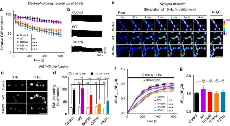
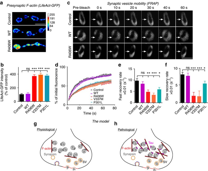
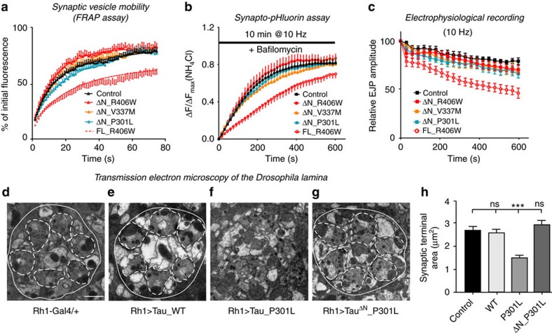
Tau is implicated in more than 20 neurodegenerative diseases, including Alzheimer’s disease. Under pathological conditions, Tau dissociates from axonal microtubules and missorts to pre- and postsynaptic terminals. Patients suffer from early synaptic dysfunction prior to Tau aggregate formation, but the underlying mechanism is unclear. Here we show that pathogenic Tau binds to synaptic vesicles via its N-terminal domain and interferes with presynaptic functions, including synaptic vesicle mobility and release rate, lowering neurotransmission in fly and rat neurons. Pathological Tau mutants lacking the vesicle binding domain still localize to the presynaptic compartment but do not impair synaptic function in fly neurons. Moreover, an exogenously applied membrane-permeable peptide that competes for Tau-vesicle binding suppresses Tau-induced synaptic toxicity in rat neurons. Our work uncovers a presynaptic role of Tau that may be part of the early pathology in various Tauopathies and could be exploited therapeutically.
Overview
The claims and statistics of this paper haven’t been checked yet.
Free: it reads the open-access text and checks the first 5 claims that cite a source we can check. Create a free account to check every claim.
Integrity signals
Registry
Cites
Statistics
Figures


Verification
Claims the paper makes
The claims and statistics of this paper haven’t been checked yet.
Free: it reads the open-access text and checks the first 5 claims that cite a source we can check. Create a free account to check every claim.
Claims other papers make about this one
Each is checked against this paper’s own text: does it say what the citing paper says it does?
The source backs part of the claim, not all of it — or backs it for a different population, condition or comparison.
In the citing paper
“The movement of vesicles and endo-lysosomal compartments along axons is a key part of amyloid fibril formation and vesicle-mediated aggregation of Tau proteins [7, 87].”
From Abnormal accumulation of extracellular vesicles in hippocampal dystrophic axons and regulation by the primary cilia in Alzheimer's disease (2023) · cites this paper as [87]
Experimental — accuracy not yet measured
Self-check
Percentages, sums and reported differences re-computed from the paper’s own numbers.
The self-check needs the paper’s full text.
Citations
Cites
Amyloid oligomers exacerbate tau pathology in a mouse model of tauopathy
Selenica, M.-L. B., Brownlow, M., Jimenez, J. P., Lee, D. C., Pena, G., Dickey, C. A., Gordon, M. N., & Morgan, D. (2013) · Neurodegenerative Diseases · doi:10.1159/000337230 · Cited by 10,359
Visualizing secretion and synaptic transmission with pH-sensitive green fluorescent proteins
Miesenböck, G., De Angelis, D. A., & Rothman, J. E. (1998) · Nature · doi:10.1038/28190 · Cited by 2,563
Tau in physiology and pathology
Wang, Y., & Mandelkow, E. (2016) · Nature Reviews Neuroscience · doi:10.1038/nrn.2015.1 · Cited by 2,251
Self-citationSynapse Loss and Microglial Activation Precede Tangles in a P301S Tauopathy Mouse Model
Yoshiyama, Y., Higuchi, M., Zhang, B., Huang, S.-M., Iwata, N., Saido, T. C., Maeda, J., Suhara, T., Trojanowski, J. Q., & Lee, V. M.-Y. (2007) · Neuron · doi:10.1016/j.neuron.2007.01.010 · Cited by 2,208
Tau-mediated neurodegeneration in Alzheimer’s disease and related disorders
Ballatore, C., Lee, V. M.-Y., & Trojanowski, J. Q. (2007) · Nature Reviews Neuroscience · doi:10.1038/nrn2194 · Cited by 2,174
Tau Suppression in a Neurodegenerative Mouse Model Improves Memory Function
SantaCruz, K., Lewis, J., Spires, T., Paulson, J., Kotilinek, L., Ingelsson, M., Guimaraes, A., DeTure, M., Ramsden, M., McGowan, E., et al. (2005) · Science · doi:10.1126/science.1113694 · Cited by 2,014
Self-citationReducing Endogenous Tau Ameliorates Amyloid ß-Induced Deficits in an Alzheimer's Disease Mouse Model
Roberson, E. D., Scearce-Levie, K., Palop, J. J., Yan, F., Cheng, I. H., Wu, T., Gerstein, H., Yu, G.-Q., & Mucke, L. (2007) · Science · doi:10.1126/science.1141736 · Cited by 1,892
Dendritic Function of Tau Mediates Amyloid-β Toxicity in Alzheimer's Disease Mouse Models
Ittner, L. M., Ke, Y. D., Delerue, F., Bi, M., Gladbach, A., van Eersel, J., Wölfing, H., Chieng, B. C., Christie, M. J., Napier, I. A., et al. (2010) · Cell · doi:10.1016/j.cell.2010.06.036 · Cited by 1,821
Hoover, B. R., Reed, M. N., Su, J., Penrod, R. D., Kotilinek, L. A., Grant, M. K., Pitstick, R., Carlson, G. A., Lanier, L. M., Yuan, L.-L., et al. (2010) · Neuron · doi:10.1016/j.neuron.2010.11.030 · Cited by 1,118
Hippocampal synaptic loss in early Alzheimer's disease and mild cognitive impairment
Scheff, S. W., Price, D. A., Schmitt, F. A., & Mufson, E. J. (2006) · Neurobiology of Aging · doi:10.1016/j.neurobiolaging.2005.09.012 · Cited by 1,017
Cited by
Items 101–200 of 473
Li, J., Du, Q., Li, N., Du, S., & Sun, Z. (2020) · Biomedicine & Pharmacotherapy · doi:10.1016/j.biopha.2020.111167 · Cited by 47
Neuroimmune Tau Mechanisms: Their Role in the Progression of Neuronal Degeneration
Cortés, N., Andrade, V., Guzmán-Martínez, L., Estrella, M., & Maccioni, R. (2018) · International Journal of Molecular Sciences · doi:10.3390/ijms19040956 · Cited by 42
“Moreover, tau is implicated in more than 20 neurodegenerative diseases [23], and therefore, it is of interest to review at least some of them.”
BackgroundInter-relationship of Histone Deacetylase-6 with Tau-cytoskeletal organization and remodeling
Balmik, A. A., & Chinnathambi, S. (2022) · European Journal of Cell Biology · doi:10.1016/j.ejcb.2022.151202 · Cited by 46
Chinnathambi, S., & Das, R. R. (2021) · Cell & Bioscience · doi:10.1186/s13578-023-01028-0 · Cited by 46
Moawad, M. H. E. D., Serag, I., Alkhawaldeh, I. M., Abbas, A., Sharaf, A., Alsalah, S., Sadeq, M. A., Shalaby, M. M. M., Hefnawy, M. T., Abouzid, M., et al. (2024) · Molecular Neurobiology · doi:10.1007/s12035-024-04468-y · Cited by 46
“Through its interaction with Synapto-gyrin-3, it was discovered to limit synaptic vesicles’ mobility and diminish neurotransmitters’ release from vesicles [64, 71, 72].”
Castrillo, J. I., Lista, S., Hampel, H. J., & Ritchie, C. W. (2018) · Methods in molecular biology · doi:10.1007/978-1-4939-7704-8_3 · Cited by 46
Hill, E., Wall, M. J., Moffat, K. G., & Karikari, T. K. (2020) · Frontiers in Molecular Neuroscience · doi:10.3389/fnmol.2020.00155 · Cited by 46
Roles of humanin and derivatives on the pathology of neurodegenerative diseases and cognition
Thiankhaw, K., Chattipakorn, K., Chattipakorn, S. C., & Chattipakorn, N. (2022) · Biochimica et Biophysica Acta (BBA) - General Subjects · doi:10.1016/j.bbagen.2022.130097 · Cited by 45
Implication of type 4 NADPH oxidase (NOX4) in tauopathy
Luengo, E., Trigo-Alonso, P., Fernández‐Mendívil, C., Núñez, Á., del Campo, M., Porrero, C., García‐Magro, N., Negredo, P., Senar, S., Sánchez‐Ramos, C., et al. (2021) · Free Radical Biology and Medicine · doi:10.1016/j.redox.2021.102210 · Cited by 45
Evidence for trans-synaptic propagation of oligomeric tau in human progressive supranuclear palsy
McGeachan, R. I., Keavey, L., Simzer, E. M., Chang, Y. Y., Rose, J. L., Spires-Jones, M. P., Gilmore, M., Holt, K., Meftah, S., Ravingerova, N., et al. (2025) · Nature Neuroscience · doi:10.1038/s41593-025-01992-5 · Cited by 44
“To test whether tau colocalizes with the presynaptic vesicle protein synaptogyrin-3 in human PSP synapses (as has been previously observed in mice and flies 8,24 ), array tomography ribbons from the frontal cortex were also immunostained for total tau, synaptogyrin-3 and SYO (Extended Data Fig.”
InfluentialSelf-citationNatale, G. D., Sabatino, G., Sciacca, M. F. M., Tosto, R., Milardi, D., & Pappalardo, G. (2022) · Molecules · doi:10.3390/molecules27165066 · Cited by 44
“Aβ is also present in the synaptic cleft region [117] while recent studies indicate that tau can be detected into the synaptic vesicles under pathological conditions [118].”
BackgroundSingle-Cell Spatial Proteomic Imaging for Human Neuropathology
Vijayaragavan, K., Cannon, B. J., Tebaykin, D., Bosse, M. M., Baranski, A., Oliveria, J. P., Bukhari, S. A., Mrdjen, D., Corces, M. R., McCaffrey, E., et al. (2022) · Acta Neuropathologica Communications · doi:10.1186/s40478-022-01465-x · Cited by 44
“Drosophila to mouse studies have shown that synaptic PHF1-TAU interferes with synaptic vesicles (Sahara et al., 2014; Tai et al., 2014; Zhou et al., 2017).”
Tau Abnormalities and the Potential Therapy in Alzheimer’s Disease
Almansoub, H. A. M. M., Tang, H. R., Wu, Y., Wang, D., Mahaman, Y. A. R., Wei, N., Almansob, Y. A. M., He, W., & Liu, D. (2018) · Journal of Alzheimer s Disease · doi:10.3233/jad-180868 · Cited by 43
Corsetti, V., Borreca, A., Latina, V., Giacovazzo, G., Pignataro, A., Krashia, P., Natale, F., Cocco, S., Rinaudo, M., Malerba, F., et al. (2020) · Brain Communications · doi:10.1093/braincomms/fcaa039 · Cited by 42
“…(Brandt et al. , 1995) and undergoes early conformational changes in human tauopathies including Alzheimer’s disease (Combs et al. , 2016, 2017)—is endowed with deleterious action(s), mainly at nerve endings (King et al. , 2006; Ittner et al. , 2010; Amadoro et al. , 2012; Zhou et al. , 2017).”
Schaler, A. W., Runyan, A. M., Clelland, C. L., Sydney, E. J., Fowler, S. L., Figueroa, H. Y., Shioda, S., Santa-Maria, I., Duff, K. E., & Myeku, N. (2021) · Science Translational Medicine · doi:10.1126/scitranslmed.aba7394 · Cited by 42
Taylor, L. W., Simzer, E. M., Pimblett, C., Lacey-Solymar, O. T. T., McGeachan, R. I., Meftah, S., Rose, J. L., Spires-Jones, M. P., Holt, K., Catterson, J. H., et al. (2024) · Acta Neuropathologica · doi:10.1007/s00401-023-02667-w · Cited by 42
“Recent work has highlighted potentially important roles of synaptic tau for both toxicity and involvement of trans-synaptic tau spread [9, 36, 47, 68].”
Self-citationCaenorhabditis elegans models of tauopathy
Pir, G. J., Choudhary, B. C., & Mandelkow�, E. (2017) · The FASEB Journal · doi:10.1096/fj.201701007 · Cited by 42
“In a recent study, tauwas reported to impair neurotransmission at the presynapse by binding and inhibiting synaptic vesicle transport and release, an effect mediated by the N-terminal domain (78).”
BackgroundInsulin Resistance as a Common Link Between Current Alzheimer’s Disease Hypotheses
Alves, S. S., da Silva-Júnior, R. M. P., Servilha‐Menezes, G., Homolak, J., Šalković‐Petrišić, M., & Garcia‐Cairasco, N. (2021) · Journal of Alzheimer s Disease · doi:10.3233/jad-210234 · Cited by 42
Prikas, E., Parić, E., Asih, P. R., Stefanoska, K., Stefen, H., Fath, T., Poljak, A., & Ittner, A. (2022) · The EMBO Journal · doi:10.15252/embj.2021110242 · Cited by 42
“…interactor proteins include microtubules and annexins in the axon (Gauthier-Kemper et al , 2018), vesicle-associated proteins such as synaptobrevin (Zhou et al , 2017), postsynaptic proteins such as postsynaptic density protein-95 (PSD-95) (Prikas et al , 2021), SynGAP (Bi et al , 2017) and…”
Glial cells and adaptive immunity in frontotemporal dementia with tau pathology
Hartnell, I. J., Blum, D., Nicoll, J. A. R., Dorothee, G., & Boche, D. (2021) · Brain · doi:10.1093/brain/awaa457 · Cited by 42
Biomarkers of synaptic degeneration in Alzheimer’s disease
Cheng, Q., Fan, Y., Zhang, P., Liu, H., Han, J., Yu, Q., Wang, X., Wu, S., & Lu, Z. (2024) · Ageing Research Reviews · doi:10.1016/j.arr.2024.102642 · Cited by 42
The interplay between age at menopause and synaptic integrity on Alzheimer’s disease risk in women
Alexander, M. W., Honer, W. G., Saloner, R., Galea, L. A. M., Bennett, D. A., Rabin, J. S., & Casaletto, K. B. (2025) · Science Advances · doi:10.1126/sciadv.adt0757 · Cited by 42
“…tangles and global cognitive decline and given the evidence that synaptic dysregulation may causally contribute to AD pathogenesis ( 20 , 37 , 49 – 52 ), we tested whether the interactions between synaptic biomarkers and age at menopause on cognitive decline were mediated by tau levels in post…”
CSF total tau levels are associated with hippocampal novelty irrespective of hippocampal volume
Düzel, E., Berron, D., Schütze, H., Cárdenas‐Blanco, A., Metzger, C. D., Betts, M. J., Ziegler, G., Chen, Y., Dobisch, L., Bittner, D., et al. (2018) · Alzheimer s & Dementia Diagnosis Assessment & Disease Monitoring · doi:10.1016/j.dadm.2018.10.003 · Cited by 41
“However, more recent discussions consider the possibility that tau can also be associated with synaptic dysfunction before neurodegeneration has occurred [14].”
BackgroundResultPotjewyd, F., & Axtman, A. D. (2021) · Frontiers in Cellular Neuroscience · doi:10.3389/fncel.2021.768655 · Cited by 40
Molecular pathology and synaptic loss in primary tauopathies: an 18F-AV-1451 and 11C-UCB-J PET study
Holland, N., Malpetti, M., Rittman, T., Mak, E. E., Passamonti, L., Kaalund, S. S., Hezemans, F. H., Jones, P. S., Savulich, G., Hong, Y. T., et al. (2022) · Brain · doi:10.1093/brain/awab282 · Cited by 39
“Animal models of tauopathy have illustrated the co-localisation of tau aggregates at the synaptic bouton, associated with synaptic dysfunction and synaptic loss 18, 30 but the tau-synapse association is yet to be determined in vivo.”
Comerota, M. M., Tumurbaatar, B., Krishnan, B., Kayed, R., & Taglialatela, G. (2019) · Molecular Neurobiology · doi:10.1007/s12035-018-1248-9 · Cited by 37
Sokolova, D., Childs, T., & Hong, S. Y. (2021) · Faculty Reviews · doi:10.12703/r/10-19 · Cited by 39
Capilla-López, M. D., Deprada-Fernandez, Á., Andrade‐Talavera, Y., Martínez‐Gallego, I., Coatl-Cuaya, H., Sotillo, P. S., Rodríguez-Álvarez, J., Moreno, A. R., Parra‐Damas, A., & Saura, C. A. (2025) · Molecular Psychiatry · doi:10.1038/s41380-025-02901-9 · Cited by 38
“A β and tau affect both presynaptic and postsynaptic mechanisms impairing excitatory glutamatergic transmission [35, 41, 42], but whether synaptic tau is responsible and/or synergize with A β to induce synapse pathology and behavioral changes is unclear.”
TDP-43 Proteinopathy and Tauopathy: Do They Have Pathomechanistic Links?
Riku, Y., Yoshida, M., Iwasaki, Y., Sobue, G., Katsuno, M., & Ishigaki, S. (2022) · International Journal of Molecular Sciences · doi:10.3390/ijms232415755 · Cited by 38
“Misfolded tau also binds to intraneuronal organelles, including mitochondria [53] and presynaptic vesicles [54].”
BackgroundGlennon, E. B. C., Lau, D. H. W., Gabriele, R. M. C., Taylor, M. F., Troakes, C., Opie-Martin, S., Elliott, C., Killick, R., Hanger, D. P., Perez‐Nievas, B. G., et al. (2020) · Brain Communications · doi:10.1093/braincomms/fcaa011 · Cited by 38
“The redistribution of highly phosphorylated and/or oligomeric tau species to pre- and post-synapses causes the disruption of synaptic vesicle mobility and neurotransmitter release (Zhou et al., 2017; McInnes et al., 2018) and excitotoxicity (Ittner et al.”
BackgroundA Conserved Cytoskeletal Signaling Cascade Mediates Neurotoxicity of FTDP-17 Tau Mutations In Vivo
Bardai, F. H., Wang, L., Mutreja, Y., Yenjerla, M., Gamblin, T. C., & Feany, M. B. (2018) · The Journal of Neuroscience · doi:10.1523/jneurosci.1550-17.2017 · Cited by 37
“A separate study by another group suggests that two additional FTDP-17 variants, P301L and V337M, likewise promote tau neurotoxicity in vivo through excess stabilization of F-actin (Zhou et al., 2017).”
BackgroundTracing synaptic loss in Alzheimer's brain with SV2A PET‐tracer UCB‐J
Kumar, A., Scarpa, M., & Nordberg, A. K. (2024) · Alzheimer s & Dementia · doi:10.1002/alz.13720 · Cited by 37
Therapeutic Targeting Strategies for Early- to Late-Staged Alzheimer’s Disease
Kang, Y. J., Diep, Y. N., Tran, M., & Cho, H. (2020) · International Journal of Molecular Sciences · doi:10.3390/ijms21249591 · Cited by 36
“In vitro studies revealed that abnormal phosphorylated tau proteins could increase the risk of AD significantly by disrupting microtubule stability and enhancing tau aggregates in the brain [88,113].”
Background3D Analysis of the Synaptic Organization in the Entorhinal Cortex in Alzheimer’s Disease
Domínguez-Álvaro, M., Montero-Crespo, M., Blazquez-Llorca, L., Plaza-Alonso, S., Cano-Astorga, N., DeFelipe, J., & Alonso-Nanclares, L. (2021) · eneuro · doi:10.1523/eneuro.0504-20.2021 · Cited by 33
“Tau protein and b -amyloid peptide in pathologic conditions could have toxic effects on synapses leading to synaptic loss and/or dysfunction in AD (Zhou et al., 2017; for review, see Henstridge et al., 2016; Rajmohan and Reddy, 2017).”
BackgroundHori, T., Eguchi, K., Wang, H.-Y., Miyasaka, T., Guillaud, L., Taoufiq, Z., Mahapatra, S., Yamada, H., Takei, K., & Takahashi, T. (2022) · eLife · doi:10.7554/elife.73542 · Cited by 35
“In these experiments, exocytic 474 capacitance change (ΔCm) represents ~ 5 times larger number of SVs (estimated from ΔCm 475 divided by Cm of single SV) than that in the immediately releasable pool by presynaptic action 476 potentials (estimated by the size of maximally evoked EPSCs divided by the size of miniature 4…”
BackgroundMethodInfluentialLi, W., Peng, X., Mei, X., Dong, M., Li, Y., & Dong, H. (2023) · ACS Applied Materials & Interfaces · doi:10.1021/acsami.3c03181 · Cited by 35
The tumour-promoting role of protein homeostasis: Implications for cancer immunotherapy
Liang, R., Tan, H., Jin, H., Wang, J., Tang, Z., & Lu, X. (2023) · Cancer Letters · doi:10.1016/j.canlet.2023.216354 · Cited by 35
Electrochemical Monitoring of Real‐Time Vesicle Dynamics Induced by Tau in a Confined Nanopipette
Chen, K., Yu, R., Zhong, C., Wang, Z., Xie, B.-K., Ma, H., Ao, M., Zheng, P., Ewing, A. G., & Long, Y. (2024) · Angewandte Chemie International Edition · doi:10.1002/anie.202406677 · Cited by 34
Brekk, O. R., Moskites, A., Isacson, O., & Hallett, P. J. (2018) · Scientific Reports · doi:10.1038/s41598-018-33474-z · Cited by 34
Sundararajan, V., Venkatasubbu, G. D., & Mohideen, S. S. (2021) · 3 Biotech · doi:10.1007/s13205-021-02706-x · Cited by 34
A comprehensive review on anticancer mechanism of bazedoxifene
Zafar, E., Maqbool, M. F., Iqbal, A., Maryam, A., Shakir, H. A., Irfan, M. I., Khan, M. N., Li, Y., & Ma, T. (2021) · Biotechnology and Applied Biochemistry · doi:10.1002/bab.2150 · Cited by 34
Teravskis, P. J., Oxnard, B. R., Miller, E. C., Kemper, L. J., Ashe, K. H., & Liao, D. (2019) · The Journal of Physiology · doi:10.1113/jp277459 · Cited by 33
“…synapses in cellular and mouse models of tauopathies (Santacruz et al. 2005; Hoover et al. 2010; Ittner et al. 2010; Min et al. 2010; Lasagna-Reeves et al. 2011; Maday et al. 2014; Sherman et al. 2016; Tracy et al. 2016; Zhao et al. 2016; Sealey et al. 2017; Zhou et al. 2017; Mclnnes et al. 2018).”
BackgroundTranscellular Spreading of Tau in Tauopathies
Demaegd, K. C., Schymkowitz, J. W. H., & Rousseau, F. (2018) · ChemBioChem · doi:10.1002/cbic.201800288 · Cited by 34
Hippocampal neurophysiology is modified by a disease-associated C-terminal fragment of tau protein
Tamagnini, F., Walsh, D. A., Brown, J. T., Bondulich, M. K., Hanger, D. P., & Randall, A. D. (2017) · Neurobiology of Aging · doi:10.1016/j.neurobiolaging.2017.07.005 · Cited by 32
“This result parallels the observation of reduced amplitude of 10 Hz evoked EPSPs in cultured rat hippocampal neurons and drosophila neuromuscular junctions, carrying mutations associated to familial FTD (i.e., P301L on tau) (Zhou et al., 2017).”
Alfaro-Ruiz, R., Martín-Belmonte, A., Aguado, C., Hernández, F., Moreno-Martínez, A. E., Ávila, J., & Luján, R. (2021) · International Journal of Molecular Sciences · doi:10.3390/ijms222011106 · Cited by 36
“Although most studies dealing with the implication of Aβ and tau in synaptic dysfunction have concentrated in postsynaptic receptors and dendritic mechanisms [36], increasing evidence suggest that deficits in presynaptic mechanisms are also present in AD [47,48].”
Trease, A. J., George, J. W., Roland, N. J., Lichter, E. Z., Emanuel, K. M., Totusek, S., Fox, H. S., & Stauch, K. L. (2022) · Frontiers in Molecular Neuroscience · doi:10.3389/fnmol.2022.852368 · Cited by 32
Lantero‐Rodriguez, J., Tissot, C., Snellman, A., Servaes, S., Benedet, A. L., Rahmouni, N., Montoliu‐Gaya, L., Therriault, J., Brum, W. S., Stevenson, J., et al. (2023) · Alzheimer s & Dementia · doi:10.1002/alz.13119 · Cited by 32
“50,51 Moreover, plasma NTA-tau could be an easily implementable tool to detect individuals at middle to late stages of AD, as well as individuals at risk of accumulating further tau.”
MethodNeurophysiological trajectories in Alzheimer’s disease progression
Kudo, K., Ranasinghe, K. G., Morise, H., Syed, F., Sekihara, K., Rankin, K. P., Miller, B. L., Kramer, J. H., Rabinovici, G. D., Vossel, K. A., et al. (2023) · eLife · doi:10.7554/elife.91044 · Cited by 32
Alternative hypotheses related to Alzheimer’s disease
Cubinkova, V., Valachova, B., Uhrinova, I., Brezováková, V., Smolek, T., Jadhav, S., & Žilka, N. (2018) · Bratislavské lekárske listy/Bratislava medical journal · doi:10.4149/bll_2018_039 · Cited by 31
“This concept has been intensely investigated in recent years (54, 55).”
The Role of Synaptic Dysfunction in Alzheimer’s Disease
Pei, Y. A., Davies, J., Zhang, M., & Zhang, H. (2020) · Journal of Alzheimer s Disease · doi:10.3233/jad-191334 · Cited by 31
Amyloid pathology but notAPOEε4 status is permissive for tau-related hippocampal dysfunction
Düzel, E., Ziegler, G., Berron, D., Maaß, A., Schütze, H., Cárdenas‐Blanco, A., Glanz, W., Metzger, C. D., Dobisch, L., Reuter, M., et al. (2021) · Brain · doi:10.1093/brain/awab405 · Cited by 31
“sistent with animal studies showing that misfolded and hyperphosphoylated tau can impair neuronal function.(39,40) Early local accumulations of pathological tau in axons leads to presynaptic”
Macromolecular Structures and Proteins Interacting with the Microtubule Associated Tau Protein
Jiménez, J. S. (2022) · Neuroscience · doi:10.1016/j.neuroscience.2022.05.023 · Cited by 31
Secker, C., Motzny, A., Kostova, S., Buntru, A., Helmecke, L., Reus, L., Steinfort, R., Brusendorf, L., Boeddrich, A., Neuendorf, N., et al. (2023) · Journal of Neurochemistry · doi:10.1111/jnc.15842 · Cited by 31
High-fat diet induces cognitive impairment through repression of SIRT1/AMPK-mediated autophagy
Yi, W., Chen, F., Yuan, M., Wang, C., Wang, S., Wen, J., Zou, Q., Pu, Y., & Cai, Z. (2023) · Experimental Neurology · doi:10.1016/j.expneurol.2023.114591 · Cited by 31
Ma, X., Zhu, Y., Lu, J., Xie, J., Li, C. V., Shin, W. S., Qiang, J., Liu, J., Dou, S., Xiao, Y., et al. (2020) · eLife · doi:10.7554/elife.51859 · Cited by 31
“It was previously described that Tau increases F-actin aggregation at synaptic terminals, promotes actin polymerization, and reduces synaptic vesicle mobility (Zhou et al., 2017).”
BackgroundDüzel, E., Costagli, M., Donatelli, G., Speck, O., & Cosottini, M. (2021) · European Radiology Experimental · doi:10.1186/s41747-021-00221-5 · Cited by 30
“show that misfolded and hyperphosphorylated tau can impair neuronal function [52].”
BackgroundMass Spectrometric Analysis of Urinary N-Glycosylation Changes in Patients with Parkinson’s Disease
Xu, M., Jin, H., Ge, W., Zhao, L., Liu, Z., Guo, Z., Wu, Z., Chen, J., Mao, C., Zhang, X., et al. (2023) · ACS Chemical Neuroscience · doi:10.1021/acschemneuro.3c00404 · Cited by 30
Kong, Y., Huang, L., Li, W., Liu, X., Zhou, Y., Liu, C., Zhang, S., Xie, F., Zhang, Z., Jiang, D., et al. (2021) · Frontiers in Cell and Developmental Biology · doi:10.3389/fcell.2021.609908 · Cited by 29
“It is known that tau cross-links synaptic vesicles, thereby slowing their mobilization and ultimately reducing synaptic transmission during intense stimulation (Zhou et al., 2017).”
BackgroundRetromer in Synaptic Function and Pathology
Brodin, L., & Shupliakov, O. (2018) · Frontiers in Synaptic Neuroscience · doi:10.3389/fnsyn.2018.00037 · Cited by 29
Synaptic sabotage: How Tau and α-Synuclein undermine synaptic health
Uytterhoeven, V., Verstreken, P., & Nachman, E. (2024) · The Journal of Cell Biology · doi:10.1083/jcb.202409104 · Cited by 29
Self-citationXie, A., Hou, T., Xiong, W., Huang, H., Zheng, J., Li, K., Man, H., Hu, Y., Han, Z., Zhang, H., et al. (2019) · Aging Cell · doi:10.1111/acel.12929 · Cited by 29
“Tau binds to synaptic vesicles and interferes with their mobility and release rate (Zhou et al., 2017).”
BackgroundDillon, G. M., Henderson, J. L., Bao, C., Joyce, J. A., Calhoun, M. E., Amaral, B., King, K. W., Bajrami, B., & Rabah, D. (2020) · PLoS ONE · doi:10.1371/journal.pone.0228771 · Cited by 29
“Several new lines of evidence show that aside from microtubule binding, the hyperphosphorylation of tau affects a variety of cellular processes including RNA translational selectivity [11], nuclear import/export [44], pre-synaptic vesicle motility [45] and post-synaptic excitotoxicity [46].”
BackgroundJin, M., Wang, S., Gao, X., Zou, Z., Hirotsune, S., & Sun, L. (2023) · Neural Regeneration Research · doi:10.4103/1673-5374.382231 · Cited by 28
“Besides its well-known roles in MT dynamics, low-level distribution of tau in synaptic terminals is considered to play important roles in cell signaling and synaptic plasticity (Ittner et al., 2010; Zhou et al., 2017).”
BackgroundJash, S., Banerjee, S., Cheng, S., Wang, B., Qiu, C., Kondo, A., Ernerudh, J., Zhou, X. Z., Lu, K. P., & Sharma, S. (2023) · Nature Communications · doi:10.1038/s41467-023-41144-6 · Cited by 27
Vesicular Axonal Transport is Modified In Vivo by Tau Deletion or Overexpression in Drosophila
Talmat-Amar, Y., Arribat, Y., & Parmentier, M. (2018) · International Journal of Molecular Sciences · doi:10.3390/ijms19030744 · Cited by 27
“Because this domain is involved in membrane association of Tau, especially in vesicles [58,59], it may be involved in accumulation of vesicles into batches observed in the presence of Tau overexpression.”
BackgroundChoudhary, B. C., Mandelkow�, E., Mandelkow, E.-M., & Pir, G. J. (2018) · Neurobiology of Disease · doi:10.1016/j.nbd.2018.06.005 · Cited by 27
The Synaptic Dysregulation in Adolescent Rats Exposed to Maternal Immune Activation
Cieślik, M., Gąssowska-Dobrowolska, M., Zawadzka, A., Frontczak‐Baniewicz, M., Gewartowska, M., Dominiak, A., Czapski, G. A., & Adamczyk, A. (2021) · Frontiers in Molecular Neuroscience · doi:10.3389/fnmol.2020.555290 · Cited by 27
Haytural, H., Jordá-Siquier, T., Winblad, B. G., Mulle, C., Tjernberg, L. O., Granholm, A., Frykman, S., & Barthet, G. (2021) · Brain Communications · doi:10.1093/braincomms/fcab079 · Cited by 26
Templated α-Synuclein Inclusion Formation Is Independent of Endogenous Tau
Stoyka, L. E., Mahoney-Crane, C. L., Thrasher, D. R., Russell, D. L., Cook, A. K., Harris, A. T., Narayanan, A., Janado, T. P., Standaert, D. G., Roberson, E. D., et al. (2021) · eNeuro · doi:10.1523/eneuro.0458-20.2021 · Cited by 26
Amyloid-β in Alzheimer’s disease – front and centre after all?
Weglinski, C., & Jeans, A. F. (2022) · Neuronal Signaling · doi:10.1042/ns20220086 · Cited by 26
Goossens, J., González, A. C., Dewit, N., Lidón, L., Fortea, J., Alcolea, D. A., Lleó, A., Belbin, O., & Vanmechelen, E. (2023) · Alzheimer s Research & Therapy · doi:10.1186/s13195-023-01336-0 · Cited by 26
“A meta-analysis of synaptic pathology on human brain tissue pointed towards a selective molecular vesicular machinery vulnerability [26] and in several AD models, defects in synaptic vesicle recycling were found to be linked to amyloid and tau toxicity in early stages [27, 28].”
BackgroundSynaptopathy: presynaptic convergence in frontotemporal dementia and amyotrophic lateral sclerosis
Clayton, E. L., Huggon, L., Cousin, M. A., & Mizielinska, S. (2024) · Brain · doi:10.1093/brain/awae074 · Cited by 26
“106 Expression of R406W, V337M or P301L mutants of tau in the Drosophila model reduced synaptic transmission, synaptic vesicle re-cycling and fusion during sustained high-frequency stimulation.”
BackgroundInfluentialWang, J., Huang, Q., He, K., Li, J., Guo, T., Yang, Y., Lin, Z., Li, S., Vanderlinden, G., Huang, Y. H., et al. (2024) · Alzheimer s & Dementia · doi:10.1002/alz.13817 · Cited by 26
CYP1B1-RMDN2 Alzheimer’s disease endophenotype locus identified for cerebral tau PET
Nho, K. T., Risacher, S. L., Apostolova, L. G., Bice, P. J., Brosch, J. R., Deardorff, R. L., Faber, K. M., Farlow, M. R., Foroud, T., Gao, S., et al. (2024) · Nature Communications · doi:10.1038/s41467-024-52298-2 · Cited by 26
Amyloid-β oligomers increase the binding and internalization of tau oligomers in human synapses
Kadamangudi, S., Marcatti, M., Zhang, W.-R., Fracassi, A., Kayed, R., Limon, A., & Taglialatela, G. (2024) · Acta Neuropathologica · doi:10.1007/s00401-024-02839-2 · Cited by 25
Astrocytic C–X–C motif chemokine ligand-1 mediates β-amyloid-induced synaptotoxicity
Perez‐Nievas, B. G., Johnson, L., Beltran-Lobo, P., Hughes, M. M., Gammallieri, L., Tarsitano, F., Myszczynska, M. A., Vázquez-Villaseñor, I., Jiménez-Sánchez, M., Troakes, C., et al. (2021) · Journal of Neuroinflammation · doi:10.1186/s12974-021-02371-0 · Cited by 25
Cyclosporine A (CsA) prevents synaptic impairment caused by truncated tau by caspase-3
Tapia-Monsalves, C., Olesen, M. A., Villavicencio-Tejo, F., & Quintanilla, R. A. (2023) · Molecular and Cellular Neuroscience · doi:10.1016/j.mcn.2023.103861 · Cited by 26
White matter tauopathy: Transient functional loss and novel myelin remodeling
Jackson, J., Bianco, G., Rosa, A. O., Cowan, K., Bond, P., Anichtchik, O. V., & Fern, R. F. (2018) · Glia · doi:10.1002/glia.23286 · Cited by 25
“For example, recent evidence shows that early mutant tau induction leads to occlusion of synaptic transmission via the direct actions of tau upon vesicle release (Zhou et al., 2017).”
BackgroundInfluentialTau Interacting Proteins: Gaining Insight into the Roles of Tau in Health and Disease
Stancu, I., Ferraiolo, M., Terwel, D., & Dewachter, I. (2019) · Advances in experimental medicine and biology · doi:10.1007/978-981-32-9358-8_13 · Cited by 25
Self-citationRiku, Y., Seilhean, D., Duyckaerts, C. J., Boluda, S., Iguchi, Y., Ishigaki, S., Iwasaki, Y., Yoshida, M., Sobue, G., & Katsuno, M. (2021) · International Journal of Molecular Sciences · doi:10.3390/ijms22083843 · Cited by 24
“It is unknown whether the aggregation of p-TDP-43 within axonal terminals demonstrates in situ synaptotoxicity, which has been reported for a tauopathy model; induction of pathogenic mutant tau bound to pre-synaptic vesicles and disrupted synaptic functions [62].”
BackgroundAlyenbaawi, H., Allison, W. T., & Mok, S. (2020) · Biomolecules · doi:10.3390/biom10111487 · Cited by 24
Keller, N., Christensen, T. A., Wanberg, E. J., Salisbury, J. L., & Trushina, E. V. (2025) · Scientific Reports · doi:10.1038/s41598-025-90925-0 · Cited by 23
“…toward the axon terminal, and Glutamate receptor 3 ( GRIA4 ), a mediator of synaptic excitatory neurotransmission, metabotropic glutamate receptors 7 and 21 ( GRMN7 and GRM2 ), which facilitate the formation of LTP, and Double C2 protein ( Doc2a ), which contributes to spontaneous excitatory and…”
Olesen, M. A., & Quintanilla, R. A. (2023) · Molecular Neurobiology · doi:10.1007/s12035-023-03434-4 · Cited by 23
“Tau has been observed in synaptic compartments of the AD brain, playing a pivotal role in the defects of neuronal communication [101, 102].”
BackgroundAlzheimer’s disease: an axonal injury disease?
Liang, D., & Zhang, Z. (2023) · Frontiers in Aging Neuroscience · doi:10.3389/fnagi.2023.1264448 · Cited by 23
“In the presynaptic terminal, pathogenic tau binds to synaptic vesicles through its N-terminal structural domain and interferes with presynaptic processes, including synaptic vesicle mobility and release rate, ultimately leading to reduced neurotransmission in fly and rat neurons (Zhou et al., 2017).”
Organellar Omics—A Reviving Strategy to Untangle the Biomolecular Complexity of the Cell
Tharkeshwar, A. K., Gevaert, K., & Annaert, W. G. (2017) · PROTEOMICS · doi:10.1002/pmic.201700113 · Cited by 23
Hanger, D. P., Goniotaki, D., & Noble, W. J. (2019) · Advances in experimental medicine and biology · doi:10.1007/978-981-32-9358-8_9 · Cited by 23
Tau-Mediated Dysregulation of Neuroplasticity and Glial Plasticity
Koller, E. J., & Chakrabarty, P. (2020) · Frontiers in Molecular Neuroscience · doi:10.3389/fnmol.2020.00151 · Cited by 23
CSF total tau as a proxy of synaptic degeneration
Soares, C., Bellaver, B., Ferreira, P. C. L., Povala, G., Aguzzoli, C. S., Ferrari‐Souza, J. P., Zalzale, H., Lussier, F. Z., Rohden, F., Abbas, S., et al. (2025) · Nature Communications · doi:10.1038/s41467-025-63545-5 · Cited by 22
Probiotics for Preventing Cognitive Impairment in Alzheimer’s Disease
Wong, C. B., Kobayashi, Y., & Xiao, J. (2018) · IntechOpen eBooks · doi:10.5772/intechopen.79088 · Cited by 22
“Hyperphosphorylation of tau is known to induce a lower grade of interaction of tau proteins with microtubules that leads to greater self-aggregation of tau proteins and consequently induces malfunction of axonal transport [23], mis-stabilization of actin [24], synaptic impairment [25], and defects in mitochondrial int…”
BackgroundThe Mystery of Chemotherapy Brain: Kynurenines, Tubulin and Biophoton Release
Sordillo, P. P., & Sordillo, L. A. (2020) · Anticancer Research · doi:10.21873/anticanres.14061 · Cited by 22
Monteiro‐Fernandes, D., Silva, J. M., Soares‐Cunha, C., Dalla, C., Kokras, N., Arnaud, F., Billiras, R., Zhuravleva, V., Waites, C. L., Bretin, S., et al. (2020) · Molecular Psychiatry · doi:10.1038/s41380-020-0794-5 · Cited by 22
“In addition to disrupted postsynaptic function, presynaptic dysregulation may also contribute to the pathogenesis of AD, with recent work indicating that Tau accumulation in the presynaptic compartment may be an underpinning mechanism [60] by impairing synaptic vesicle dynamics and therefore, neurotransmission [61, 62…”
BackgroundPenny, L. K., Lofthouse, R. A., Arastoo, M., Porter, A., Palliyil, S., Harrington, C. R., & Wischik, C. M. (2024) · Translational Neurodegeneration · doi:10.1186/s40035-024-00417-w · Cited by 21
“A further consideration is that the tau oligomer load in synapses and subsequent dysfunction play a critical role in AD pathophysiology and clinical decline [77–79].”
BackgroundJang, J., Yeo, S., Baek, S., Jung, H. J., Lee, M. S., Choi, S. H., & Choe, Y. (2023) · Acta Neuropathologica Communications · doi:10.1186/s40478-023-01637-3 · Cited by 20
“The movement of vesicles and endo-lysosomal compartments along axons is a key part of amyloid fibril formation and vesicle-mediated aggregation of Tau proteins [7, 87].”
BackgroundBowles, K. R., Pedicone, C., Pugh, D. A., Oja, L.-M., Sousa, F. H., Keavey, L. K., Fulton-Howard, B., Weitzman, S. A., Liu, Y., Chen, J. L., et al. (2024) · Cell Reports · doi:10.1016/j.celrep.2024.115013 · Cited by 20
Somatic Mutations and Alzheimer’s Disease
Downey, J. S., Lam, J. C. K., Li, V. O. K., & Gozes, I. (2022) · Journal of Alzheimer s Disease · doi:10.3233/jad-220643 · Cited by 20
“Hyperphosphorylation, however, is well-established to disrupt tau-association with MTs, resulting in aberrant localization [101, 102] and, whether or not NFTs are themselves pathogenic, there is a strong evidence that aggregate-fomenting forms of tau contribute to AD neuropathology [103].”
Tyrosyl-tRNA synthetase has a noncanonical function in actin bundling
Ermanoska, B., Asselbergh, B., Morant, L., Erfurth, M., Hosseinibarkooie, S., Leitão-Gonçalves, R., Almeida-Souza, L., Bervoets, S., Sun, L., Lee, L., et al. (2023) · Nature Communications · doi:10.1038/s41467-023-35908-3 · Cited by 20
Self-citationLi, C., Wang, Q., Wu, L., Hu, J.-Y., Gao, Q.-C., Jiao, X.-L., Zhang, Y., Tang, S., Qi, Y. M., & He, P. F. (2024) · Scientific Reports · doi:10.1038/s41598-024-75377-2 · Cited by 20
Longitudinal synaptic loss versus tau Braak staging in amnestic mild cognitive impairment
Vanderlinden, G., Koole, M. J. B., Michiels, L., Lemmens, R., Vandenbulcke, M., & Laere, K. V. (2024) · Alzheimer s & Dementia · doi:10.1002/alz.14412 · Cited by 20
“8 Soluble tau oligomers are believed to be the toxic species leading to synaptic degeneration, even in the absence of mature NFTs. 9,10 More specifically, Taddei et al. 11 recently showed that the presence of tau oligomers in synapses leads to increased synapse elimination by microglia and…”
Puliatti, G., Puma, D. D. L., Aceto, G., Lazzarino, G., Acquarone, E., Mangione, R., D'adamio, L., Ripoli, C., Arancio, O., Piacentini, R., et al. (2023) · Progress in Neurobiology · doi:10.1016/j.pneurobio.2023.102482 · Cited by 19
Tau: a biomarker of Huntington’s disease
Lepinay, E., & Cicchetti, F. (2023) · Molecular Psychiatry · doi:10.1038/s41380-023-02230-9 · Cited by 19
“As per its location, tau is essential to the formation/stabilization of neuronal microtubule networks, nuclear stability, axonal/mitochondrial transport [9] as well as synaptic activity, including vesicular release [10, 11] and function of glutamatergic NMDA (N-methyl-D-aspartate) receptors [12].”
Metadata
- Authors
- Zhou, L., McInnes, J., Wierda, K., Holt, M., Herrmann, A. G., Jackson, R. J., Wang, Y.-C., Swerts, J., Beyens, J., Miskiewicz, K., Vilain, S., Dewachter, I., Moechars, D., De Strooper, B., Spires-Jones, T. L., De Wit, J., & Verstreken, P.
- Affiliations
- VIB-KU Leuven Center for Brain & Disease Research (BE)
- Euan MacDonald Centre for Motor Neuron Disease Research (GB)
- Hasselt University (BE)
- Janssen (Belgium) (BE)
- Corresponding author
- Verstreken, Patrik
- ORCID
- Venue
- Nature Communications
- Published
- 2017-05-11
- Volume
- 8
- Issue
- 1
- Pages
- 15295-15295
- ISSN
- 2041-1723 (ISSN-L)
- PMC
- PMC5437271
- OpenAlex
- W2613492737
- Cited by
- 434
- References
- 72
- Citations by year
- 2026: 40 · 2025: 47 · 2024: 61 · 2023: 54 · 2022: 35 · 2021: 68 · 2020: 49 · 2019: 31 · 2018: 40 · 2017: 8
- FWCI
- 25.89
- Type
- journal article
- Language
- en
- Open access
- gold
- doi.org
- Licence
- cc-by
- creativecommons.org/licenses/by/4.0 (tdm)
- creativecommons.org/licenses/by/4.0 (vor)
- Locations
- Nature Communications (publishedVersion, cc-by)
- Europe PMC (PubMed Central) (publishedVersion, cc-by)
- PubMed Central (publishedVersion, cc-by)
- PubMed (publishedVersion)
- Discovery Research Portal (University of Dundee) (publishedVersion, other-oa)
- Edinburgh Research Explorer (University of Edinburgh) (publishedVersion, cc-by)
- Lirias (submittedVersion, cc-by)
- RePEc: Research Papers in Economics (submittedVersion)
- DOAJ (DOAJ: Directory of Open Access Journals) (submittedVersion, cc-by-sa)
- Document Server@UHasselt (UHasselt) (submittedVersion, cc-by)
- UCL Discovery (University College London) (submittedVersion)
- Funders
- Belgian American Educational Foundation
- Belgian Federal Science Policy Office
- KU Leuven
- Vlaamse regering
- Medical Research Council (MC_PC_17116)
- National Institute on Aging (P50AG005134)
- H2020 European Research Council (646671)
- FP7 Ideas: European Research Council (260678)
- Topics
- Life Sciences › Neuroscience › Cellular and Molecular Neuroscience › Alzheimer's disease research and treatments
- Life Sciences › Neuroscience › Cellular and Molecular Neuroscience › Neuroscience and Neuropharmacology Research
- Life Sciences › Neuroscience › Neurology › Neuroinflammation and Neurodegeneration Mechanisms
- Keywords
- tau protein, synaptic vesicles, presynaptic dysfunction, tauopathy, Alzheimer's disease, neurotransmission, peptide competition, synaptic toxicity, Drosophila neurons, rat neurons
- MeSH terms
- Protein Domains, Actins, Animals, Drosophila melanogaster, Hippocampus, Humans, Mutation, Synaptic Transmission, Neurons, Synaptic Vesicles, tau Proteins, Presynaptic Terminals, Protein Transport, Rats
About these checks
Registry, retraction, Cites and statistics results are computed, not judged, with one exception: a printed reference that no index lists is read once by a model before it counts as a citation error. Claims are extracted by a model and kept only when their quote appears verbatim in the text; each cited source is then checked, with a model’s help, for whether it says what the claim relies on. Those verdicts can be wrong — read the claim’s quote and the cited source before relying on one.
Statistics and arithmetic are recomputed, not judged.
Confidence bands are shown only for fields calibrated on our annotated test set. None is calibrated yet, so no band is shown.
What counts as a claim ›How we measure confidence ›How we check this ›
Are you an author, or do you see a problem with this page? Request a review
Data sources
Bibliographic data: Semantic Scholar, OpenAlex, Crossref.
Contains information from the Semantic Scholar Academic Graph Datasets, made available under the ODC Attribution License (ODC-BY 1.0).
Semantic Scholar’s data is described in: Kinney, R., et al. (2023). The Semantic Scholar Open Data Platform. arXiv:2301.10140.
