

Tau association with synaptic vesicles causes presynaptic dysfunction
Open accessThe DOI matches its Crossref record; 72 of the 72 references it cites were found in a registry or an index.
Abstract
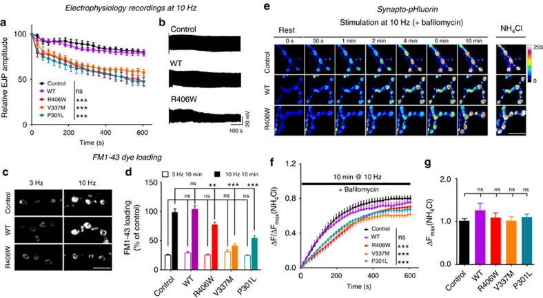
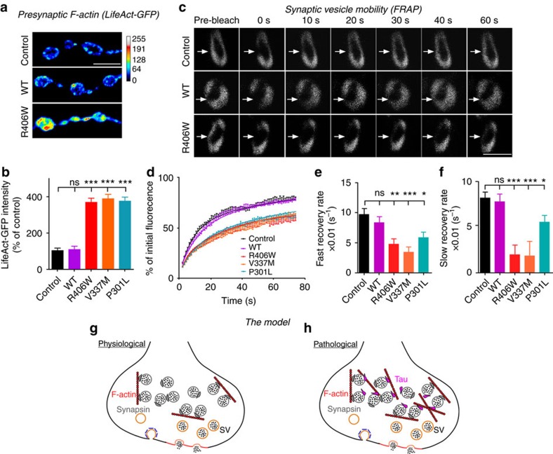
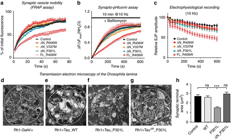
Tau is implicated in more than 20 neurodegenerative diseases, including Alzheimer’s disease. Under pathological conditions, Tau dissociates from axonal microtubules and missorts to pre- and postsynaptic terminals. Patients suffer from early synaptic dysfunction prior to Tau aggregate formation, but the underlying mechanism is unclear. Here we show that pathogenic Tau binds to synaptic vesicles via its N-terminal domain and interferes with presynaptic functions, including synaptic vesicle mobility and release rate, lowering neurotransmission in fly and rat neurons. Pathological Tau mutants lacking the vesicle binding domain still localize to the presynaptic compartment but do not impair synaptic function in fly neurons. Moreover, an exogenously applied membrane-permeable peptide that competes for Tau-vesicle binding suppresses Tau-induced synaptic toxicity in rat neurons. Our work uncovers a presynaptic role of Tau that may be part of the early pathology in various Tauopathies and could be exploited therapeutically.
Overview
The claims and statistics of this paper haven’t been checked yet.
Free: it reads the open-access text and checks the first 5 claims that cite a source we can check. Create a free account to check every claim.
Integrity signals
Registry
Cites
Statistics
Figures


Verification
Claims the paper makes
The claims and statistics of this paper haven’t been checked yet.
Free: it reads the open-access text and checks the first 5 claims that cite a source we can check. Create a free account to check every claim.
Claims other papers make about this one
Each is checked against this paper’s own text: does it say what the citing paper says it does?
The source backs part of the claim, not all of it — or backs it for a different population, condition or comparison.
In the citing paper
“The movement of vesicles and endo-lysosomal compartments along axons is a key part of amyloid fibril formation and vesicle-mediated aggregation of Tau proteins [7, 87].”
From Abnormal accumulation of extracellular vesicles in hippocampal dystrophic axons and regulation by the primary cilia in Alzheimer's disease (2023) · cites this paper as [87]
Experimental — accuracy not yet measured
Self-check
Percentages, sums and reported differences re-computed from the paper’s own numbers.
The self-check needs the paper’s full text.
Citations
Cites
Amyloid oligomers exacerbate tau pathology in a mouse model of tauopathy
Selenica, M.-L. B., Brownlow, M., Jimenez, J. P., Lee, D. C., Pena, G., Dickey, C. A., Gordon, M. N., & Morgan, D. (2013) · Neurodegenerative Diseases · doi:10.1159/000337230 · Cited by 10,359
Visualizing secretion and synaptic transmission with pH-sensitive green fluorescent proteins
Miesenböck, G., De Angelis, D. A., & Rothman, J. E. (1998) · Nature · doi:10.1038/28190 · Cited by 2,563
Tau in physiology and pathology
Wang, Y., & Mandelkow, E. (2016) · Nature Reviews Neuroscience · doi:10.1038/nrn.2015.1 · Cited by 2,251
Self-citationSynapse Loss and Microglial Activation Precede Tangles in a P301S Tauopathy Mouse Model
Yoshiyama, Y., Higuchi, M., Zhang, B., Huang, S.-M., Iwata, N., Saido, T. C., Maeda, J., Suhara, T., Trojanowski, J. Q., & Lee, V. M.-Y. (2007) · Neuron · doi:10.1016/j.neuron.2007.01.010 · Cited by 2,208
Tau-mediated neurodegeneration in Alzheimer’s disease and related disorders
Ballatore, C., Lee, V. M.-Y., & Trojanowski, J. Q. (2007) · Nature Reviews Neuroscience · doi:10.1038/nrn2194 · Cited by 2,174
Tau Suppression in a Neurodegenerative Mouse Model Improves Memory Function
SantaCruz, K., Lewis, J., Spires, T., Paulson, J., Kotilinek, L., Ingelsson, M., Guimaraes, A., DeTure, M., Ramsden, M., McGowan, E., et al. (2005) · Science · doi:10.1126/science.1113694 · Cited by 2,014
Self-citationReducing Endogenous Tau Ameliorates Amyloid ß-Induced Deficits in an Alzheimer's Disease Mouse Model
Roberson, E. D., Scearce-Levie, K., Palop, J. J., Yan, F., Cheng, I. H., Wu, T., Gerstein, H., Yu, G.-Q., & Mucke, L. (2007) · Science · doi:10.1126/science.1141736 · Cited by 1,892
Dendritic Function of Tau Mediates Amyloid-β Toxicity in Alzheimer's Disease Mouse Models
Ittner, L. M., Ke, Y. D., Delerue, F., Bi, M., Gladbach, A., van Eersel, J., Wölfing, H., Chieng, B. C., Christie, M. J., Napier, I. A., et al. (2010) · Cell · doi:10.1016/j.cell.2010.06.036 · Cited by 1,821
Hoover, B. R., Reed, M. N., Su, J., Penrod, R. D., Kotilinek, L. A., Grant, M. K., Pitstick, R., Carlson, G. A., Lanier, L. M., Yuan, L.-L., et al. (2010) · Neuron · doi:10.1016/j.neuron.2010.11.030 · Cited by 1,118
Hippocampal synaptic loss in early Alzheimer's disease and mild cognitive impairment
Scheff, S. W., Price, D. A., Schmitt, F. A., & Mufson, E. J. (2006) · Neurobiology of Aging · doi:10.1016/j.neurobiolaging.2005.09.012 · Cited by 1,017
Cited by
Items 201–300 of 473
Higham, J. P., Hidalgo, S., Buhl, E., & Hodge, J. J. L. (2019) · Frontiers in Cellular Neuroscience · doi:10.3389/fncel.2019.00409 · Cited by 19
“Furthermore, during development at the larval neuromuscular junction, motor neurons overexpressing human 0N3R or 0N4R caused a reduction in size and irregular and abnormally shaped synaptic terminals, a reduction in endocytosis and exocytosis and a reduction in high frequency synaptic transmission (Chee et al., 2005;…”
BackgroundChi, H., Lee, S., Shiu, R.-H., Han, R., Hsieh, C.-P., Wei, T.-M., Lo, C., Chang, H.-Y., & Sang, T. (2020) · Scientific Reports · doi:10.1038/s41598-020-70423-1 · Cited by 19
Chen, S., Yoo, H., Li, C. H., Park, C., Park, G., Tan, L. Y., Jung, S., & Park, H. (2021) · iScience · doi:10.1016/j.isci.2021.103181 · Cited by 19
Decoding the role of microtubules: a trafficking road for vesicle
Qu, J., Li, J., Wang, H., Lan, J., Huo, Z., & Li, X. (2025) · Theranostics · doi:10.7150/thno.110120 · Cited by 18
“This alteration caused vesicles to cross-link with presynaptic actin, limiting vesicle trafficking [88].”
Borreca, A., Latina, V., Corsetti, V., Middei, S., Piccinin, S., Valle, F. D., Bussani, R., Ammassari‐Teule, M., Nisticò, R., Calissano, P., et al. (2018) · Molecular Neurobiology · doi:10.1007/s12035-018-0974-3 · Cited by 18
LOOF, A. D., & Schoofs, L. (2019) · OBM Neurobiology · doi:10.21926/obm.neurobiol.1904046 · Cited by 18
Prediction of longitudinal synaptic loss in Alzheimer's disease using tau PET and plasma biomarkers
Wang, J., Huang, Q., Chen, X., You, Z., He, K., Mao, X., Huang, Y. H., Franzmeier, N., Schöll, M., Guo, T., et al. (2025) · Alzheimer s & Dementia · doi:10.1002/alz.70333 · Cited by 17
Proteostasis as a fundamental principle of Tau immunotherapy
Cruz, E., Nisbet, R. M., Padmanabhan, P., Waardenberg, A. J., Graham, M. E., Nkajja, G., Tapaswi, S., Connor, B. J., Robinson, P., & Götz, J. (2024) · Brain · doi:10.1093/brain/awae254 · Cited by 17
Emerging biophysical techniques for probing synaptic transmission in neurodegenerative disorders
Kale, M. B., Wankhede, N. L., Bishoyi, A. K., Ballal, S., Kalia, R., Arya, R., Kumar, S., Khalid, M., Gulati, M., Umare, M. D., et al. (2024) · Neuroscience · doi:10.1016/j.neuroscience.2024.11.055 · Cited by 18
Orchestrating vesicular and nonvesicular membrane dynamics by intrinsically disordered proteins
Sigrist, S. J., & Haucke, V. (2023) · EMBO Reports · doi:10.15252/embr.202357758 · Cited by 17
“Elegant work in flies and rat neurons has shown that pathogenic Tau binds to synaptic vesicles via its N-terminal domain and interferes with presynaptic functions, including synaptic vesicle mobility and release rate, resulting in impaired pre-synaptic neurotransmission (Zhou et al , 2017).”
BackgroundKe, Y. D., Chan, G., Stefanoska, K., Au, C. G., Bi, M., Müller, J., Przybyla, M., Feiten, A., Prikas, E., Halliday, G. M., et al. (2019) · Journal of Biological Chemistry · doi:10.1074/jbc.ra118.005263 · Cited by 17
DRG2 Deficient Mice Exhibit Impaired Motor Behaviors with Reduced Striatal Dopamine Release
Lim, H. R., Vo, M., Kim, D. J., Lee, U. H., Yoon, J. H., Kim, H. J., Kim, J., Kim, S. R., Lee, J. Y., Yang, C. H., et al. (2019) · International Journal of Molecular Sciences · doi:10.3390/ijms21010060 · Cited by 17
“DRG1 interacts with MTs and promotes MT polymerization [16], while DRG2 interacts with Tau and regulates MT polymerization [17].”
BackgroundInfluentialKudo, T., Takuwa, H., Takahashi, M., Urushihata, T., Shimojo, M., Sampei, K., Yamanaka, M., Tomita, Y., Sahara, N., Suhara, T., et al. (2023) · iScience · doi:10.1016/j.isci.2023.106342 · Cited by 16
“In addition, mutated tau (P301L) binds to synaptic vesicles and reduces the efficacy of synaptic transmission during high-frequency stimulation.(75) Consistently, excitatory synaptic dysfunctions have been reported in the hippocampus of rTg4510 mice at age 6–7 months.”
BackgroundRho GTPase signaling and mDia facilitate endocytosis via presynaptic actin
Oevel, K., Hohensee, S., Kumar, A., Rosas-Brugada, I., Bartolini, F., Soykan, T., & Haucke, V. (2023) · eLife · doi:10.7554/elife.92755 · Cited by 16
Mammeri, N. E., Duan, P., & Hong, M. (2025) · Journal of the American Chemical Society · doi:10.1021/jacs.4c13772 · Cited by 16
Therapies for Tau-associated neurodegenerative disorders: targeting molecules, synapses, and cells
Robbins, M. (2023) · Neural Regeneration Research · doi:10.4103/1673-5374.373670 · Cited by 16
“SV protein synaptogyrin-3 has also been suggested as a therapeutic target for tauopathies, as reducing its expression rescues synaptic dysfunction in tauopathy mouse models (Zhou et al., 2017; McInnes et al., 2018; Largo-Barrientos et al., 2021a).”
A Connected Network of Interacting Proteins Is Involved in Human-Tau Toxicity in Drosophila
Feuillette, S., Charbonnier, C., Frébourg, T., Campion, D., & Lecourtois, M. (2020) · Frontiers in Neuroscience · doi:10.3389/fnins.2020.00068 · Cited by 16
“Under pathological conditions, Tau localizes with both pre- and post-synaptic terminals (Tai et al., 2012, 2014; Zhou et al., 2017; McInnes et al., 2018), suggesting that Tau function at the presynapse may also contribute to disease pathogenesis.”
BackgroundInfluentialBeyond microtubule regulation: the multifaceted roles of tau in neuronal function and dysfunction
Bakota, L., Tulva, K., Trushina, N. I., & Brandt, R. (2026) · Translational Psychiatry · doi:10.1038/s41398-026-03947-1 · Cited by 15
“Indeed, tau has since been found to be localized to the neuronal plasma membrane [9, 10], the nucleus [11, 12], the presynapse [13], and postsynaptic spines [14].”
Presynaptic Pathophysiology Encoded in Different Domains of Tau – Hyper-Versus Hypoexcitability?
Decker, J. M., & Mandelkow, E. (2019) · Advances in experimental medicine and biology · doi:10.1007/978-981-32-9358-8_8 · Cited by 15
Glial phagocytosis for synapse and toxic proteins in neurodegenerative diseases
Choi, Y., & Chung, W. (2025) · Molecular Neurodegeneration · doi:10.1186/s13024-025-00870-9 · Cited by 16
“Indeed, tau is closely associated with synaptic components across various models, including in vitro primary murine neurons [143], flies [155], mice [47, 156], and humans [50, 157], where it has been shown to induce synaptic dysfunction [155, 156, 158].”
Dai, B., Ma, X., Tang, Y., Xu, L., Guo, S., Chen, X., Lu, S., Wang, G., & Liu, Y. (2020) · Bioorganic & Medicinal Chemistry · doi:10.1016/j.bmc.2020.115891 · Cited by 15
Warpechowski, M., Warpechowski, J. J., Kulczyńska‐Przybik, A., & Mroczko, B. (2022) · Medical Science Monitor · doi:10.12659/msm.938826 · Cited by 15
“In addition, some researchers suggest that hyperphosphorylation of tau lowers its affinity to microtubules, resulting in localization of tau in dendrites and presynaptic terminals [86], which impairs motility of synaptic vesicles, contributing to synaptic dysfunction [87,88].”
Yang, F., Zhao, J., Chen, G., Han, H., Hu, S., Wang, N., Wang, J., Chen, Y., Zhou, Z., Dai, B., et al. (2023) · Bioorganic Chemistry · doi:10.1016/j.bioorg.2023.106432 · Cited by 15
Lemke, N., Melis, V., Lauer, D., Magbagbeolu, M., Neumann, B., Harrington, C. R., Riedel, G., Wischik, C. M., Theuring, F., & Schwab, K. (2020) · Journal of Biological Chemistry · doi:10.1074/jbc.ra120.014890 · Cited by 15
“These vesicle-tau interactions hamper transmitter release through polymerization of Nterminal tau domains linking presynaptic F-actin with vesicle membranes, thereby making the presynaptic compartment more rigid (29).”
BackgroundResultInfluentialA Meta-Analysis on Presynaptic Changes in Alzheimer’s Disease
Anschuetz, A., Schwab, K., Harrington, C. R., Wischik, C. M., & Riedel, G. (2023) · Journal of Alzheimer s Disease · doi:10.3233/jad-231034 · Cited by 14
Activity-Induced Cortical Glutamatergic Neuron Nascent Proteins
Schiapparelli, L., Xie, Y., Sharma, P., McClatchy, D. B., Ma, Y., Yates, J. R., Maximov, A., & Cline, H. T. (2022) · Journal of Neuroscience · doi:10.1523/jneurosci.0707-22.2022 · Cited by 14
Tripartite Relationship Among Synaptic, Amyloid, and Tau Proteins: An In Vivo and Postmortem Study
Casaletto, K. B., Zetterberg, H., Blennow, K., Brinkmalm, A., Honer, W. G., Schneider, J. A., Bennett, D. A., Djukic, N. A., You, M. Y., Weiner‐Light, S., et al. (2021) · Neurology · doi:10.1212/wnl.0000000000012145 · Cited by 14
“Additionally, tau hyperphosphorylation may be propagated by and further propagate the severity and spread of aberrant synaptic firing and ultimately lead to widespread neuronal cell loss 15–17 .”
BackgroundTruncating tau reveals different pathophysiological actions of oligomers in single neurons
Hill, E., Karikari, T. K., Lantero‐Rodriguez, J., Zetterberg, H., Blennow, K., Richardson, M. J. E., & Wall, M. J. (2021) · Communications Biology · doi:10.1038/s42003-021-02791-x · Cited by 13
“277 278 The N-terminal truncation (C) at position 123, has been identified as a major N-terminal cleavage 279 site in tau found in human CSF (21) and a truncated tau construct at a nearby position has been used 280 to study effects on synaptic transmission (31).”
Kong, Y., Zhang, S., Huang, L., Zhang, C., Xie, F., Zhang, Z., Huang, Q., Jiang, D., Li, J., Zhou, W., et al. (2021) · Frontiers in Aging Neuroscience · doi:10.3389/fnagi.2021.731114 · Cited by 13
“, 2015); on the other hand, it is speculated that phosphorylated tau interacts with SV2A to affect the movement and release rate of synaptic vesicles, and cause the abnormal secretion of GABA and glutamate neurotransmitters (Zhou et al., 2017; Vanoye-Carlo and Gómez-Lira, 2019).”
BackgroundModulation of tau protein aggregation using ‘Trojan’ sequences
Pandey, G., Morla, S., Kumar, S., & Ramakrishnan, V. (2020) · Biochimica et Biophysica Acta (BBA) - General Subjects · doi:10.1016/j.bbagen.2020.129569 · Cited by 13
Unravelling the mechanotransduction pathways in Alzheimer’s disease
Donnaloja, F., Limonta, E., Mancosu, C., Morandi, F., Boeri, L., Albani, D., & Raimondi, M. T. (2023) · Journal of Biological Engineering · doi:10.1186/s13036-023-00336-w · Cited by 13
Khan, R. I., Nirzhor, S. S. R., & Rashid, B. (2018) · Brain Sciences · doi:10.3390/brainsci8090162 · Cited by 13
“This distinct progression sequence suggested a strong link between the observed clinical symptoms and relevant affected areas, thereby underlining its pivotal role in synaptic dysfunction [94,126,127].”
BackgroundHole, K. L., Zhu, B., Huggon, L., Brown, J. T., Mason, J. M., & Williams, R. J. (2024) · Cell Death and Disease · doi:10.1038/s41419-024-06815-2 · Cited by 12
“Presynap-tically, the association of tau with vesicles hinders neurotransmitter release [17, 18, 20], while at dendritic spines, mutant tau impairs AMPA receptor internalisation and long-term potentiation (LTP) [21 – 24].”
Unknown roles of tau pathology in neurological disorders. Challenges and new perspectives
Olesen, M. A., Villavicencio-Tejo, F., Cuevas-Espinoza, V., & Quintanilla, R. A. (2024) · Ageing Research Reviews · doi:10.1016/j.arr.2024.102594 · Cited by 12
Nussbaumer, J., Barve, A., Zufferey, V., Espourteille, J., Kirabali, T., Konietzko, U., Razansky, D., Rominger, A., Nordberg, A. K., Buée, L., et al. (2025) · Translational Neurodegeneration · doi:10.1186/s40035-025-00508-2 · Cited by 12
MARATOVICH, B. A., Al-Yozbaki, M., Ashirbekov, Y., Satken, K., Abaildayev, A., Yeleussizov, A., Jainakbayev, N. T., Sharipov, K. O., & Wilson, C. M. (2025) · Frontiers in Cell and Developmental Biology · doi:10.3389/fcell.2025.1676530 · Cited by 13
Owens, L., Benedetto, A., Dawson, N., Gaffney, C. J., & Parkin, E. T. (2021) · Brain Research · doi:10.1016/j.brainres.2020.147264 · Cited by 12
Chu, J., Wang, J., Cui, L., Liu, S., An, N., Han, J., Che, X., Wu, C., & Yang, J. (2021) · Mechanisms of Ageing and Development · doi:10.1016/j.mad.2021.111496 · Cited by 12
Taipala, E., Pfitzer, J. C., Hellums, M., Reed, M. N., & Gramlich, M. W. (2022) · Frontiers in Synaptic Neuroscience · doi:10.3389/fnsyn.2022.925546 · Cited by 12
“Further, cultured hippocampal neuron studies used transfection with a htau mutation to show a significant reduction in presynaptic transmission (Zhou et al., 2017; McInnes et al., 2018; Siano et al., 2019); however, it has not been shown how acute introduction of tau is directly related to the normal time-course of ne…”
BackgroundMethodResultInfluentialLu, Y., & Initiative, A. D. N. (2022) · Aging Clinical and Experimental Research · doi:10.1007/s40520-022-02202-z · Cited by 12
“Tau pathology triggering synapse degeneration might explain their correlations in CSF [38].”
BackgroundAdvancing medicine for Alzheimer’s disease: A plasma neural exosome platform
Goetzl, E. J. (2020) · The FASEB Journal · doi:10.1096/fj.202001655 · Cited by 13
A 3-Gene-Based Diagnostic Signature in Alzheimer’s Disease
Wang, H., Zhang, Y., Zheng, C., Yang, S., Chen, X., Wang, H., & Gao, S. (2021) · European Neurology · doi:10.1159/000518727 · Cited by 12
Clayton, E. L., Bonnycastle, K., Isaacs, A. M., Cousin, M. A., & Schorge, S. (2021) · Journal of Neurochemistry · doi:10.1111/jnc.15551 · Cited by 12
Neurophysiological trajectories in Alzheimer’s disease progressionPreprint
Kudo, K., Ranasinghe, K. G., Morise, H., Syed, F., Sekihara, K., Rankin, K. P., Miller, B. L., Kramer, J. H., Rabinovici, G. D., Vossel, K. A., et al. (2024) · eLife · doi:10.7554/elife.91044.2 · Cited by 12
Diverse influences on tau aggregation and implications for disease progression
Alstyne, M. V., Pratt, J., & Parker, R. (2025) · Genes & Development · doi:10.1101/gad.352551.124 · Cited by 11
“In cultured neuronal models, tau interacts with synaptic vesicle proteins, and tau with FTD-associated mutations can slow synaptic vesicle mobility (Zhou et al. 2017; Tracy et al. 2022).”
Extracellular PHF-tau modulates astrocyte mitochondrial dynamics and mediates neuronal connectivity
Zufferey, V., Barve, A., Parietti, E., Belinga, L. N., Bringaud, A., Varisco, Y., Fabbri, K., Capotosti, F., Bezzi, P., Déglon, N., et al. (2025) · Translational Neurodegeneration · doi:10.1186/s40035-025-00474-9 · Cited by 11
“However, more subtle alterations in signaling pathways, notably those involved in phosphatase, protease and mitochondrial metabolic activities, were detected, aligning with the conclusions of previous studies [74, 80].”
Sanchez-Todo, R., Mercadal, B., Lopez-Sola, E., Guasch-Morgades, M., Deco, G., & Ruffini, G. (2026) · Human Brain Mapping · doi:10.1002/hbm.70428 · Cited by 11
Behroozi, Z., Askarpour, H., & Baghcheghi, Y. (2026) · Journal of Alzheimer s Disease Reports · doi:10.1177/25424823261415807 · Cited by 12
Current molecular approaches to investigate pre‐synaptic dysfunction
Harper, C. B., & Smillie, K. J. (2021) · Journal of Neurochemistry · doi:10.1111/jnc.15316 · Cited by 11
“…activity- dependent trafficking of synaptotagmin; (Harper et al. 2020)), Parkinson's disease (disrupted SV clustering; (Nemani et al. 2010)) and Alzheimer's Disease (multiple stages of SV lifecycle impacted; (Lazarevic et al. 2017; Park et al. 2013; Zhou et al. 2017)) to highlight a few examples.”
BackgroundSchwab, K., Melis, V., Harrington, C. R., Wischik, C. M., Magbagbeolu, M., Theuring, F., & Riedel, G. (2021) · Cells · doi:10.3390/cells10082162 · Cited by 11
“Furthermore, tau binding promotes presynaptic actin polymerisation to crosslink synaptic vesicles, which strongly restricts their mobilisation and affects docking, transport and fusion of synaptic vesicles and impairs neurotransmitter release [88].”
BackgroundDysfunction of the glutamatergic photoreceptor synapse in the P301S mouse model of tauopathy
Arouche-Delaperche, L., Cadoni, S., Joffrois, C., Labernède, G., Valet, M., Cesar, Q., Dégardin, J., Girardon, S., Gabriel, C., Krantic, S. M., et al. (2023) · Acta Neuropathologica Communications · doi:10.1186/s40478-022-01489-3 · Cited by 11
“However, this enhanced transmitter release in the context of pathological tau protein expression conflicts with the reported inhibition of vesicular release by tau binding to the vesicles [69].”
BackgroundMetabotropic Glutamate Receptors Modulate Exocytotic Tau Release and Propagation
Mazzo, F., Butnaru, I., Grubisha, O., Ficulle, E., Sanger, H., Fitzgerald, G. J., Pan, F., Pasqui, F., Murray, T. K., Monn, J. A., et al. (2022) · Journal of Pharmacology and Experimental Therapeutics · doi:10.1124/jpet.122.001307 · Cited by 11
Hahn, L., Eickhoff, S. B., Mueller, K., Schilbach, L., Barthel, H., Fassbender, K. C., Fließbach, K., Kornhuber, J., Prudlo, J., Synofzik, M., et al. (2023) · eLife · doi:10.7554/elife.86085 · Cited by 11
“For example, tau can cause presynaptic dysfunction prior to loss of function or cell death ( Zhou et al., 2017 ), whereas overexpression of TDP-43 leads to impairment of presynaptic integrity ( Heyburn and Moussa, 2016 ).”
Backgroundα-Synuclein and Tau: Mitochondrial Kill Switches
Pech, U., & Verstreken, P. (2018) · Neuron · doi:10.1016/j.neuron.2017.12.024 · Cited by 11
Self-citationArnés, M., Kim, Y. A., Lannes, J., Alaniz, M. E., Cho, J. D., McCabe, B. D., & Santa‐María, I. (2019) · bioRxiv (Cold Spring Harbor Laboratory) · doi:10.1101/607176 · Cited by 11
Neuronal hypofunction and network dysfunction in a mouse model at an early stage of tauopathy
Ji, C., Yang, X., Eleish, M., Jiang, Y., Tetlow, A. M., Song, S. C., Martín‐Ávila, A., Wu, Q., Zhou, Y., Gan, W., et al. (2024) · Alzheimer s & Dementia · doi:10.1002/alz.14273 · Cited by 10
Actin Cytoskeleton at the Synapse: An Alzheimer's Disease Perspective
Haseena, P. A., Basavaraju, N., Gupta, A., & Kommaddi, R. P. (2025) · Cytoskeleton · doi:10.1002/cm.21993 · Cited by 10
“This leads to cross-linking of synaptic vesicles and F-actin at presynapse thereby restraining the release of vesicles due to decreased mobilization further lowering synaptic transmission (Zhou et al. 2017).”
Chen, H., Fan, L., Guo, Q., Wong, M. Y., Zhu, J., Foxe, N. R., Wang, W. T., Nessim, A., Carling, G., Liu, B., et al. (2025) · Molecular Neurodegeneration · doi:10.1186/s13024-025-00903-3 · Cited by 10
Targeting tau in Alzheimer’s Disease: rationale, approach and challenges
Aslanyan, A., Foiani, M. S., Giovannucci, T. A., McDade, E., Duff, K., Mummery, C. J., Schöll, M., Wildsmith, K. R., & Paterson, R. W. (2026) · Molecular Neurodegeneration · doi:10.1186/s13024-026-00925-5 · Cited by 10
“This is different from beta amyloid (Aβ) synaptotoxicity which is thought to involve interaction with cell membrane receptors [69, 79–81].”
Tau‐mediated synaptic dysfunction is coupled with HCN channelopathy
Goniotaki, D., Tamagnini, F., Biasetti, L., Rumpf, S.-L., Troakes, C., Pollack, S. J., Ukwesa, S., Sun, H., Kraev, I., Serpell, L. C., et al. (2024) · Alzheimer s & Dementia · doi:10.1002/alz.14074 · Cited by 10
“, 2018) and vesicular proteins, including synapsin-I, α-synuclein, synaptogyrin-3, synaptotagmin (Liu et al., 2016; McInnes et al., 2018; Zhou et al., 2017), and potassium ion channels (Hall et al.”
BackgroundThe Gαq/phospholipase Cβ signaling system represses tau aggregation
Garwain, O., Yerramilli, V. S., Romero, K., & Scarlata, S. (2020) · Cellular Signalling · doi:10.1016/j.cellsig.2020.109620 · Cited by 10
“Tau interacts with microtubules that stabilize the cell cytoskeleton including neuronal synapses [45].”
BackgroundGaudreault, R., Hervé, V., van de Ven, T. G. M., Mousseau, N., & Ramassamy, C. (2021) · Journal of Alzheimer s Disease · doi:10.3233/jad-201549 · Cited by 10
Prikas, E., Ahel, H. I., Stefanoska, K., Asih, P. R., Volkerling, A. M., Ittner, L. M., & Ittner, A. (2021) · Biochemistry and Cell Biology · doi:10.1139/bcb-2020-0604 · Cited by 10
Jackson, J., Johnson, J. D., Meftah, S., Murray, T. K., Ahmed, Z., Fasiolo, M., Hutton, M. L., Isaac, J. T. R., O’Neill, M. J., & Ashby, M. C. (2020) · bioRxiv (Cold Spring Harbor Laboratory) · doi:10.1101/2020.04.29.067629 · Cited by 10
Soma-localized Rab39 inhibits synaptic autophagy by controlling trafficking of Atg9 vesicles
Kilic, A., Özturan, G., Vandekerkhove, D., Kuenen, S., Swerts, J., Pedrazo, E. M., Calatayud, C., Escamilla‐Ayala, A., Corthout, N., Hernández-Varas, P., et al. (2025) · The EMBO Journal · doi:10.1038/s44318-025-00536-8 · Cited by 9
Self-citationIgLON5 autoimmune antibodies activate Tau via neuronal hyperactivity
Askin, B., Kilic, C., Gómez, C. C., Duong, S. L.-L., Dominguez‐Baquero, A., Goihl, A., Nalbach, K., Petushi, J., Grundschöttel, P., Wagner, J., et al. (2026) · Science Advances · doi:10.1126/sciadv.aec2042 · Cited by 9
“This is in line with previous reports that identified (pre)synaptic Tau is phosphorylated at pS396/pS404 ( 89 , 90 ), an epitope targeted by multiple kinases, including GSK3 β , CDK5, and ERK ( 91 , 92 ).”
Botulinum neurotoxin A modulates the axonal release of pathological tau in hippocampal neurons
Panzi, C., Surana, S., La‐Rocque, S. D., Moretto, E., Lazo, O. M., & Schiavo, G. (2023) · Toxicon · doi:10.1016/j.toxicon.2023.107110 · Cited by 9
“The association of tau with exosomes (Wang et al., 2017), synaptic vesicles (McInnes et al., 2018; Zhou et al., 2017) as well as the soluble N-ethylmaleimide-sensitive factor attachment protein receptor (SNARE) proteins (Lee et al., 2021; Pilliod et al., 2020; Tracy et al., 2022; Xu, 2021) suggests…”
BackgroundPoon, A. D., McGill, S. H., Sunkesula, S. R. B., Burgess, Z. S., Dunne, P. J., Kang, E. E., & Bittner, G. D. (2018) · Journal of Neuroscience Research · doi:10.1002/jnr.24232 · Cited by 9
“Other targets of CaMKII might also affect plasmalemmal sealing, such as microtubule-associated protein Tau (Yoshimura, Ichinose, & Yamauchi, 2003; Zhou et al., 2017), synaptotagmin (Hilfiker, Pieribone, Nordstedt, Greengard, & Czernik, 1999; Detrait et al.”
BackgroundInfluentialAnschuetz, A., Schwab, K., Harrington, C. R., Wischik, C. M., & Riedel, G. (2025) · Journal of Alzheimer s Disease · doi:10.1177/13872877251362212 · Cited by 8
Lu, S., Qiu, S., Yu, G., Zhang, A., & Zhao, Q. (2026) · Ageing Research Reviews · doi:10.1016/j.arr.2026.103097 · Cited by 8
Kedia, S., Fertan, E., Wu, Y., Zhang, Y. P., Meisl, G., Lam, J. Y. L., Wiseman, F. K., McEwan, W. A., Quaegebeur, A., Spillantini, M. G., et al. (2025) · Cell chemical biology · doi:10.1016/j.chembiol.2025.01.001 · Cited by 8
“14 More-over, tau can spread between neurons via synaptic transmission 15 and synaptic accumulation of oligomeric tau, in the absence of fibrillar species, has been reported in early AD, suggesting the involvement of these small-soluble aggregates in synaptotoxic processes.”
Gui, S., Zeng, F., Wu, Z., Nonaka, S., Sano, T., Ni, J., Nakanishi, H., Moriyama, M., & Kanematsu, T. (2025) · Journal of Alzheimer s Disease · doi:10.1177/13872877251326879 · Cited by 8
“8,54 This possibility is supported by the present study, where AL-MCM exposure in N2a cells upregulated phosphorylated tau (Ser 396) expression, and the level of Ser 396 was negatively correlated with SYN1 and PSD-95 expression in N2a neurons.”
Fibre density and cross-section associate with hallmark pathology in early Alzheimer’s disease
Vanderlinden, G., Radwan, A., Christiaens, D., Blommaert, J., Sunaert, S. G., Vandenbulcke, M., Koole, M. J. B., & Laere, K. V. (2025) · Alzheimer s Research & Therapy · doi:10.1186/s13195-025-01710-0 · Cited by 8
“Tau binding to presynaptic vesicles causes presynaptic dysfunction by lowering neurotransmission and ultimately results in synaptic loss [12, 13].”
The Role of Tau in the Post-synapse
Regan, P. L., & Cho, K. (2019) · Advances in experimental medicine and biology · doi:10.1007/978-981-32-9358-8_10 · Cited by 8
Chakravorty, A., Sharma, A., Sheeba, V., & Manjithaya, R. R. (2022) · Frontiers in Molecular Neuroscience · doi:10.3389/fnmol.2022.842772 · Cited by 8
Cranston, A., Kraev, I., Stewart, M. G., Horsley, D., Santos, R. X., Robinson, L., Dreesen, E., Armstrong, P., Palliyil, S., Harrington, C. R., et al. (2024) · Cellular Signalling · doi:10.1016/j.cellsig.2024.111269 · Cited by 8
“Increased tau in synaptosomal preparations is consistent with recent work by Zhou and co-workers [38] who found in mutant tau models (0N4R tau carrying R406W, V337M and P301L mutations) of both Drosophila and mouse that pathological tau mislocalises to pre-synaptic terminals where it promotes…”
Ibrahim, A. N., Attallah, M. I., & El‐Naggar, R. (2018) · Egyptian Journal of Basic and Clinical Pharmacology · doi:10.11131/2018/101369 · Cited by 8
Pareja-Navarro, K. A., King, C. D., Kauwe, G., Ngwala, Y. Y., Lokitiyakul, D., Wong, I., Vira, A., Liu, Y., Chen, J. H., Sharma, M., et al. (2026) · Molecular Neurodegeneration · doi:10.1186/s13024-026-00928-2 · Cited by 7
“Notably, the expression of tau carrying mutations that cause familial frontotemporal dementia inhibits pre-synaptic function by hindering synaptic vesicle mobility [103, 104].”
Jones, A. A., Ramos‐Miguel, A., Gicas, K. M., Petyuk, V., Leurgans, S. E., Jager, P. L. D., Schneider, J. A., Bennett, D., Honer, W. G., & Casaletto, K. B. (2024) · Alzheimer s & Dementia · doi:10.1002/alz.14286 · Cited by 7
Le, J., Xia, C., Xu, J., Cai, J., Hu, C., Bai, Y., Chen, H., Rong, W., Jiang, Y., Wu, X., et al. (2024) · CNS Neuroscience & Therapeutics · doi:10.1111/cns.70100 · Cited by 7
Amabebe, E., Huang, Z., Jash, S., Krishnan, B., Cheng, S., Nakashima, A., Li, Y., Li, Z., Wang, R., Menon, R., et al. (2024) · Biomedicines · doi:10.3390/biomedicines13010029 · Cited by 7
Olesen, M. A., Villavicencio-Tejo, F., Johnson, G. V. W., Porter, G. A., & Quintanilla, R. A. (2025) · Free Radical Biology and Medicine · doi:10.1016/j.freeradbiomed.2025.02.035 · Cited by 7
Calcineurin inhibition may prevent Alzheimer disease in people with Down syndrome
Dohl, J., Treadwell, Z., Norris, C. M., & Head, E. (2025) · Alzheimer s & Dementia · doi:10.1002/alz.70034 · Cited by 7
Wilson, P., Kim, N., Cotter, R., Parkes, M., Cmelak, L., Reed, M. N., & Gramlich, M. W. (2025) · Cell Reports · doi:10.1016/j.celrep.2025.115410 · Cited by 7
Tryptophan-tyrosine dipeptide improves tau-related symptoms in tauopathy mice
Ano, Y., Takaichi, Y., Ohya, R., Uchida, K., Nakayama, H., & Takashima, A. (2022) · Nutritional Neuroscience · doi:10.1080/1028415x.2022.2090075 · Cited by 7
Tulloch, J., Netsyk, O., Pickett, E. K., Herrmann, A. G., Jain, P., Stevenson, A. J., Oren, I., Hardt, O. M., & Spires‐Jones, T. L. (2020) · European Journal of Neuroscience · doi:10.1111/ejn.14918 · Cited by 7
“Our previous work in the APP/PS1 + Tau mouse line and human post-mortem tissue indicates that both Aβ and tau accumulate in synapses, where it may contribute to synaptic dysfunction and downstream cognitive decline (Hesse et al., 2019; Jackson et al., 2016, 2019; McInnes et al., 2018; Pickett et al., 2019; Zhou et al.…”
BackgroundSelf-citationRole of vitamin D3 in mitigating sodium arsenite-induced neurotoxicity in male rats
Abdou, H. M., Saad, A., Elkader, H. A. E. A., & Essawy, A. E. (2024) · Toxicology Research · doi:10.1093/toxres/tfae203 · Cited by 6
Axonal tau reduction ameliorates tau and amyloid pathology in a mouse model of Alzheimer’s disease
Zare, A., Salehi, S., Bader, J. M., Wiessler, A.-L., Prokesch, M., Albrecht, V., Villmann, C., Mann, M., Briese, M., & Sendtner, M. (2025) · Translational Neurodegeneration · doi:10.1186/s40035-025-00499-0 · Cited by 6
“In presynaptic compartments, pathological tau associates with synaptic vesicles via Synaptogyrin-3, impairing neurotransmitter release [24, 25].”
Prefoldin 5 is a microtubule-associated protein that suppresses Tau aggregation and neurotoxicity
Bisht, A., Pippadpally, S., Majumder, S., Gopi, A. T., Das, A., Sahi, C., Ramaswami, M., & Kumar, V. (2024) · eLife · doi:10.7554/elife.104691.3 · Cited by 6
Wal, P., Dutta, A., Jawaid, T., Rahman, M. U., Gowri, K., Jyothi, S. R., Kaur, I., Al-Hasnaawei, S., Sinha, A., Maharana, L., et al. (2026) · Biogerontology · doi:10.1007/s10522-026-10394-4 · Cited by 7
“Reductions in SV2A, a protein associated with neurotransmitter release probability, in the brains of individuals with AD are strongly associated with cognitive decline (Zhou et al. 2017).”
Stone, A., Cujic, O., Rowlett, A., Aderhold, S., Savage, E., Graham, B. P., & Steinert, J. R. (2023) · Frontiers in Synaptic Neuroscience · doi:10.3389/fnsyn.2023.1124061 · Cited by 6
“Further evidence for synaptic dysfunction stems from studies reporting that glycated tau binds to synaptic vesicles via its N-terminal domain which interferes with presynaptic functions, including synaptic vesicle mobility and release rate resulting in compromised neurotransmission in fly and rat neurons (Zhou et al.,…”
BackgroundShen, Z., Zhang, H., Du, L., He, X.-Y., & Sun, X. (2023) · Chaos An Interdisciplinary Journal of Nonlinear Science · doi:10.1063/5.0154322 · Cited by 6
Synaptic alterations associated with disrupted sensory encoding in a mouse model of tauopathy
Meftah, S., Cavallini, A., Murray, T. K., Jankowski, Ł., Bose, S., Ashby, M. C., Brown, J. T., & Witton, J. (2024) · Brain Communications · doi:10.1093/braincomms/fcae134 · Cited by 6
Schwab, K., Lauer, D., Magbagbeolu, M., Theuring, F., Gąsiorowska, A., Zadrożny, M., Harrington, C. R., Wischik, C. M., Niewiadomska, G., & Riedel, G. (2024) · Brain Research Bulletin · doi:10.1016/j.brainresbull.2024.110955 · Cited by 6
LOOF, A. D., & Schoofs, L. (2019) · Frontiers in Endocrinology · doi:10.3389/fendo.2019.00885 · Cited by 6
Minimalistic in vitro systems for investigating tau pathology
Hallinan, G. I., Pitera, A. P., Patel, P., West, J. J., & Deinhardt, K. (2018) · Journal of Neuroscience Methods · doi:10.1016/j.jneumeth.2018.09.032 · Cited by 6
Array tomography: 15 years of synaptic analysis
Avila, A. S., & Henstridge, C. M. (2022) · Neuronal Signaling · doi:10.1042/ns20220013 · Cited by 6
Foiani, M. S., Bourdenx, M., Lukas, K., Nirujogi, R., Angela, Y., Hannah, D., Saisha, P., Damoc, L.-S., Mitchener, L., Jaunmuktane, Z., et al. (2026) · bioRxiv (Cold Spring Harbor Laboratory) · doi:10.64898/2026.04.26.720932 · Cited by 6
“The enrichment of presynaptic proteins is in line with reports of tau accumulation at presynaptic terminals and subsequent synapse loss (Zhou et al. 2017; Kiani 2023; Fertan et al. 2026).”
Tau protein aggregation: A therapeutic target for neurodegenerative diseases
Duggal, A., Mahindru, D., Baghel, K., Mehrotra, S., & Prajapati, V. K. (2025) · Advances in protein chemistry and structural biology · doi:10.1016/bs.apcsb.2024.11.012 · Cited by 5
Early synaptic pathology is associated with small tau aggregates in Alzheimer’s disease
Fertan, E., Kedia, S., Nolan, G., Meisl, G., Cotton, M. W., Müller, K. H., Zhang, Z., Mureşan, L., Quaegebeur, A., Spillantini, M. G., et al. (2026) · Acta Neuropathologica · doi:10.1007/s00401-026-02977-9 · Cited by 5
Metadata
- Authors
- Zhou, L., McInnes, J., Wierda, K., Holt, M., Herrmann, A. G., Jackson, R. J., Wang, Y.-C., Swerts, J., Beyens, J., Miskiewicz, K., Vilain, S., Dewachter, I., Moechars, D., De Strooper, B., Spires-Jones, T. L., De Wit, J., & Verstreken, P.
- Affiliations
- VIB-KU Leuven Center for Brain & Disease Research (BE)
- Euan MacDonald Centre for Motor Neuron Disease Research (GB)
- Hasselt University (BE)
- Janssen (Belgium) (BE)
- Corresponding author
- Verstreken, Patrik
- ORCID
- Venue
- Nature Communications
- Published
- 2017-05-11
- Volume
- 8
- Issue
- 1
- Pages
- 15295-15295
- ISSN
- 2041-1723 (ISSN-L)
- PMC
- PMC5437271
- OpenAlex
- W2613492737
- Cited by
- 434
- References
- 72
- Citations by year
- 2026: 40 · 2025: 47 · 2024: 61 · 2023: 54 · 2022: 35 · 2021: 68 · 2020: 49 · 2019: 31 · 2018: 40 · 2017: 8
- FWCI
- 25.89
- Type
- journal article
- Language
- en
- Open access
- gold
- doi.org
- Licence
- cc-by
- creativecommons.org/licenses/by/4.0 (tdm)
- creativecommons.org/licenses/by/4.0 (vor)
- Locations
- Nature Communications (publishedVersion, cc-by)
- Europe PMC (PubMed Central) (publishedVersion, cc-by)
- PubMed Central (publishedVersion, cc-by)
- PubMed (publishedVersion)
- Discovery Research Portal (University of Dundee) (publishedVersion, other-oa)
- Edinburgh Research Explorer (University of Edinburgh) (publishedVersion, cc-by)
- Lirias (submittedVersion, cc-by)
- RePEc: Research Papers in Economics (submittedVersion)
- DOAJ (DOAJ: Directory of Open Access Journals) (submittedVersion, cc-by-sa)
- Document Server@UHasselt (UHasselt) (submittedVersion, cc-by)
- UCL Discovery (University College London) (submittedVersion)
- Funders
- Belgian American Educational Foundation
- Belgian Federal Science Policy Office
- KU Leuven
- Vlaamse regering
- Medical Research Council (MC_PC_17116)
- National Institute on Aging (P50AG005134)
- H2020 European Research Council (646671)
- FP7 Ideas: European Research Council (260678)
- Topics
- Life Sciences › Neuroscience › Cellular and Molecular Neuroscience › Alzheimer's disease research and treatments
- Life Sciences › Neuroscience › Cellular and Molecular Neuroscience › Neuroscience and Neuropharmacology Research
- Life Sciences › Neuroscience › Neurology › Neuroinflammation and Neurodegeneration Mechanisms
- Keywords
- tau protein, synaptic vesicles, presynaptic dysfunction, tauopathy, Alzheimer's disease, neurotransmission, peptide competition, synaptic toxicity, Drosophila neurons, rat neurons
- MeSH terms
- Protein Domains, Actins, Animals, Drosophila melanogaster, Hippocampus, Humans, Mutation, Synaptic Transmission, Neurons, Synaptic Vesicles, tau Proteins, Presynaptic Terminals, Protein Transport, Rats
About these checks
Registry, retraction, Cites and statistics results are computed, not judged, with one exception: a printed reference that no index lists is read once by a model before it counts as a citation error. Claims are extracted by a model and kept only when their quote appears verbatim in the text; each cited source is then checked, with a model’s help, for whether it says what the claim relies on. Those verdicts can be wrong — read the claim’s quote and the cited source before relying on one.
Statistics and arithmetic are recomputed, not judged.
Confidence bands are shown only for fields calibrated on our annotated test set. None is calibrated yet, so no band is shown.
What counts as a claim ›How we measure confidence ›How we check this ›
Are you an author, or do you see a problem with this page? Request a review
Data sources
Bibliographic data: Semantic Scholar, OpenAlex, Crossref.
Contains information from the Semantic Scholar Academic Graph Datasets, made available under the ODC Attribution License (ODC-BY 1.0).
Semantic Scholar’s data is described in: Kinney, R., et al. (2023). The Semantic Scholar Open Data Platform. arXiv:2301.10140.
