

Tau association with synaptic vesicles causes presynaptic dysfunction
Open accessThe DOI matches its Crossref record; 72 of the 72 references it cites were found in a registry or an index.
Abstract
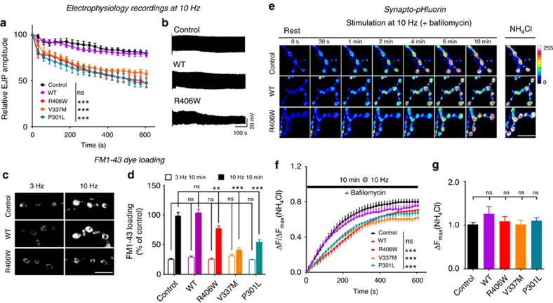
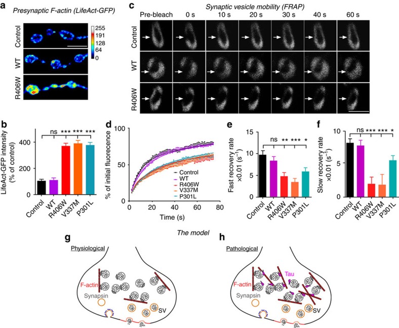
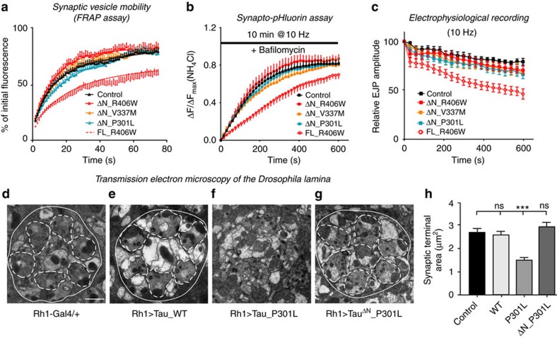
Tau is implicated in more than 20 neurodegenerative diseases, including Alzheimer’s disease. Under pathological conditions, Tau dissociates from axonal microtubules and missorts to pre- and postsynaptic terminals. Patients suffer from early synaptic dysfunction prior to Tau aggregate formation, but the underlying mechanism is unclear. Here we show that pathogenic Tau binds to synaptic vesicles via its N-terminal domain and interferes with presynaptic functions, including synaptic vesicle mobility and release rate, lowering neurotransmission in fly and rat neurons. Pathological Tau mutants lacking the vesicle binding domain still localize to the presynaptic compartment but do not impair synaptic function in fly neurons. Moreover, an exogenously applied membrane-permeable peptide that competes for Tau-vesicle binding suppresses Tau-induced synaptic toxicity in rat neurons. Our work uncovers a presynaptic role of Tau that may be part of the early pathology in various Tauopathies and could be exploited therapeutically.
Overview
The claims and statistics of this paper haven’t been checked yet.
Free: it reads the open-access text and checks the first 5 claims that cite a source we can check. Create a free account to check every claim.
Integrity signals
Registry
Cites
Statistics
Figures


Verification
Claims the paper makes
The claims and statistics of this paper haven’t been checked yet.
Free: it reads the open-access text and checks the first 5 claims that cite a source we can check. Create a free account to check every claim.
Claims other papers make about this one
Each is checked against this paper’s own text: does it say what the citing paper says it does?
The source backs part of the claim, not all of it — or backs it for a different population, condition or comparison.
In the citing paper
“The movement of vesicles and endo-lysosomal compartments along axons is a key part of amyloid fibril formation and vesicle-mediated aggregation of Tau proteins [7, 87].”
From Abnormal accumulation of extracellular vesicles in hippocampal dystrophic axons and regulation by the primary cilia in Alzheimer's disease (2023) · cites this paper as [87]
Experimental — accuracy not yet measured
Self-check
Percentages, sums and reported differences re-computed from the paper’s own numbers.
The self-check needs the paper’s full text.
Citations
Cites
Amyloid oligomers exacerbate tau pathology in a mouse model of tauopathy
Selenica, M.-L. B., Brownlow, M., Jimenez, J. P., Lee, D. C., Pena, G., Dickey, C. A., Gordon, M. N., & Morgan, D. (2013) · Neurodegenerative Diseases · doi:10.1159/000337230 · Cited by 10,359
Visualizing secretion and synaptic transmission with pH-sensitive green fluorescent proteins
Miesenböck, G., De Angelis, D. A., & Rothman, J. E. (1998) · Nature · doi:10.1038/28190 · Cited by 2,563
Tau in physiology and pathology
Wang, Y., & Mandelkow, E. (2016) · Nature Reviews Neuroscience · doi:10.1038/nrn.2015.1 · Cited by 2,251
Self-citationSynapse Loss and Microglial Activation Precede Tangles in a P301S Tauopathy Mouse Model
Yoshiyama, Y., Higuchi, M., Zhang, B., Huang, S.-M., Iwata, N., Saido, T. C., Maeda, J., Suhara, T., Trojanowski, J. Q., & Lee, V. M.-Y. (2007) · Neuron · doi:10.1016/j.neuron.2007.01.010 · Cited by 2,208
Tau-mediated neurodegeneration in Alzheimer’s disease and related disorders
Ballatore, C., Lee, V. M.-Y., & Trojanowski, J. Q. (2007) · Nature Reviews Neuroscience · doi:10.1038/nrn2194 · Cited by 2,174
Tau Suppression in a Neurodegenerative Mouse Model Improves Memory Function
SantaCruz, K., Lewis, J., Spires, T., Paulson, J., Kotilinek, L., Ingelsson, M., Guimaraes, A., DeTure, M., Ramsden, M., McGowan, E., et al. (2005) · Science · doi:10.1126/science.1113694 · Cited by 2,014
Self-citationReducing Endogenous Tau Ameliorates Amyloid ß-Induced Deficits in an Alzheimer's Disease Mouse Model
Roberson, E. D., Scearce-Levie, K., Palop, J. J., Yan, F., Cheng, I. H., Wu, T., Gerstein, H., Yu, G.-Q., & Mucke, L. (2007) · Science · doi:10.1126/science.1141736 · Cited by 1,892
Dendritic Function of Tau Mediates Amyloid-β Toxicity in Alzheimer's Disease Mouse Models
Ittner, L. M., Ke, Y. D., Delerue, F., Bi, M., Gladbach, A., van Eersel, J., Wölfing, H., Chieng, B. C., Christie, M. J., Napier, I. A., et al. (2010) · Cell · doi:10.1016/j.cell.2010.06.036 · Cited by 1,821
Hoover, B. R., Reed, M. N., Su, J., Penrod, R. D., Kotilinek, L. A., Grant, M. K., Pitstick, R., Carlson, G. A., Lanier, L. M., Yuan, L.-L., et al. (2010) · Neuron · doi:10.1016/j.neuron.2010.11.030 · Cited by 1,118
Hippocampal synaptic loss in early Alzheimer's disease and mild cognitive impairment
Scheff, S. W., Price, D. A., Schmitt, F. A., & Mufson, E. J. (2006) · Neurobiology of Aging · doi:10.1016/j.neurobiolaging.2005.09.012 · Cited by 1,017
Cited by
Items 301–400 of 473
Weber, A. J., Ng, B., Greathouse, K. M., Bennett, D. A. A., Tasaki, S., Gaiteri, C., & Herskowitz, J. H. (2026) · Neuron · doi:10.1016/j.neuron.2026.03.001 · Cited by 5
Widespread synaptic density loss in schizophrenia follows molecular and network architecture
Chopra, S., Worhunsky, P. D., Naganawa, M., Zhang, X.-H., Segal, A., Labache, L., Orchard, E. R., Cropley, V., Wood, S. J., Angarita, G. A., et al. (2026) · Molecular Psychiatry · doi:10.1038/s41380-026-03717-x · Cited by 5
Drosophila appear resistant to trans-synaptic tau propagation
Catterson, J. H., Mouofo, E. N., Soler, I. L. D. T., Lean, G., Dlamini, S., Liddell, P., Voong, G., Katsinelos, T., Wang, Y., Schoovaerts, N., et al. (2024) · Brain Communications · doi:10.1093/braincomms/fcae256 · Cited by 5
Self-citationSánchez‐Aced, É., Moya‐Llamas, B., Escabias, J. A., Torres, S., Colom‐Cadena, M., Pegueroles, J., de Quintana-Schmidt, C. J., Bayés, À., Molina‐Porcel, L., Aldecoa, I., et al. (2025) · Neuropathology and Applied Neurobiology · doi:10.1111/nan.70010 · Cited by 5
Self-citationP2RX7 drives inflammatory microglia and pathogenic extracellular vesicle secretion in tauopathy
Bodart‐Santos, V., Ravula, A. R., You, Y., Abdullah, M., Ruan, Z., Ellison, J. W., Radhakishun, S., Ray, N. B., Melvin, B. C., Kurti, A., et al. (2026) · Neuron · doi:10.21203/rs.3.rs-6771517/v1 · Cited by 7
“As tau pathology is often associated with synaptic loss 32-34 , we assessed synaptic integrity by quantifying vesicular glutamate transporter 1 (VGLUT1, a presynaptic marker) and postsynaptic density 95 (PSD-95, a postsynaptic marker) puncta in the CA1 region.”
Luan, Y., Huang, Q., Wang, J., Guan, Y., Li, B., Franzmeier, N., Xie, F., & Ewers, M. (2025) · Brain · doi:10.1093/brain/awaf425 · Cited by 5
Electrochemical Monitoring of Real‐Time Vesicle Dynamics Induced by Tau in a Confined Nanopipette
Chen, K., Yu, R., Zhong, C., Wang, Z., Xie, B.-K., Ma, H., Ao, M., Zheng, P., Ewing, A. G., & Long, Y. (2024) · Angewandte Chemie · doi:10.1002/ange.202406677 · Cited by 5
Tau Abnormalities and Autophagic Defects in Neurodegenerative Disorders; A Feed-forward Cycle
Samimi, N., Asada, A., & Ando, K. (2020) · Galen Medical Journal · doi:10.31661/gmj.v9i0.1681 · Cited by 5
“It has been reported that tau in the presynaptic terminal reduces vesicle mobility and release rates via structural changes, Ca2+ dysregulation [62], or direct association with synaptic vesicles [63, 64].”
BackgroundNie, S., Tan, Y., Zhang, Z., Chen, G., Xiong, J., Hu, D., Ye, K., Zhang, Y., Cao, X., Chen, L., et al. (2018) · Frontiers in Aging Neuroscience · doi:10.3389/fnagi.2018.00303 · Cited by 5
Hernández-Díaz, S., Martinez-Olondo, P., Sanchez-Mirasierra, I., Montecinos-Oliva, C., Ghimire, S., Sutherland, J. D., Barrio, R., & Soukup, S. (2023) · bioRxiv (Cold Spring Harbor Laboratory) · doi:10.1101/2023.07.04.547658 · Cited by 5
Bouwman, M. M. A., Frigerio, I., Graaf, Y. G.-D., van de Berg, W. D. J., & Jonkman, L. E. (2026) · Brain Pathology · doi:10.1111/bpa.70081 · Cited by 4
“Moreover, already at early AD stages, intra-cellular oligomeric tau protein interacts directly with synaptic vesicles [23] and accumulates at the presynaptic terminal [24], affecting synaptic functioning and eventually leading to synaptic loss [25].”
Regulation of presynaptic homeostatic plasticity by glial signalling in Alzheimer's disease
Cai, Y., & Wang, T. (2024) · The Journal of Physiology · doi:10.1113/jp286751 · Cited by 4
“Recent studies reveal that human pathogenic mutant tau impairs synaptic transmission, vesicle cycling and release during sustained high-frequency stimulation at the Drosophila NMJ and in cultured mouse hippocampal neurons (McInnes et al., 2018; Zhou et al., 2017).”
Leveraging Proteomics to Explore the Molecular Mechanisms of Primary Tauopathies
Nementzik, L. R., Kavanagh, T., Genoud, S., & Drummond, E. (2025) · Journal of Neurochemistry · doi:10.1111/jnc.70270 · Cited by 4
“Studies in mouse and Drosophila tauopathy models show that the interaction of tau with SYNGR3 impairs synaptic vesicle motility, leading to reduced neurotransmission during neuronal stimulation (McInnes et al. 2018; Zhou et al. 2017).”
Insights to develop tau‐directed therapeutics to protect the synaptic integrity for tauopathies
Cha, E., Kim, H. J., & Lim, S. M. (2023) · Bulletin of the Korean Chemical Society · doi:10.1002/bkcs.12792 · Cited by 4
Schwab, K., Robinson, L., Annschuetz, A., Dreesen, E., Magbagbeolu, M., Melis, V., Theuring, F., Harrington, C. R., Wischik, C. M., & Riedel, G. (2024) · Brain Research Bulletin · doi:10.1016/j.brainresbull.2024.111172 · Cited by 4
“Importantly, we have shown that oligomeric/aggregated tau is enriched in the synapses of L66 mice (Lemke et al., 2020), where it can bind to synaptic vesicles and cause synaptic dysfunction, in agreement with human AD (Henkins et al., 2012; Sokolow et al., 2015; Zhou et al., 2017).”
A comprehensive review on novel opportunities for Alzheimer therapy by targeting BIN1
Saha, T., & Karati, D. (2025) · Egyptian Journal of Medical Human Genetics · doi:10.1186/s43042-025-00759-8 · Cited by 4
“In AD brains, BIN1 may have an impact on Ca (v) 1.2 trafficking and selectivity of calcium channel blockers, which may reduce tau load and enhance autophagy function-a feature that may also be essential for successful AD treatment [90].”
InfluentialCA1 and CA3 pyramidal cell morphologies under Alzheimer’s disease Amyloid beta interactionPreprint
Rodrigues, Y. E., & Freitas, J. D. S. (2017) · bioRxiv (Cold Spring Harbor Laboratory) · doi:10.1101/187476 · Cited by 4
“For instance, the CA1 region which shows to be highly affected by tau protein toxic build-up in early stages of AD has scarce tau's electrophysiology measurements suitable for virtualization [11] [4].”
BackgroundBielska, E., Matuszewska, M., Wójcik, P., Wilkaniec, A., Cieślik, M., Gąssowska-Dobrowolska, M., Sulejczak, D., Czapski, G. A., & Adamczyk, A. (2025) · International Journal of Molecular Sciences · doi:10.3390/ijms262110778 · Cited by 3
“These interactions may influence clathrin-mediated endocytosis and activity-dependent vesicle recycling, ultimately shaping release probability and neurotransmitter output [5–7].”
Pahal, S., Ganne, A., Balasubramaniam, M., Griffin, S. T., Reis, R. J. S., & Ayyadevara, S. (2026) · Frontiers in Aging Neuroscience · doi:10.3389/fnagi.2025.1713391 · Cited by 3
Xie, S., Liang, Y., Yang, T., Sheng, D., Ding, L., & Jia, J. (2026) · The Journal of Prevention of Alzheimer s Disease · doi:10.1016/j.tjpad.2026.100514 · Cited by 3
“This pattern supports a model of synergistic injury: A β accumulation may initiate a low-level, chronic presynaptic stress, whereas the subsequent onset of tau pathology likely precipitates widespread synaptic dysfunction [36 – 39].”
Zhu, Y., Zhao, L., Li, Y., Tian, M., Liao, Y., Huang, J.-L., Guo, P., & Xie, Y. (2026) · International Journal of Molecular Sciences · doi:10.3390/ijms27073089 · Cited by 3
“Similarly, tau protein, traditionally regarded as a stabilizer of microtubules, has been identified at presynaptic sites where it binds to SVs under pathological conditions, consequently impeding their mobilization [15].”
Controlled Tau Cleavage in Cells Reveals Abnormal Localizations of Tau Fragments
Fourest‐Lieuvin, A., Vinit, A., Blot, B., Perrot, A., Denarier, É., Saudou, F., & Arnal, I. (2022) · Neuroscience · doi:10.1016/j.neuroscience.2022.08.016 · Cited by 3
ELEMENTARY PATHOPHYSIOLOGY, TREATMENT, RISK FACTOR OF ALZHEIMER’S DISEASE
(2022) · International Research Journal of Modernization in Engineering Technology and Science · doi:10.56726/irjmets30873 · Cited by 3
Research Progress on the Relationship between Intestinal Flora and Alzheimer’s Disease
魏, 佳. (2019) · Advances in Clinical Medicine · doi:10.12677/acm.2019.95107 · Cited by 3
Zhang, Y., Liu, J., Mao, X., Fan, H., Li, F., Wang, S., Li, J., Li, M., & Zuo, X. (2023) · Advanced Materials · doi:10.1002/adma.202308344 · Cited by 3
McInnes, J., Zhou, L., & Verstreken, P. (2018) · BIO-PROTOCOL · doi:10.21769/bioprotoc.3043 · Cited by 3
Self-citationSmall, C., Martínez‐Mármol, R., Wallis, T. P., Gormal, R. S., Götz, J., & Meunier, F. A. (2020) · bioRxiv (Cold Spring Harbor Laboratory) · doi:10.1101/2020.09.04.282855 · Cited by 3
Bell-Simons, M., Buchholz, S., Klimek, J., & Zempel, H. (2024) · bioRxiv (Cold Spring Harbor Laboratory) · doi:10.1101/2024.06.28.601286 · Cited by 3
Cholinergic synaptic plasticity shapes resilience and vulnerability to tauPreprint
Onuska, K. M., Shanks, H. R. C., Devito, L. A., Qi, Q., Crooks, A. M., Germán-Castelán, L., Ngo, G. N., Haast, R. A. M., Andrews, T. S., Williams, K. M., et al. (2025) · bioRxiv (Cold Spring Harbor Laboratory) · doi:10.1101/2025.05.27.656174 · Cited by 3
Martínez, P., Patel, H., You, Y., Lopes, D., Amaro, A., Jury, N., Min, Y., Redding‐Ochoa, J., Dutta, S., Rochet, J., et al. (2025) · bioRxiv (Cold Spring Harbor Laboratory) · doi:10.1101/2025.06.17.660179 · Cited by 3
Jordá-Siquier, T., Petrel, M., Kouskoff, V., Cordelières, F. P., Frykman, S., Müller, U. C., Mulle, C., & Barthet, G. (2020) · bioRxiv (Cold Spring Harbor Laboratory) · doi:10.1101/2020.10.16.342196 · Cited by 3
Examining the vulnerability of adult neuron subtypes to tau-mediated toxicity in Drosophila
Sivanantharajah, L., Mudher, A. K., & Shepherd, D. (2025) · Translational Psychiatry · doi:10.1038/s41398-025-03342-2 · Cited by 2
Bell‐Simons, M., Breuer, H., Wunderlich, L., Chmes, H., Adam, D., Klimek, J., Buchholz, S., & Zempel, H. (2025) · Aging Cell · doi:10.1111/acel.70215 · Cited by 2
Thapa, R., Adhikari, N., Gautam, S., Sun, M., McOmie, S., Davydzenka, V., SMITH, D. R., Syring, J., Kaligis, H., Kosmicki, J. M., et al. (2025) · iScience · doi:10.1016/j.isci.2025.113745 · Cited by 2
“30–34 Excessive levels of Ca 2+ influx followed by a sustained intracellular calcium overload in neurons can lead to excitotoxicity and activation of caspases important for apoptosis and autophagy in multiple models of neurodegenerative disorders.”
Brain-derived synaptic vesicles have an intrinsic ability to sequester tubulin
Mimoso, T., Коробейников, А., Stein, A., Milovanović, D., Rizzoli, S. O., Köster, S., & Reshetniak, S. (2025) · BMC Biology · doi:10.1186/s12915-025-02464-9 · Cited by 2
“…least in pathological conditions, by binding to the SV membrane proteins synaptogyrin-3 (McInnes et al. , 2018), where it seems to alter actin and microtubule assembly, by causing a local de novo polymerization, with deleterious effects on synaptic function (Zhou et al. , 2017; Hori et al. , 2022).”
The Liquid State of RIM1α and RBP Condensates is Maintained by Lipids
Fischer, C. M., Toprakcioglu, Z., de Csilléry, E., Schierle, G. S. K., & Knowles, T. P. J. (2025) · ACS Nano · doi:10.1021/acsnano.5c07661 · Cited by 2
Selective vulnerability of human synapses to soluble tau oligomers
Kadamangudi, S., Sánchez-Sánchez, L., Kayed, R., Limón, A., & Taglialatela, G. (2026) · Journal of Alzheimer s Disease · doi:10.1177/13872877261416539 · Cited by 2
“61 This interaction disrupts vesicle mobility and impairs neurotransmitter release, mirroring the role of Synapsins, which also tether vesicles to the actin cytoskeleton to maintain reserve pools.”
Justus, J. S., Rodolphi, M. S., Kopczynski, A., Strogulski, N. R., Herasinczuk, G. C. S., Valdameri, B., Limberger, C., Geller, C. A., Vinadé, L., Dal-Belo, C., et al. (2026) · Neurochemical Research · doi:10.1007/s11064-026-04729-3 · Cited by 2
“In this context, kinases such as cyclin-dependent kinase 5 (CDK5) become overactivated promoting Tau hyperphosphorylation followed by its accumulation in axons and synaptic terminals [4, 19, 38, 39].”
Galizzi, G. (2026) · Marine Drugs · doi:10.3390/md24070224 · Cited by 2
“In addition to their neurotoxic effects, both A β aggregates and pathological tau modifications interfere with synaptic function and plasticity, ultimately impairing neuronal communication and accelerating cognitive decline [106–108].”
Hahn, L., Eickhoff, S. B., Otto, M., Dukart, J., & Schroeter, M. L. (2021) · Biological Psychiatry · doi:10.1016/j.biopsych.2021.02.451 · Cited by 2
Wu, G., Luo, Y., Guo, Q., Yang, M., Mahaman, Y. A. R., Liu, Y., Wang, J., Liu, R., Gao, X., & Wang, X. (2024) · Journal of Alzheimer s Disease · doi:10.1177/13872877241307341 · Cited by 2
“26,27 To investigate the effects of these peptides on synapses, primary neurons were treated with R1-pS262 and R1-nS262, respectively.”
Berry, J. K., & Cox, D. L. (2021) · Physical review. E · doi:10.1103/physreve.104.024405 · Cited by 2
“release[46] and increased synaptic junction distance[20] in the presence of elevated Aβ or phosphorylated tau.”
BackgroundRobbins, M., & Clayton, E. L. (2022) · Neural Regeneration Research · doi:10.4103/1673-5374.343905 · Cited by 2
“This crosslinking was shown to reduce synaptic transmission during prolonged neuronal activity (Zhou et al., 2017).”
BackgroundPathogenic tau inhibits synaptic plasticity by blocking eIF4B-mediated local protein synthesisPreprint
Kauwe, G., Lokitiyakul, D., Wong, I. L., Schneider, K., Sridhar, V., Sellegounder, D., Ngwala, Y. Y., Yao, L., Chen, J. H., Pareja-Navarro, K. A., et al. (2025) · bioRxiv (Cold Spring Harbor Laboratory) · doi:10.1101/2025.09.11.675671 · Cited by 2
Biomarkers for Alzheimer's Disease: Early Diagnosis
Panwar, D., Sharma, P., Sharma, S., Malik, M., & Kaur, J. (2024) · Advances in bioinformatics and biomedical engineering book series · doi:10.4018/979-8-3693-6442-0.ch003 · Cited by 1
Kara, B., Beck, J. S., Fu, Z., Hickey, S. L., Kanaan, N. M., Mufson, E. J., Ginsberg, S. D., & Counts, S. (2025) · Acta Neuropathologica Communications · doi:10.1186/s40478-025-02080-2 · Cited by 1
Regional correlates of tau pathology and synaptic function in primary age-related tauopathy
Kadamangudi, S., Sánchez-Sánchez, L., Limón, A., & Taglialatela, G. (2026) · Journal of Alzheimer s Disease · doi:10.1177/13872877261416524 · Cited by 1
“13,18,82,109,110 Notably, a large-scale meta-analysis of human AD postmortem studies reported that presynaptic vesicle components are among the most consistently downregulated elements across tauopathic disease cohorts.”
Sohn, C., Pardo, S., Molleur, D., Paduri, S. R., Lambert, M., Uttke, Z., Sohn, E. J., Thomas, M. G., Weintraub, S. T., & Frost, B. E. (2026) · Nucleus · doi:10.1080/19491034.2026.2697135 · Cited by 1
“Proteins involved in the oxidative stress response and neuronal compartments, including the axon, terminal bouton, and neuron projection terminus, are depleted in iTau cells, also consistent with previous work [35–37].”
Böken, D., Beltran-Lobo, P., Wu, Y., Rowe, L. A., Fertan, E., Croft, C. L., Jiménez-Sánchez, M., Cox, D., & Klenerman, D. (2026) · Advanced Science · doi:10.1002/advs.77822 · Cited by 1
“Particularly the smaller, earlier aggregate species are implicated in synaptic dysfunction [ 2 ], proteostasis failure [ 3 ], and neuronal toxicity [ 4 ], yet the precise molecular mechanism(s) linking tau aggregation to neurodegeneration remain inadequately understood.”
A Missing Origin of the Tau Protein Aggregation Pathway Triggered by Thermal and Biological Forces
Lu, H. P. (2023) · Journal of Integrative Neuroscience · doi:10.31083/j.jin2206145 · Cited by 1
Hesse, R., & Henstridge, C. M. (2018) · European Journal of Neuroscience · doi:10.1111/ejn.14059 · Cited by 1
“Of note, UK DRI researchers already have collaborative publications with other FENS- Kavli Network of Excellence scholars examining synapse degeneration in dementia models (McInnes et al., 2018; Zhou et al., 2017).”
Tau, Tangles and Tauopathies: Insights from Drosophila Disease Models
Nisha, N., Pragati, Tandon, S., Aqsa, A., Aggarwal, P., & Sarkar, S. (2019) · Springer eBooks · doi:10.1007/978-981-13-2218-1_8 · Cited by 1
Novel Therapeutic Targets for Treating Alzheimer’s Disease
Varshini, M. S., Kiran, A. V. V. V. R., Garikapati, K. K., Krishnamurthy, P. T., Patil, V. M., & Khaydarov, R. R. (2023) · Springer eBooks · doi:10.1007/978-981-99-2657-2_2 · Cited by 1
α-synuclein disrupts neuron network rhythmic activity when overexpressed in cultured neurons
Leite, K. (2021) · eDiss (Georg-August-Universität Göttingen) · doi:10.53846/goediss-8468 · Cited by 1
THE ROLE OF MICROTUBULE-ASSOCIATED PROTEIN TAU IN NEURONAL EXCITABILITY AND EPILEPTOGENESIS
Cloyd, R. (2021) · UKnowledge (University of Kentucky) · doi:10.13023/etd.2021.271 · Cited by 1
Vrillon, A. (2022) · theses.fr (ABES) · doi:10.70675/58f64870z23eaz429bz9f78z6d92e769384b · Cited by 1
Early synaptic pathology is associated with small tau aggregates in Alzheimer’s diseasePreprint
Fertan, E., Kedia, S., Meisl, G., Cotton, M. W., Quaegebeur, A., Spillantini, M. G., & Klenerman, D. (2025) · bioRxiv (Cold Spring Harbor Laboratory) · doi:10.1101/2025.09.13.676039 · Cited by 1
“Synaptic tau aggregates can interact with pre-synaptic vesicles to cause neuronal communication deficits 53 and alter the synaptic proteome 54 , leading to synaptic dysfunction and loss, chiefly regulated by microglia 17 .”
Jiang, N. (2019) · theses.fr (ABES) · doi:10.70675/e788b268zc8e3z491az8d02z52080880e988 · Cited by 1
Lam, S. (2021) · theses.fr (ABES) · doi:10.70675/60b353ffz56fcz41a2z9e53zf0d9ac078167 · Cited by 1
Unifying experimental heterogeneity in a geometrical synaptic plasticity model
Rodrigues, Y. E. (2021) · theses.fr (ABES) · doi:10.70675/62ca5e06zd2c0z4133za51ez04d3f2ee6023 · Cited by 1
Molecular Aspects of Concussion and Chronic Traumatic Encephalopathy
Farooqui, A. A. (2018) · Elsevier eBooks · doi:10.1016/b978-0-12-813596-9.00008-0
Tau pathology : secretion and internalization as the key for understanding protein propagation
Brunello, C. A. (2018) · Helda - University of Helsinki Institutional Repository
Sartori, M. (2018) · theses.fr (ABES) · doi:10.70675/505633b5zb239z45edza4a7zab6e6c7b36ac
Neurodegenerative tauopathies disrupt neuronal function and circadian behaviour in Drosophila
Jaciuch, D. (2019) · White Rose eTheses Online (University of Leeds, The University of Sheffield, University of York)
Investigating the mechanisms underlying synaptic and cognitive deficits in alpha-synucleinopathies
Singh, B. (2019) · University of Minnesota Digital Conservancy (University of Minnesota)
Tau proteolysis mechanisms and relevance for tauopathies
Quinn, J. P. (2019) · Research Explorer (The University of Manchester)
Effects of anle138b treatment on amyloid-β neurotoxicity
Thomas, C. (2020) · eDiss (Georg-August-Universität Göttingen) · doi:10.53846/goediss-8233
Babcock, I. W., Bamburg, J. R., Di Pietro, S. M., & Zabel, M. D. (2020) · Digital Collections of Colorado (Colorado State University) · doi:10.25675/3.04888
Study of Tau and Synaptogyrin-3 interaction - A drug discovery approach
de Oliveira, A. F. M. (2019) · Portuguese National Funding Agency for Science, Research and Technology (RCAAP Project by FCT)
Modulation of neuronal function by tau, alpha synuclein or carbon dioxide
Hill, E. (2026) · Warwick Research Archive Portal (University of Warwick) · doi:10.82444/warw.33830401
Exercise-Mediated Autophagy and Alzheimer’s Disease
Kou, X., Zhang, M., Zhang, H., Kirberger, M., & Chen, N. (2021) · Springer eBooks · doi:10.1007/978-981-16-4525-9_7
実験核物理領域,理論核物理領域「LHCとRHICの競演が拓く新世代のクォーク・グルーオン・プラズマ物性」(2012年秋季大会シンポジウムの報告,学会報告)
佐藤, 進. (2013) · 日本物理學會誌
Zampar, S. (2021) · eDiss (Georg-August-Universität Göttingen) · doi:10.53846/goediss-9004
Deep trans-omic network fusion reveals altered synaptic network in Alzheimer’s DiseasePreprint
Xie, L., Raj, Y., Varathan, P., He, B., Nho, K. T., Risacher, S. L., Salama, P., Saykin, A. J., & Yan, J. (2022) · bioRxiv (Cold Spring Harbor Laboratory) · doi:10.1101/2022.05.02.490336
“Data from animal models found that Tau binds to synapse vesicle and interferes with presynaptic functions, like synaptic vesicle mobility and neurotransmitter release rate [72].”
BackgroundAlzheimer’s disease as a syndrome of overloaded amyloidic synaptic tagging in memory networksPreprint
Murphy, N. (2021) · OSF Preprints (OSF Preprints) · doi:10.31219/osf.io/7gsaz
Nairn, A. C., & Leslie, S. N. (2018) · Faculty Opinions – Post-Publication Peer Review of the Biomedical Literature · doi:10.3410/f.733655368.793550061
Faculty Opinions recommendation of Matter over mind: Liquid phase separation and neurodegeneration.
Lowe, J. A., III (2019) · Faculty Opinions – Post-Publication Peer Review of the Biomedical Literature · doi:10.3410/f.735402399.793559698
Cotter, R., Hellums, M., Gray, D., Batista, D., Pfitzer, J. C., Reed, M. N., & Gramlich, M. W. (2023) · bioRxiv (Cold Spring Harbor Laboratory) · doi:10.1101/2023.06.30.547132
“Synaptic communication decreases caused by reduced presynaptic glutamate release due to P301L tau binding to synaptic vesicles to inhibit vesicle mobility and exocytosis.(6,7) More recent studies on the P301L tau mutation have shown that early in the disease process there is an increase in presynaptic glutamate releas…”
BackgroundeLife Assessment: Neurophysiological trajectories in Alzheimer’s disease progression
Herrmann, B. (2023) · eLife · doi:10.7554/elife.91044.1.sa2
Neurophysiological trajectories in Alzheimer’s disease progressionPreprint
Kudo, K., Ranasinghe, K. G., Morise, H., Syed, F., Sekihara, K., Rankin, K. P., Miller, B. L., Kramer, J. H., Rabinovici, G. D., Vossel, K. A., et al. (2023) · eLife · doi:10.7554/elife.91044.1
Rho GTPase signaling and mDia facilitate endocytosis via presynaptic actinPreprint
Oevel, K., Hohensee, S., Kumar, A., Rosas-Brugada, I., Bartolini, F., Soykan, T., & Haucke, V. (2023) · eLife · doi:10.7554/elife.92755.1
Author Response: Neurophysiological trajectories in Alzheimer’s disease progression
Kudo, K., Ranasinghe, K. G., Morise, H., Syed, F., Sekihara, K., Rankin, K. P., Miller, B. L., Kramer, J. H., Rabinovici, G. D., Vossel, K. A., et al. (2024) · eLife · doi:10.7554/elife.91044.2.sa3
Zufferey, V., Parietti, E., Barve, A., Espourteille, J., Varisco, Y., Fabbri, K., Capotosti, F., Preitner, N., & Richetin, K. (2024) · bioRxiv (Cold Spring Harbor Laboratory) · doi:10.1101/2024.03.19.585791
“The mechanism involves the binding of tau to synaptic vesicles via its N-terminal domain, which disrupts presynaptic functions, including synaptic vesicle mobility and release rate, effectively reducing neurotransmission [24].”
BackgroundeLife assessment: Neurophysiological trajectories in Alzheimer’s disease progression
Herrmann, B. (2024) · eLife · doi:10.7554/elife.91044.3.sa0
Caenorhabditis elegans models of tauopathy
Hady, B. H. A. E., & Mohsen, M. (2024) · Elsevier eBooks · doi:10.1016/b978-0-443-16128-5.00015-3
Prefoldin 5 is a microtubule-associated protein that suppresses Tau-aggregation and neurotoxicityPreprint
Bisht, A., Pippadpally, S., Majumder, S., Gopi, A. T., Das, A., Sahi, C., Ramaswami, M., & Kumar, V. (2024) · eLife · doi:10.7554/elife.104691.1
Chan, D., Spires‐Jones, T. L., Provenzano, F. A., Glennon, E. B. C., & Duff, K. (2025) · Oxford University Press eBooks · doi:10.1093/med/9780190065324.003.0020
Self-citationSabec, M. H., Adam, O. R., & Ashby, M. C. (2025) · bioRxiv (Cold Spring Harbor Laboratory) · doi:10.1101/2025.05.02.651902
Berrouschot, V., Sonntag, M., Reimann, K., Jäger, C., Martin, S., Holzer, M., Stieler, J., & Morawski, M. (2025) · Scientific Reports · doi:10.1038/s41598-025-14942-9
“For instance, p-tau could impair synaptic transmission by decelerating axonal transport of synaptic vesicles to the presynapse as has been previously reported 45,106 .”
Gauvrit, T., Benderradji, H., Pelletier, A., Carvalho, K., Aboulouard, S., Faivre, É., Chatelain, E., Cannafarina, H., Labous, L., Launay, A., et al. (2025) · Brain · doi:10.1093/brain/awaf417
Prefoldin 5 is a microtubule-associated protein that suppresses Tau-aggregation and neurotoxicityPreprint
Bisht, A., Pippadpally, S., Majumder, S., Gopi, A. T., Das, A., Sahi, C., Ramaswami, M., & Kumar, V. (2025) · eLife · doi:10.7554/elife.104691.2
Cancelliero, G. S. (2025) · Digital Library of Theses and Dissertations (Universidade de São Paulo) · doi:10.11606/d.17.2025.tde-18092025-155434
Contribution of the rostral thalamus to tauopathy
Sárkány, B. (2024) · Oxford University Research Archive (ORA) (University of Oxford) · doi:10.5287/ora-zgej8jg7b
Goossens, J., González, A. C., Dewit, N., Lidón, L., Fortea, J., Alcolea, D. A., Lleó, A., Belbin, O., & Vanmechelen, E. (2024) · Figshare · doi:10.6084/m9.figshare.26633556
Jang, J., Yeo, S., Baek, S., Jung, H. J., Lee, M. S., Choi, S. H., & Choe, Y. (2024) · Figshare · doi:10.6084/m9.figshare.26614915
Arouche-Delaperche, L., Cadoni, S., Joffrois, C., Labernède, G., Valet, M., Cesar, Q., Dégardin, J., Girardon, S., Gabriel, C., Krantic, S., et al. (2024) · Figshare · doi:10.6084/m9.figshare.26560694
Goossens, J., González, A. C., Dewit, N., Lidón, L., Fortea, J., Alcolea, D. A., Lleó, A., Belbin, O., & Vanmechelen, E. (2024) · Figshare · doi:10.6084/m9.figshare.26633550
Metadata
- Authors
- Zhou, L., McInnes, J., Wierda, K., Holt, M., Herrmann, A. G., Jackson, R. J., Wang, Y.-C., Swerts, J., Beyens, J., Miskiewicz, K., Vilain, S., Dewachter, I., Moechars, D., De Strooper, B., Spires-Jones, T. L., De Wit, J., & Verstreken, P.
- Affiliations
- VIB-KU Leuven Center for Brain & Disease Research (BE)
- Euan MacDonald Centre for Motor Neuron Disease Research (GB)
- Hasselt University (BE)
- Janssen (Belgium) (BE)
- Corresponding author
- Verstreken, Patrik
- ORCID
- Venue
- Nature Communications
- Published
- 2017-05-11
- Volume
- 8
- Issue
- 1
- Pages
- 15295-15295
- ISSN
- 2041-1723 (ISSN-L)
- PMC
- PMC5437271
- OpenAlex
- W2613492737
- Cited by
- 434
- References
- 72
- Citations by year
- 2026: 40 · 2025: 47 · 2024: 61 · 2023: 54 · 2022: 35 · 2021: 68 · 2020: 49 · 2019: 31 · 2018: 40 · 2017: 8
- FWCI
- 25.89
- Type
- journal article
- Language
- en
- Open access
- gold
- doi.org
- Licence
- cc-by
- creativecommons.org/licenses/by/4.0 (tdm)
- creativecommons.org/licenses/by/4.0 (vor)
- Locations
- Nature Communications (publishedVersion, cc-by)
- Europe PMC (PubMed Central) (publishedVersion, cc-by)
- PubMed Central (publishedVersion, cc-by)
- PubMed (publishedVersion)
- Discovery Research Portal (University of Dundee) (publishedVersion, other-oa)
- Edinburgh Research Explorer (University of Edinburgh) (publishedVersion, cc-by)
- Lirias (submittedVersion, cc-by)
- RePEc: Research Papers in Economics (submittedVersion)
- DOAJ (DOAJ: Directory of Open Access Journals) (submittedVersion, cc-by-sa)
- Document Server@UHasselt (UHasselt) (submittedVersion, cc-by)
- UCL Discovery (University College London) (submittedVersion)
- Funders
- Belgian American Educational Foundation
- Belgian Federal Science Policy Office
- KU Leuven
- Vlaamse regering
- Medical Research Council (MC_PC_17116)
- National Institute on Aging (P50AG005134)
- H2020 European Research Council (646671)
- FP7 Ideas: European Research Council (260678)
- Topics
- Life Sciences › Neuroscience › Cellular and Molecular Neuroscience › Alzheimer's disease research and treatments
- Life Sciences › Neuroscience › Cellular and Molecular Neuroscience › Neuroscience and Neuropharmacology Research
- Life Sciences › Neuroscience › Neurology › Neuroinflammation and Neurodegeneration Mechanisms
- Keywords
- tau protein, synaptic vesicles, presynaptic dysfunction, tauopathy, Alzheimer's disease, neurotransmission, peptide competition, synaptic toxicity, Drosophila neurons, rat neurons
- MeSH terms
- Protein Domains, Actins, Animals, Drosophila melanogaster, Hippocampus, Humans, Mutation, Synaptic Transmission, Neurons, Synaptic Vesicles, tau Proteins, Presynaptic Terminals, Protein Transport, Rats
About these checks
Registry, retraction, Cites and statistics results are computed, not judged, with one exception: a printed reference that no index lists is read once by a model before it counts as a citation error. Claims are extracted by a model and kept only when their quote appears verbatim in the text; each cited source is then checked, with a model’s help, for whether it says what the claim relies on. Those verdicts can be wrong — read the claim’s quote and the cited source before relying on one.
Statistics and arithmetic are recomputed, not judged.
Confidence bands are shown only for fields calibrated on our annotated test set. None is calibrated yet, so no band is shown.
What counts as a claim ›How we measure confidence ›How we check this ›
Are you an author, or do you see a problem with this page? Request a review
Data sources
Bibliographic data: Semantic Scholar, OpenAlex, Crossref.
Contains information from the Semantic Scholar Academic Graph Datasets, made available under the ODC Attribution License (ODC-BY 1.0).
Semantic Scholar’s data is described in: Kinney, R., et al. (2023). The Semantic Scholar Open Data Platform. arXiv:2301.10140.
