

Tau association with synaptic vesicles causes presynaptic dysfunction
Open accessThe DOI matches its Crossref record; 72 of the 72 references it cites were found in a registry or an index.
Abstract
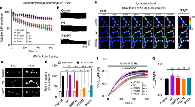
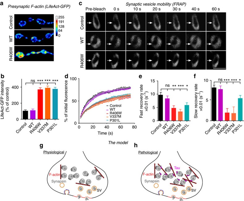
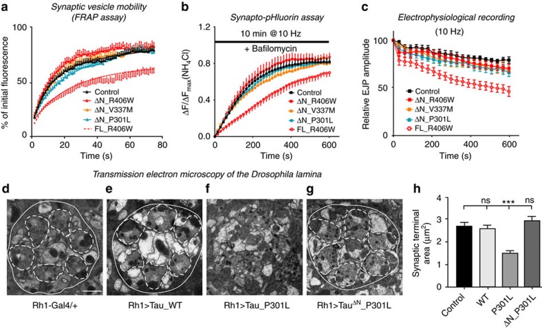
Tau is implicated in more than 20 neurodegenerative diseases, including Alzheimer’s disease. Under pathological conditions, Tau dissociates from axonal microtubules and missorts to pre- and postsynaptic terminals. Patients suffer from early synaptic dysfunction prior to Tau aggregate formation, but the underlying mechanism is unclear. Here we show that pathogenic Tau binds to synaptic vesicles via its N-terminal domain and interferes with presynaptic functions, including synaptic vesicle mobility and release rate, lowering neurotransmission in fly and rat neurons. Pathological Tau mutants lacking the vesicle binding domain still localize to the presynaptic compartment but do not impair synaptic function in fly neurons. Moreover, an exogenously applied membrane-permeable peptide that competes for Tau-vesicle binding suppresses Tau-induced synaptic toxicity in rat neurons. Our work uncovers a presynaptic role of Tau that may be part of the early pathology in various Tauopathies and could be exploited therapeutically.
Overview
The claims and statistics of this paper haven’t been checked yet.
Free: it reads the open-access text and checks the first 5 claims that cite a source we can check. Create a free account to check every claim.
Integrity signals
Registry
Cites
Statistics
Figures


Verification
Claims the paper makes
The claims and statistics of this paper haven’t been checked yet.
Free: it reads the open-access text and checks the first 5 claims that cite a source we can check. Create a free account to check every claim.
Claims other papers make about this one
Each is checked against this paper’s own text: does it say what the citing paper says it does?
The source backs part of the claim, not all of it — or backs it for a different population, condition or comparison.
In the citing paper
“The movement of vesicles and endo-lysosomal compartments along axons is a key part of amyloid fibril formation and vesicle-mediated aggregation of Tau proteins [7, 87].”
From Abnormal accumulation of extracellular vesicles in hippocampal dystrophic axons and regulation by the primary cilia in Alzheimer's disease (2023) · cites this paper as [87]
Experimental — accuracy not yet measured
Self-check
Percentages, sums and reported differences re-computed from the paper’s own numbers.
The self-check needs the paper’s full text.
Citations
Cites
Amyloid oligomers exacerbate tau pathology in a mouse model of tauopathy
Selenica, M.-L. B., Brownlow, M., Jimenez, J. P., Lee, D. C., Pena, G., Dickey, C. A., Gordon, M. N., & Morgan, D. (2013) · Neurodegenerative Diseases · doi:10.1159/000337230 · Cited by 10,359
Visualizing secretion and synaptic transmission with pH-sensitive green fluorescent proteins
Miesenböck, G., De Angelis, D. A., & Rothman, J. E. (1998) · Nature · doi:10.1038/28190 · Cited by 2,563
Tau in physiology and pathology
Wang, Y., & Mandelkow, E. (2016) · Nature Reviews Neuroscience · doi:10.1038/nrn.2015.1 · Cited by 2,251
Self-citationSynapse Loss and Microglial Activation Precede Tangles in a P301S Tauopathy Mouse Model
Yoshiyama, Y., Higuchi, M., Zhang, B., Huang, S.-M., Iwata, N., Saido, T. C., Maeda, J., Suhara, T., Trojanowski, J. Q., & Lee, V. M.-Y. (2007) · Neuron · doi:10.1016/j.neuron.2007.01.010 · Cited by 2,208
Tau-mediated neurodegeneration in Alzheimer’s disease and related disorders
Ballatore, C., Lee, V. M.-Y., & Trojanowski, J. Q. (2007) · Nature Reviews Neuroscience · doi:10.1038/nrn2194 · Cited by 2,174
Tau Suppression in a Neurodegenerative Mouse Model Improves Memory Function
SantaCruz, K., Lewis, J., Spires, T., Paulson, J., Kotilinek, L., Ingelsson, M., Guimaraes, A., DeTure, M., Ramsden, M., McGowan, E., et al. (2005) · Science · doi:10.1126/science.1113694 · Cited by 2,014
Self-citationReducing Endogenous Tau Ameliorates Amyloid ß-Induced Deficits in an Alzheimer's Disease Mouse Model
Roberson, E. D., Scearce-Levie, K., Palop, J. J., Yan, F., Cheng, I. H., Wu, T., Gerstein, H., Yu, G.-Q., & Mucke, L. (2007) · Science · doi:10.1126/science.1141736 · Cited by 1,892
Dendritic Function of Tau Mediates Amyloid-β Toxicity in Alzheimer's Disease Mouse Models
Ittner, L. M., Ke, Y. D., Delerue, F., Bi, M., Gladbach, A., van Eersel, J., Wölfing, H., Chieng, B. C., Christie, M. J., Napier, I. A., et al. (2010) · Cell · doi:10.1016/j.cell.2010.06.036 · Cited by 1,821
Hoover, B. R., Reed, M. N., Su, J., Penrod, R. D., Kotilinek, L. A., Grant, M. K., Pitstick, R., Carlson, G. A., Lanier, L. M., Yuan, L.-L., et al. (2010) · Neuron · doi:10.1016/j.neuron.2010.11.030 · Cited by 1,118
Hippocampal synaptic loss in early Alzheimer's disease and mild cognitive impairment
Scheff, S. W., Price, D. A., Schmitt, F. A., & Mufson, E. J. (2006) · Neurobiology of Aging · doi:10.1016/j.neurobiolaging.2005.09.012 · Cited by 1,017
Cited by
Items 401–473 of 473
Jang, J., Yeo, S., Baek, S., Jung, H. J., Lee, M. S., Choi, S. H., & Choe, Y. (2024) · Figshare · doi:10.6084/m9.figshare.26614924
Jang, J., Yeo, S., Baek, S., Jung, H. J., Lee, M. S., Choi, S. H., & Choe, Y. (2024) · Figshare · doi:10.6084/m9.figshare.26614909
Arouche-Delaperche, L., Cadoni, S., Joffrois, C., Labernède, G., Valet, M., Cesar, Q., Dégardin, J., Girardon, S., Gabriel, C., Krantic, S., et al. (2024) · Figshare · doi:10.6084/m9.figshare.26560682
Arouche-Delaperche, L., Cadoni, S., Joffrois, C., Labernède, G., Valet, M., Cesar, Q., Dégardin, J., Girardon, S., Gabriel, C., Krantic, S., et al. (2024) · Figshare · doi:10.6084/m9.figshare.26560691
Arouche-Delaperche, L., Cadoni, S., Joffrois, C., Labernède, G., Valet, M., Cesar, Q., Dégardin, J., Girardon, S., Gabriel, C., Krantic, S., et al. (2024) · Figshare · doi:10.6084/m9.figshare.26560679
Shi, H., Ge, X., Ma, X., Zheng, M., Cui, X., Pan, W., Zheng, P., Yang, X., Zhang, P., Hu, M., et al. (2021) · Figshare · doi:10.6084/m9.figshare.16986964
Prins, N., Harrison, J., Chu, H.-M., Blackburn, K., Alam, J. J., & Scheltens, P. (2021) · Figshare · doi:10.6084/m9.figshare.14691721
Coomans, E. M., Schoonhoven, D. N., Tuncel, H., Verfaillie, S. C. J., Wolters, E. E., Boellaard, R., Ossenkoppele, R., den Braber, A., Scheper, W., Schober, P. R., et al. (2021) · Figshare · doi:10.6084/m9.figshare.13725141
Goossens, J., González, A. C., Dewit, N., Lidón, L., Fortea, J., Alcolea, D. A., Lleó, A., Belbin, O., & Vanmechelen, E. (2024) · Figshare · doi:10.6084/m9.figshare.26633553
Arouche-Delaperche, L., Cadoni, S., Joffrois, C., Labernède, G., Valet, M., Cesar, Q., Dégardin, J., Girardon, S., Gabriel, C., Krantic, S., et al. (2024) · Figshare · doi:10.6084/m9.figshare.26560697
Jang, J., Yeo, S., Baek, S., Jung, H. J., Lee, M. S., Choi, S. H., & Choe, Y. (2024) · Figshare · doi:10.6084/m9.figshare.26614918
Targeting the M1 muscarinic acetylcholine receptor in neurodegeneration
Hesse, S. (2023) · Enlighten: Theses (The University of Glasgow) · doi:10.5525/gla.thesis.83466
Jang, J., Yeo, S., Baek, S., Jung, H. J., Lee, M. S., Choi, S. H., & Choe, Y. (2024) · Figshare · doi:10.6084/m9.figshare.26614921
Prins, N. D., Harrison, J., Chu, H.-M., Blackburn, K., Alam, J. J., & Scheltens, P. (2021) · Figshare · doi:10.6084/m9.figshare.14691724
Arouche-Delaperche, L., Cadoni, S., Joffrois, C., Labernède, G., Valet, M., Cesar, Q., Dégardin, J., Girardon, S., Gabriel, C., Krantic, S., et al. (2024) · Figshare · doi:10.6084/m9.figshare.26560685
Chinnathambi, S., & Das, R. (2024) · Figshare · doi:10.6084/m9.figshare.26591393
Jang, J., Yeo, S., Baek, S., Jung, H. J., Lee, M. S., Choi, S. H., & Choe, Y. (2024) · Figshare · doi:10.6084/m9.figshare.26614927
DEVELOPMENT OF AN ACCELERATED ALZHEIMER’S DISEASE IN VITRO MODEL WITH THE ADDITION OF PROGERIN
Xue, H. (2023) · University Libraries (University of Maryland) · doi:10.13016/dspace/gfhu-xnen
Arouche-Delaperche, L., Cadoni, S., Joffrois, C., Labernède, G., Valet, M., Cesar, Q., Dégardin, J., Girardon, S., Gabriel, C., Krantic, S., et al. (2024) · Figshare · doi:10.6084/m9.figshare.26560688
Prins, N. D., Harrison, J., Chu, H.-M., Blackburn, K., Alam, J. J., & Scheltens, P. (2021) · Figshare · doi:10.6084/m9.figshare.14691727
Additional file 1 of Synaptic tau: A pathological or physiological phenomenon?
Robbins, M., Clayton, E. L., & Schierle, G. S. K. (2021) · Figshare · doi:10.6084/m9.figshare.16599510
Jang, J., Yeo, S., Baek, S., Jung, H. J., Lee, M. S., Choi, S. H., & Choe, Y. (2024) · Figshare · doi:10.6084/m9.figshare.26614906
Jang, J., Yeo, S., Baek, S., Jung, H. J., Lee, M. S., Choi, S. H., & Choe, Y. (2024) · Figshare · doi:10.6084/m9.figshare.26614912
Ozer, E. (2025) · Kölner Universitäts PublikationsServer (Universität zu Köln)
Badillos-Rodríguez, R. (2023) · Dipòsit Digital de Documents de la UAB (Universitat Autònoma de Barcelona)
Characterization of preclinical transgenic rat models for Alzheimer’s disease-like pathology
Emmerson, J. T. (2023) · eScholarship@McGill (McGill) · doi:10.82308/166
Bellozi, P. M. Q. (2018) · LA Referencia (Red Federada de Repositorios Institucionales de Publicaciones Científicas)
Schreurs, A. (2020) · Lirias
Ao, O. (2024) · eScholarship@McGill (McGill) · doi:10.82308/40971
Functional networks in aging and Alzheimer’s disease: Contributions and consequences
Ziontz, J. (2024) · eScholarship (California Digital Library)
Modulation of the spread of pathological tau in vitro and in vivo
Panzi, C. (2025) · UCL Discovery (University College London)
Oliveira, A. C. (2023) · Portuguese National Funding Agency for Science, Research and Technology (RCAAP Project by FCT) · doi:10.34626/gr6r-ka67
ポルフィロモナス・ジンジバリス由来リポ多糖は、アルツハイマー病の低Aβ濃度環境において、ニューロンのGSK3β依存性シナプス障害を間接的に誘導し、認知機能低下を引き起こす
Gui, S. (2025) · Kyushu University Institutional Repository (QIR) (Kyushu University)
Batchelor, B. (2023) · ePrints Soton (University of Southampton)
Ma, C. C. (2024) · Digital Access to Scholarship at Harvard (DASH) (Harvard University)
Neu, M., Lichtenthaler, S., & München, T. U. (2025)
Biomolecular Condensates: A New Phase of Epilepsy Research?
Trevelyan, A. J., Marshall, A. C., & Forsyth, R. J. (2026) · Epiliepsy currents/Epilepsy currents · doi:10.1177/15357597251408788
“11 All these results point to an important role for biomolecular conden-sates in synaptic transmission, but one which may also predispose toward neurodegenerative changes.”
Implications of virus-induced stress granules in tauopathies
Sharma, S., Vandenakker, A., Cortes-Pérez, C. M., Milne, S., & Douville, R. N. (2026) · Translational Neurodegeneration · doi:10.1186/s40035-026-00538-4
“Under pathological conditions, tau detached from axonal microtubules can abnormally localize to presynaptic and postsynaptic compartments of the neuron and induce synaptic dysfunction through reduced synaptic mobility and altered release of presynaptic vesicles [53, 54].”
Tau regulates epithelial morphogenesis through vesicle trafficking–dependent Notch activationPreprint
Tiwari, N., Sharma, K., & Tapadia, M. G. (2026) · bioRxiv (Cold Spring Harbor Laboratory) · doi:10.64898/2026.03.23.713835
Physiological and pathological roles of the amyloid precursor protein at the presynapse
Jordá-Siquier, T. (2021) · theses.fr (ABES) · doi:10.70675/3bff9a77z3935z4d28zb02bz5b454bcbc77e
Étude biophysique de l'interaction protéine tau - zinc
Rocca, R. L. (2020) · theses.fr (ABES) · doi:10.70675/c975b708z273cz4f8ez9703zc6efc7ce3b5c
Interaction entre autophagie et exosomes au niveau synaptique dans la maladie de Parkinson
Sanchez-Mirasierra, I. (2023) · theses.fr (ABES) · doi:10.70675/7104d6d5z945ez494eza917zda9e6be1eea6
Huo, M., Chen, Y., & Wang, H. (2026) · International Journal of Simulation and Process Modelling · doi:10.1504/ijspm.2026.153267
Oksman, I. (2019) · Lithuanian University of Health Sciences
Role of T-cell immunity in the pathophysiology of Alzheimer’s disease and other Tauopathies
Matta, K. (2024) · theses.fr (ABES) · doi:10.70675/cea4fe5ez67c3z4445z92f7z39af62ba059b
Giordano, A., Mandrioli, J., Cerri, F., Lunetta, C., Saebfar, H., Catania, M., Battipaglia, C., Leone, L., Trojsi, F., Vizziello, M., et al. (2026) · International Journal of Molecular Sciences · doi:10.3390/ijms27104208
“Experimental data also indicate that neuronal activity can regulate extracellular Tau levels, and that Tau participates in synaptic physiology and dysfunction, providing a plausible biological framework for Tau changes that are not fully mirrored by neurofilament dynamics [42–44].”
Mangiarotti, A., Ambroggio, E. E., & Celej, M. S. (2026) · Frontiers in Biophysics · doi:10.3389/frbis.2026.1860065
Kedia, S., Fertan, E., Paul, A., Davì, V., Nolan, G., Baranes, K., Wu, Y., Kim, J. E., Quaegebeur, A., Kotter, M., et al. (2026) · bioRxiv (Cold Spring Harbor Laboratory) · doi:10.64898/2026.05.30.728997
“As a microtubule associated protein concentrated on the axons, tau can translocate to the pre-synaptic terminals under pathological conditions and bind to neurotransmitter vesicles, contributing to the synaptic pathology 64,65 .”
A SIK1-led presynaptic phosphorylation network gates the circadian control of sleep-wake timingPreprint
Jagannath, A., Wakaf, Z., Ru, Y., Taylor, L., Ferrari, M., Dang, Q., Li, S., Demyanenko, Y., Hasan, S., CLARK, K., et al. (2026) · Research Square · doi:10.21203/rs.3.rs-8738572/v1
Structures and Dynamics of Tau Assemblies from Solid-State NMR
Mammeri, N. E., & Hong, M. (2026) · Accounts of Chemical Research · doi:10.1021/acs.accounts.6c00208
Mostowfi, N., Foreman, R. P., Zheng, Z., Ma, Q., 王金良, Khoury, R., Albanese, A., Ma, Q. L., Cohn, W., Wang, Z., et al. (2026) · ACS Omega · doi:10.64898/2026.06.09.731185
SynCAR platform for capturing neuronal synaptic proteopathic seeds
Sebastian, R. M., Kupp, R., Nanavati, D. M., Parfentev, I., Krzystek, T. J., Romanul, N., Yanamandra, K., Preiss, C. N., Manos, J. D., Wu, H.-Y., et al. (2026) · iScience · doi:10.1016/j.isci.2026.116362
“42 Likewise, impacts on post-synaptic function via disrupted localization of the post-synaptic kinase Fyn can be attributed to synaptic tau.”
New Insights into Drug Development and Immunotherapy for Alzheimer’s Disease
Gayen, S., Roy, S., Uddin, M. A., & Sukul, D. (2026) · BENTHAM SCIENCE PUBLISHERS eBooks · doi:10.2174/9798898815646126010014
Tau-mediated Mechanisms in Alzheimerʼs Disease Pathogenesis
Narayanappa, T. S., Ananthanarayana, V., & Chinnathambi, S. (2026) · Molecular Neurobiology · doi:10.1007/s12035-026-06057-7
“…is associated with decreased synaptic vesicle mobility and release rate, defective neuronal endocytosis, and disruption of calcium homeostasis [67, 68] Along with a rise in the intracellular reactive oxygen species and calcium levels, it impairs the morphology and density of dendritic spines.”
Subramaniam, A. G., Gensas, L. K. D. A., Gradus, T., & Perlson, E. (2026) · BIO-PROTOCOL · doi:10.21769/bioprotoc.5801
Cipriano, G. L., Anchesi, I., Floramo, A., Argento, V., Spinelli, S., Astorino, M. F., Calabrò, M., & Artimagnella, O. (2026) · Cells · doi:10.3390/cells15161433
“The N-terminal domain of hyperphosphorylated Tau binds abnormally to the vesicular protein Synaptogyrin-3 (Syngr3), forming a pathological complex that restricts SV mobility and rapidly exhausts neurotransmitter release during sustained depolarization [116].”
Overlapping pathologies in different dementia-causing diseases
Spires‐Jones, T. L. (2026) · Oxford University Press eBooks · doi:10.1093/9780198995296.003.0004
Self-citationSegen, V., Bernard, T. B., Navarro, G. C., Bahrd, P., Behrenbruch, N., Schumann‐Werner, B., Schwarck, S., Garcia‐Garcia, B., Barthel, H., Sabri, O., et al. (2026) · bioRxiv (Cold Spring Harbor Laboratory) · doi:10.64898/2026.08.26.747192
“…that pathological tau can interfere directly with synaptic function 58 , including presynaptic vesicle mobility, release rate, and neurotransmission 59 , and with human evidence linking entorhinal tau deposition to lower hippocampal synaptic density in older individuals with normal cognition and…”
Alqahtani, S. M. (2026) · Discovery Medicine · doi:10.24976/discov.med.202638212.212
Validation of MID1 as a drug target
Kettunen, A. (2023) · UEF eRepo (University of Eastern Finland)
Ji, C., Zhu, Y., Erdjument‐Bromage, H., Song, S. C., Chang, N., Liu, A. R., Lee, A. R., DeGulis, O. A., Schoppik, D., Neubert, T. A., et al. (2026) · bioRxiv (Cold Spring Harbor Laboratory) · doi:10.64898/2026.10.05.756807
“Presynaptic accumulation of pathological tau was reported in AD and FTD patient brains [45–47], and tau association with synaptic vesicles caused presynaptic dysfunction in a Drosophila tauopathy model [48].”
Synaptic tau pathology in Alzheimer's Disease
Sánchez‐Aced, É. (2026) · Tesis Doctorals en Xarxa (Consorci de Serveis Universitaris de Catalunya)
Aumont‐Rodrigue, G., Picard, C., Sarty, I. M., Savard, M., & Poirier, J. (2026) · Neurobiology of Disease · doi:10.1016/j.nbd.2026.107641
Tau phosphorylation from neuronal adaptation to Alzheimer’s disease
Trouvé, R., Verstreken, P., & Hoekstra, M. M. (2026) · Neuron · doi:10.1016/j.neuron.2026.08.032
Self-citationMatei, C., Palade, N. A., Dobri, A.-M., Dumitriu, D. O., Mutu, C., Popescu, A., Pintea, A. L., & Atasie, D. (2026) · Life · doi:10.3390/life16101749
“Consequently, tau dysregulation contributes directly to synaptic dysfunction [81].”
Sun, J., Gao, G., Wang, S., Li, S., Tang, J., Liu, H., & Tang, T.-S. (2026) · Signal Transduction and Targeted Therapy · doi:10.1038/s41392-026-02776-4
“…via synaptogyrin-3, thereby hindering the mobilization of synaptic vesicles during repetitive stimulations Tau-expressing in Drosophila larvae [35] Munc18-1 Munc18-1 KO induced the early Golgi defect, a clinical characteristic of AD Mouse CNS and DRG neurons [42, 44] SNAP-25 SNAP-25 KO…”
InfluentialLiu, J., Xiao, B., Mai, X., Wang, Y., Fei, Y., Yang, W., & Hou, X. S. (2026) · Alzheimer s & Dementia · doi:10.1002/alz.71926
Self-citationBisht, A., Pippadpally, S., & Kumar, V. (2026) · Molecular Neurobiology · doi:10.1007/s12035-026-06310-z
“…is tightly regulated by multiple factors, including developmentally controlled isoform expression [111], region-specific distribution in the brain [112–115], extensive post-translational modifications [114, 116], and disease-associated mutations in the MAPT (micro-tubule-associated protein tau)…”
Tau Regulates Synaptic Plasticity in a Sex- and Species-Dependent Manner
Ralph, L. T. (2024) · TSpace (University of Toronto)
Lai, C., Chen, Z., Ding, Y., Chen, Q., Su, S., Liu, H., Ni, R., & Tang, Z. (2022) · doi:10.3389/fimmu.2022.782434.s002
Metadata
- Authors
- Zhou, L., McInnes, J., Wierda, K., Holt, M., Herrmann, A. G., Jackson, R. J., Wang, Y.-C., Swerts, J., Beyens, J., Miskiewicz, K., Vilain, S., Dewachter, I., Moechars, D., De Strooper, B., Spires-Jones, T. L., De Wit, J., & Verstreken, P.
- Affiliations
- VIB-KU Leuven Center for Brain & Disease Research (BE)
- Euan MacDonald Centre for Motor Neuron Disease Research (GB)
- Hasselt University (BE)
- Janssen (Belgium) (BE)
- Corresponding author
- Verstreken, Patrik
- ORCID
- Venue
- Nature Communications
- Published
- 2017-05-11
- Volume
- 8
- Issue
- 1
- Pages
- 15295-15295
- ISSN
- 2041-1723 (ISSN-L)
- PMC
- PMC5437271
- OpenAlex
- W2613492737
- Cited by
- 434
- References
- 72
- Citations by year
- 2026: 40 · 2025: 47 · 2024: 61 · 2023: 54 · 2022: 35 · 2021: 68 · 2020: 49 · 2019: 31 · 2018: 40 · 2017: 8
- FWCI
- 25.89
- Type
- journal article
- Language
- en
- Open access
- gold
- doi.org
- Licence
- cc-by
- creativecommons.org/licenses/by/4.0 (tdm)
- creativecommons.org/licenses/by/4.0 (vor)
- Locations
- Nature Communications (publishedVersion, cc-by)
- Europe PMC (PubMed Central) (publishedVersion, cc-by)
- PubMed Central (publishedVersion, cc-by)
- PubMed (publishedVersion)
- Discovery Research Portal (University of Dundee) (publishedVersion, other-oa)
- Edinburgh Research Explorer (University of Edinburgh) (publishedVersion, cc-by)
- Lirias (submittedVersion, cc-by)
- RePEc: Research Papers in Economics (submittedVersion)
- DOAJ (DOAJ: Directory of Open Access Journals) (submittedVersion, cc-by-sa)
- Document Server@UHasselt (UHasselt) (submittedVersion, cc-by)
- UCL Discovery (University College London) (submittedVersion)
- Funders
- Belgian American Educational Foundation
- Belgian Federal Science Policy Office
- KU Leuven
- Vlaamse regering
- Medical Research Council (MC_PC_17116)
- National Institute on Aging (P50AG005134)
- H2020 European Research Council (646671)
- FP7 Ideas: European Research Council (260678)
- Topics
- Life Sciences › Neuroscience › Cellular and Molecular Neuroscience › Alzheimer's disease research and treatments
- Life Sciences › Neuroscience › Cellular and Molecular Neuroscience › Neuroscience and Neuropharmacology Research
- Life Sciences › Neuroscience › Neurology › Neuroinflammation and Neurodegeneration Mechanisms
- Keywords
- tau protein, synaptic vesicles, presynaptic dysfunction, tauopathy, Alzheimer's disease, neurotransmission, peptide competition, synaptic toxicity, Drosophila neurons, rat neurons
- MeSH terms
- Protein Domains, Actins, Animals, Drosophila melanogaster, Hippocampus, Humans, Mutation, Synaptic Transmission, Neurons, Synaptic Vesicles, tau Proteins, Presynaptic Terminals, Protein Transport, Rats
About these checks
Registry, retraction, Cites and statistics results are computed, not judged, with one exception: a printed reference that no index lists is read once by a model before it counts as a citation error. Claims are extracted by a model and kept only when their quote appears verbatim in the text; each cited source is then checked, with a model’s help, for whether it says what the claim relies on. Those verdicts can be wrong — read the claim’s quote and the cited source before relying on one.
Statistics and arithmetic are recomputed, not judged.
Confidence bands are shown only for fields calibrated on our annotated test set. None is calibrated yet, so no band is shown.
What counts as a claim ›How we measure confidence ›How we check this ›
Are you an author, or do you see a problem with this page? Request a review
Data sources
Bibliographic data: Semantic Scholar, OpenAlex, Crossref.
Contains information from the Semantic Scholar Academic Graph Datasets, made available under the ODC Attribution License (ODC-BY 1.0).
Semantic Scholar’s data is described in: Kinney, R., et al. (2023). The Semantic Scholar Open Data Platform. arXiv:2301.10140.
