

The DOI matches its Crossref record; 72 of the 72 references it cites were found in a registry or an index.
Tau association with synaptic vesicles causes presynaptic dysfunction
Open accessAbstract
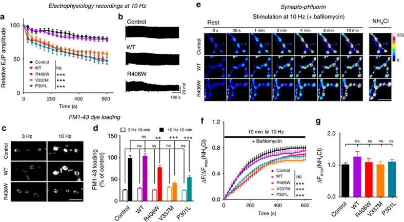
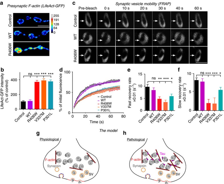
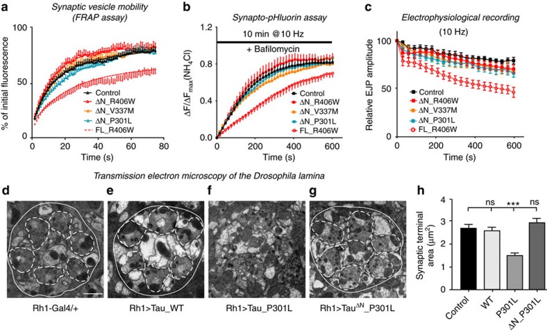
Tau is implicated in more than 20 neurodegenerative diseases, including Alzheimer’s disease. Under pathological conditions, Tau dissociates from axonal microtubules and missorts to pre- and postsynaptic terminals. Patients suffer from early synaptic dysfunction prior to Tau aggregate formation, but the underlying mechanism is unclear. Here we show that pathogenic Tau binds to synaptic vesicles via its N-terminal domain and interferes with presynaptic functions, including synaptic vesicle mobility and release rate, lowering neurotransmission in fly and rat neurons. Pathological Tau mutants lacking the vesicle binding domain still localize to the presynaptic compartment but do not impair synaptic function in fly neurons. Moreover, an exogenously applied membrane-permeable peptide that competes for Tau-vesicle binding suppresses Tau-induced synaptic toxicity in rat neurons. Our work uncovers a presynaptic role of Tau that may be part of the early pathology in various Tauopathies and could be exploited therapeutically.
Overview
The claims and statistics of this paper haven’t been checked yet.
Free: it reads the open-access text and checks the first 5 claims that cite a source we can check. Create a free account to check every claim.
Integrity signals
Registry
Cites
Statistics
Figures


Verification
Claims the paper makes
The claims and statistics of this paper haven’t been checked yet.
Free: it reads the open-access text and checks the first 5 claims that cite a source we can check. Create a free account to check every claim.
Claims other papers make about this one
Each is checked against this paper’s own text: does it say what the citing paper says it does?
The source backs part of the claim, not all of it — or backs it for a different population, condition or comparison.
In the citing paper
“The movement of vesicles and endo-lysosomal compartments along axons is a key part of amyloid fibril formation and vesicle-mediated aggregation of Tau proteins [7, 87].”
From Abnormal accumulation of extracellular vesicles in hippocampal dystrophic axons and regulation by the primary cilia in Alzheimer's disease (2023) · cites this paper as [87]
Experimental — accuracy not yet measured
Self-check
Percentages, sums and reported differences re-computed from the paper’s own numbers.
The self-check needs the paper’s full text.
Citations
Cites
Amyloid oligomers exacerbate tau pathology in a mouse model of tauopathy
Selenica, M.-L. B., Brownlow, M., Jimenez, J. P., Lee, D. C., Pena, G., Dickey, C. A., Gordon, M. N., & Morgan, D. (2013) · Neurodegenerative Diseases · doi:10.1159/000337230 · Cited by 10,359
Visualizing secretion and synaptic transmission with pH-sensitive green fluorescent proteins
Miesenböck, G., De Angelis, D. A., & Rothman, J. E. (1998) · Nature · doi:10.1038/28190 · Cited by 2,563
Tau in physiology and pathology
Wang, Y., & Mandelkow, E. (2016) · Nature Reviews Neuroscience · doi:10.1038/nrn.2015.1 · Cited by 2,251
Self-citationSynapse Loss and Microglial Activation Precede Tangles in a P301S Tauopathy Mouse Model
Yoshiyama, Y., Higuchi, M., Zhang, B., Huang, S.-M., Iwata, N., Saido, T. C., Maeda, J., Suhara, T., Trojanowski, J. Q., & Lee, V. M.-Y. (2007) · Neuron · doi:10.1016/j.neuron.2007.01.010 · Cited by 2,208
Tau-mediated neurodegeneration in Alzheimer’s disease and related disorders
Ballatore, C., Lee, V. M.-Y., & Trojanowski, J. Q. (2007) · Nature Reviews Neuroscience · doi:10.1038/nrn2194 · Cited by 2,174
Tau Suppression in a Neurodegenerative Mouse Model Improves Memory Function
SantaCruz, K., Lewis, J., Spires, T., Paulson, J., Kotilinek, L., Ingelsson, M., Guimaraes, A., DeTure, M., Ramsden, M., McGowan, E., et al. (2005) · Science · doi:10.1126/science.1113694 · Cited by 2,014
Self-citationReducing Endogenous Tau Ameliorates Amyloid ß-Induced Deficits in an Alzheimer's Disease Mouse Model
Roberson, E. D., Scearce-Levie, K., Palop, J. J., Yan, F., Cheng, I. H., Wu, T., Gerstein, H., Yu, G.-Q., & Mucke, L. (2007) · Science · doi:10.1126/science.1141736 · Cited by 1,892
Dendritic Function of Tau Mediates Amyloid-β Toxicity in Alzheimer's Disease Mouse Models
Ittner, L. M., Ke, Y. D., Delerue, F., Bi, M., Gladbach, A., van Eersel, J., Wölfing, H., Chieng, B. C., Christie, M. J., Napier, I. A., et al. (2010) · Cell · doi:10.1016/j.cell.2010.06.036 · Cited by 1,821
Hoover, B. R., Reed, M. N., Su, J., Penrod, R. D., Kotilinek, L. A., Grant, M. K., Pitstick, R., Carlson, G. A., Lanier, L. M., Yuan, L.-L., et al. (2010) · Neuron · doi:10.1016/j.neuron.2010.11.030 · Cited by 1,118
Hippocampal synaptic loss in early Alzheimer's disease and mild cognitive impairment
Scheff, S. W., Price, D. A., Schmitt, F. A., & Mufson, E. J. (2006) · Neurobiology of Aging · doi:10.1016/j.neurobiolaging.2005.09.012 · Cited by 1,017
Cited by
Knopman, D. S., Amieva, H., Petersen, R. C., Chételat, G., Holtzman, D. M., Hyman, B. T., Nixon, R. A., & Jones, D. T. (2021) · Nature Reviews Disease Primers · doi:10.1038/s41572-021-00269-y · Cited by 2,606
Molecular and cellular mechanisms underlying the pathogenesis of Alzheimer’s disease
Guo, T., Zhang, D., Zeng, Y., Huang, T. Y., Xu, H., & Zhao, Y. (2020) · Molecular Neurodegeneration · doi:10.1186/s13024-020-00391-7 · Cited by 994
Amyloid β-based therapy for Alzheimer’s disease: Challenges, successes and future
Zhang, Y., Chen, H., Li, R., Sterling, K., & Song, W. (2023) · Signal Transduction and Targeted Therapy · doi:10.1038/s41392-023-01484-7 · Cited by 988
Reconsideration of Amyloid Hypothesis and Tau Hypothesis in Alzheimer's Disease
Kametani, F., & Hasegawa, M. (2018) · Frontiers in Neuroscience · doi:10.3389/fnins.2018.00025 · Cited by 836
“Aberrant interaction of stabilized tau with filamentous actin induces mis-stabilization of actin (Fulga et al., 2007), synaptic impairment (Cabrales Fontela et al., 2017; Zhou et al., 2017; Bardai et al., 2018), and defects in mitochondrial integrity (DuBoff et al., 2012).”
BackgroundPhosphorylated Tau in Alzheimer’s Disease and Other Tauopathies
Rawat, P., Sehar, U., Bisht, J., Selman, A., Culberson, J., & Reddy, P. H. (2022) · International Journal of Molecular Sciences · doi:10.3390/ijms232112841 · Cited by 509
“Under pathological conditions, tau detached from an axonal compartment of the microtubule abnormally localizes into a presynaptic and postsynaptic compartment of the neuron and induces synaptic dysfunction in various ways, such as reduced mobility, the release of a presynaptic vesicle [187,188].”
BackgroundSynaptic degeneration in Alzheimer disease
Tzioras, M., McGeachan, R. I., Durrant, C. S., & Spires-Jones, T. L. (2023) · Nature Reviews Neurology · doi:10.1038/s41582-022-00749-z · Cited by 504
Self-citationKent, S. A., Spires-Jones, T. L., & Durrant, C. S. (2020) · Acta Neuropathologica · doi:10.1007/s00401-020-02196-w · Cited by 446
Self-citationMicroglia modulate neurodegeneration in Alzheimer’s and Parkinson’s diseases
Bartels, T., De Schepper, S., & Hong, S. (2020) · Science · doi:10.1126/science.abb8587 · Cited by 412
Amyloid-β and tau complexity — towards improved biomarkers and targeted therapies
Polanco, J. C., Li, C., Bodea, L.-G., Martinez-Marmol, R., Meunier, F. A., & Götz, J. (2018) · Nature Reviews Neurology · doi:10.1038/nrneurol.2017.162 · Cited by 378
The complexity of tau in Alzheimer’s disease
Naseri, N. N., Wang, H., Guo, J., Sharma, M., & Luo, W. (2019) · Neuroscience Letters · doi:10.1016/j.neulet.2019.04.022 · Cited by 373
“The outcome of this pathogenic association may cluster F-actin at the presynapse and physically impede synaptic vesicle release, resulting in decreased neurotransmission [93].”
BackgroundTau interactome maps synaptic and mitochondrial processes associated with neurodegeneration
Tracy, T. E., Madero-Pérez, J., Swaney, D. L., Chang, T. S., Moritz, M., Konrad, C., Ward, M. E., Stevenson, E., Hüttenhain, R., Kauwe, G., et al. (2022) · Cell · doi:10.1016/j.cell.2021.12.041 · Cited by 342
“Consistent with studies showing that Tau can bind directly to synaptic vesicles (McInnes et al., 2018; Zhou et al., 2017), we identified biotinylation on 5 integral synaptic vesicle proteins that span the vesicle membrane.”
The clinical promise of biomarkers of synapse damage or loss in Alzheimer’s disease
the Synaptic Health Endpoints Working Group, Colom-Cadena, M., Spires-Jones, T., Zetterberg, H., Blennow, K., Caggiano, A., DeKosky, S. T., Fillit, H., Harrison, J. E., Schneider, L. S., et al. (2020) · Alzheimer's Research & Therapy · doi:10.1186/s13195-020-00588-4 · Cited by 328
Self-citationMechanisms of secretion and spreading of pathological tau protein
Brunello, C. A., Merezhko, M., Uronen, R.-L., & Huttunen, H. J. (2020) · Cellular and Molecular Life Sciences · doi:10.1007/s00018-019-03349-1 · Cited by 304
Synaptic Tau Seeding Precedes Tau Pathology in Human Alzheimer’s Disease Brain
DeVos, S. L., Corjuc, B. T., Oakley, D. H., Nobuhara, C. K., Bannon, R. N., Chase, A., Commins, C., Gonzalez, J. A., Dooley, P. M., Frosch, M. P., et al. (2018) · Frontiers in Neuroscience · doi:10.3389/fnins.2018.00267 · Cited by 285
“We and others have previously shown that phosphorylated-tau oligomers are enriched in the synapses of human AD brain tissue, as compared to control, while total tau remains the same between the synaptic and cytosolic fractions (Henkins et al., 2012; Tai et al., 2012, 2014; Perez-Nievas et al., 2013; Sokolow et al., 20…”
BackgroundDendritic Tau in Alzheimer’s Disease
Ittner, A., & Ittner, L. M. (2018) · Neuron · doi:10.1016/j.neuron.2018.06.003 · Cited by 278
Molecular signatures underlying neurofibrillary tangle susceptibility in Alzheimer’s disease
Otero-Garcia, M., Mahajani, S. U., Wakhloo, D., Tang, W., Xue, Y.-Q., Morabito, S., Pan, J., Oberhauser, J., Madira, A. E., Shakouri, T., et al. (2022) · Neuron · doi:10.1016/j.neuron.2022.06.021 · Cited by 262
Synaptic dysfunction in Alzheimer's disease: Mechanisms and therapeutic strategies
Chen, Y., Fu, A. K. Y., & Ip, N. Y. (2019) · Pharmacology & Therapeutics · doi:10.1016/j.pharmthera.2018.11.006 · Cited by 258
Matter over mind: Liquid phase separation and neurodegeneration
Elbaum-Garfinkle, S. (2019) · Journal of Biological Chemistry · doi:10.1074/jbc.rev118.001188 · Cited by 249
“These include tau (40-42,73,74), FMRP (43,72), and Huntington protein (44).”
BackgroundShi, H., Ge, X., Ma, X., Zheng, M., Cui, X., Pan, W., Zheng, P., Yang, X., Zhang, P., Hu, M., et al. (2021) · Microbiome · doi:10.1186/s40168-021-01172-0 · Cited by 247
Ghadami, S., & Dellinger, K. (2023) · Frontiers in Molecular Biosciences · doi:10.3389/fmolb.2023.1198044 · Cited by 246
“Pathologic tau can increase the level of F-actin molecules in presynaptic terminals, leading to decreased synaptic vesicle mobility (Zhou et al., 2017).”
BackgroundMaestú, F., de Haan, W., Busche, M. A., & DeFelipe, J. (2021) · Ageing Research Reviews · doi:10.1016/j.arr.2021.101372 · Cited by 244
Synaptogyrin-3 Mediates Presynaptic Dysfunction Induced by Tau
McInnes, J., Wierda, K., Snellinx, A., Bounti, L., Wang, Y.-C., Stancu, I.-C., Apóstolo, N., Gevaert, K., Dewachter, I., Spires-Jones, T. L., et al. (2018) · Neuron · doi:10.1016/j.neuron.2018.01.022 · Cited by 242
“The N-terminal domain of Tau is required for binding to SVs (Zhou et al., 2017), and thus the interactor of Tau on SVs should bind full-length Tau (TauFL) but not N-terminally truncated Tau (Tau∆N).”
BackgroundMethodInfluentialSelf-citationThe role of pathological tau in synaptic dysfunction in Alzheimer’s diseases
Wu, M., Zhang, M., Yin, X., Chen, K., Hu, Z., Zhou, Q., Cao, X., Chen, Z., & Liu, D. (2021) · Translational Neurodegeneration · doi:10.1186/s40035-021-00270-1 · Cited by 241
“Pathological tau plays a vital role in impairing mitochondrial transport, thereby reducing the number of presynaptic mitochondria and hampering the release of synaptic vesicles [35, 114, 115].”
BackgroundCellular and pathological functions of tau
Parra Bravo, C., Naguib, S. A., & Gan, L. (2024) · Nature Reviews Molecular Cell Biology · doi:10.1038/s41580-024-00753-9 · Cited by 238
“At the pre-synaptic terminal, tau decreases the release of neuro-transmitter-containing synaptic vesicles by reducing their mobility in D. melanogaster neuromuscular junction terminals 242 .”
Neuronal cell death mechanisms in Alzheimer’s disease: An insight
Goel, P., Chakrabarti, S., Goel, K., Bhutani, K., Chopra, T., & Bali, S. (2022) · Frontiers in Molecular Neuroscience · doi:10.3389/fnmol.2022.937133 · Cited by 234
“Furthermore, evidence from several studies indicate that tau pathology affected several systems within the cell including signaling system (Cabrales Fontela et al., 2017; Zhou et al., 2017; Bardai et al., 2018), transport system, cytoskeletal system (Fulga et al.”
BackgroundColom-Cadena, M., Davies, C., Sirisi, S., Lee, J.-E., Simzer, E. M., Tzioras, M., Querol-Vilaseca, M., Sánchez-Aced, É., Chang, Y. Y., Holt, K., et al. (2023) · Neuron · doi:10.1016/j.neuron.2023.04.020 · Cited by 224
Self-citationDysfunctional Mitochondria and Mitophagy as Drivers of Alzheimer’s Disease Pathogenesis
Chakravorty, A., Jetto, C. T., & Manjithaya, R. (2019) · Frontiers in Aging Neuroscience · doi:10.3389/fnagi.2019.00311 · Cited by 219
Synthesis of Carbon Nanotubes by Floating Catalyst Chemical Vapor Deposition and Their Applications
Hou, P., Zhang, F., Zhang, L., Liu, C., & Cheng, H. (2022) · Advanced Functional Materials · doi:10.1002/adfm.202108541 · Cited by 213
Meftah, S., & Gan, J. (2023) · Frontiers in Synaptic Neuroscience · doi:10.3389/fnsyn.2023.1129036 · Cited by 205
“Some of the mechanisms implicated in tau-associated pre-synaptic dysfunction include tau binding to synaptic vesicles, increases in cytosolic calcium, and disrupted mitochondrial trafficking leading to disrupted synaptic transmission (Decker et al., 2015; Moreno et al., 2016; Zhou et al., 2017; McInnes et al., 2018; W…”
BackgroundSynapse pathology in Alzheimer’s disease
Griffiths, J., & Grant, S. G. N. (2023) · Seminars in Cell & Developmental Biology · doi:10.1016/j.semcdb.2022.05.028 · Cited by 198
Tau Pathology in Parkinson's Disease
Zhang, X., Gao, F., Wang, D., Li, C., Fu, Y., He, W., & Zhang, J. (2018) · Frontiers in Neurology · doi:10.3389/fneur.2018.00809 · Cited by 195
“Meanwhile, the FTDP-17 tau mutant, which is known to be associated with PDD, was shown to interfere with synaptic vesicles in presynaptic terminals, causing the dysfunction of vesicle traffic and presynaptic activity (58).”
BackgroundTargeting synapse function and loss for treatment of neurodegenerative diseases
Dejanovic, B., Sheng, M., & Hanson, J. E. (2023) · Nature reviews. Drug discovery · doi:10.1038/s41573-023-00823-1 · Cited by 157
Gene-based therapies for neurodegenerative diseases
Sun, J., & Roy, S. (2021) · Nature Neuroscience · doi:10.1038/s41593-020-00778-1 · Cited by 187
Pickett, E. K., Herrmann, A. G., McQueen, J., Abt, K., Dando, O., Tulloch, J., Jain, P., Dunnett, S., Sohrabi, S., Fjeldstad, M. P., et al. (2019) · Cell Reports · doi:10.1016/j.celrep.2019.11.044 · Cited by 179
“Tau has been shown to bind to presynaptic vesicles in human AD and Drosophila models, where it impairs neurotransmitter release (Zhou et al., 2017; McInnes et al., 2018).”
BackgroundSelf-citationDeregulated mitochondrial microRNAs in Alzheimer's disease: Focus on synapse and mitochondria
Gowda, P., Reddy, P. H., & Kumar, S. (2022) · Ageing Research Reviews · doi:10.1016/j.arr.2021.101529 · Cited by 163
Tau-mediated synaptic and neuronal dysfunction in neurodegenerative disease
Tracy, T. E., & Gan, L. (2018) · Current Opinion in Neurobiology · doi:10.1016/j.conb.2018.04.027 · Cited by 162
Harris, S. S., Wolf, F., De Strooper, B., & Busche, M. A. (2020) · Neuron · doi:10.1016/j.neuron.2020.06.005 · Cited by 161
Pickett, E. K., Rose, J. L., McCrory, C., McKenzie, C.-A., King, D., Smith, C., Gillingwater, T. H., Henstridge, C. M., & Spires‐Jones, T. L. (2018) · Acta Neuropathologica · doi:10.1007/s00401-018-1903-2 · Cited by 138
Self-citationLiquid–liquid phase separation of tau: From molecular biophysics to physiology and disease
Rai, S. K., Savastano, A., Singh, P., Mukhopadhyay, S., & Zweckstetter, M. (2021) · Protein Science · doi:10.1002/pro.4093 · Cited by 135
Synaptic tau: A pathological or physiological phenomenon?
Robbins, M., Clayton, E. L., & Schierle, G. S. K. (2021) · Acta Neuropathologica Communications · doi:10.1186/s40478-021-01246-y · Cited by 133
The effect of insomnia on development of Alzheimer’s disease
Sadeghmousavi, S., Eskian, M., Rahmani, F., & Rezaei, N. (2020) · Journal of Neuroinflammation · doi:10.1186/s12974-020-01960-9 · Cited by 132
Ranasinghe, K. G., Verma, P., Cai, C., Xie, X., Kudo, K., Gao, X., Lerner, H., Mizuiri, D., Strom, A., Iaccarino, L., et al. (2022) · eLife · doi:10.7554/elife.77850 · Cited by 127
Puangmalai, N., Bhatt, N. N., Montalbano, M., Sengupta, U., Gaikwad, S., Ventura, F. L., McAllen, S., Ellsworth, A., García, S., & Kayed, R. (2020) · Cell Death and Disease · doi:10.1038/s41419-020-2503-3 · Cited by 123
Possible Mechanisms of Tau Spread and Toxicity in Alzheimer’s Disease
Zhang, H., Cao, Y., Ma, L., Wei, Y., & Li, H. (2021) · Frontiers in Cell and Developmental Biology · doi:10.3389/fcell.2021.707268 · Cited by 123
for the REVERSE-SD Study Investigators, Prins, N. D., Harrison, J., Chu, H., Blackburn, K., Alam, J. J., & Scheltens, P. (2021) · Alzheimer s Research & Therapy · doi:10.1186/s13195-021-00843-2 · Cited by 120
Propagation of Tau via Extracellular Vesicles
Pérez, M., Avila, J., & Hernández, F. (2019) · Frontiers in Neuroscience · doi:10.3389/fnins.2019.00698 · Cited by 118
“2016) and presynaptic compartments (Zhou et al., 2017).”
Gonçalves, R. A., Wijesekara, N., Fraser, P. E., & Felice, F. G. D. (2019) · Frontiers in Cellular Neuroscience · doi:10.3389/fncel.2019.00017 · Cited by 117
“In addition, due to its scaffolding property, Tau can bind to a variety of proteins, therefore impacting multiple physiological functions (Meier et al., 2015; Suzuki and Kimura, 2017; Zhou et al., 2017; Stefanoska et al., 2018).”
BackgroundEstes, R., Lin, B. C., Khera, A., & Davis, M. Y. (2021) · Frontiers in Molecular Neuroscience · doi:10.3389/fnmol.2021.788695 · Cited by 109
“Though accumulation of hyperphosphorylated tau in NFTs is a hallmark of AD, soluble, non-aggregated forms of tau appear to be the main toxic element inducing early synaptic deficits (Ahmed et al., 2014; Zhou et al., 2017).”
BackgroundIn vivo tau pathology is associated with synaptic loss and altered synaptic function
Coomans, E. M., Schoonhoven, D. N., Tuncel, H., Verfaillie, S. C. J., Wolters, E. E., Boellaard, R., Ossenkoppele, R., den Braber, A., Scheper, W., Schober, P. R., et al. (2021) · Alzheimer s Research & Therapy · doi:10.1186/s13195-021-00772-0 · Cited by 108
“effects on synaptic structure and function [13, 14].”
Brandt, R., Trushina, N. I., & Bakota, L. (2020) · Frontiers in Neurology · doi:10.3389/fneur.2020.590059 · Cited by 107
The Evolution of Tau Phosphorylation and Interactions
Trushina, N. I., Bakota, L., Mulkidjanian, A. Y., & Brandt, R. (2019) · Frontiers in Aging Neuroscience · doi:10.3389/fnagi.2019.00256 · Cited by 103
Montero‐Crespo, M., Domínguez-Álvaro, M., Alonso‐Nanclares, L., DeFelipe, J., & Blázquez‐Llorca, L. (2020) · Brain · doi:10.1093/brain/awaa406 · Cited by 99
“There are numerous studies providing a wealth of evidence for amyloid-b peptides and abnormally phosphorylated tau proteins generating toxic effects under pathological conditions, leading to synaptic loss and neurotransmitter release dysfunction (Gylys et al., 2004; Lacor et al., 2007; Merino-Serrais et al., 2011, 201…”
BackgroundCIP2A Causes Tau/APP Phosphorylation, Synaptopathy, and Memory Deficits in Alzheimer’s Disease
Shentu, Y.-P., Huo, Y., Feng, X.-L., Gilbert, J., Zhang, Q., Liuyang, Z.-Y., Wang, X.-L., Wang, G., Zhou, H., Wang, X.-C., et al. (2018) · Cell Reports · doi:10.1016/j.celrep.2018.06.009 · Cited by 90
The Neurotoxic Role of Extracellular Tau Protein
Sebastián‐Serrano, Á., de Diego-García, L., & Díaz-Hernández, M. (2018) · International Journal of Molecular Sciences · doi:10.3390/ijms19040998 · Cited by 90
“In this respect, a study has shown that tau protein may bind to synaptic vesicles via its N-terminal domain, interfering in this way with presynaptic functions, including synaptic vesicle mobility and release rate [32].”
BackgroundIn vivo synaptic density loss is related to tau deposition in amnestic mild cognitive impairment
Vanhaute, H., Ceccarini, J., Michiels, L., Koole, M., Sunaert, S., Lemmens, R., Triau, E., Emsell, L., Vandenbulcke, M., & Van Laere, K. (2020) · Neurology · doi:10.1212/wnl.0000000000009818 · Cited by 88
Hill, E., Karikari, T. K., Moffat, K. G., Richardson, M. J. E., & Wall, M. J. (2019) · eNeuro · doi:10.1523/eneuro.0166-19.2019 · Cited by 87
Kim, H., Pyun, K. R., Lee, M., Lee, H., & Ko, S. H. (2021) · Advanced Functional Materials · doi:10.1002/adfm.202110535 · Cited by 87
Mueller, R. L., Combs, B., Alhadidy, M. M., Brady, S. T., Morfini, G. A., & Kanaan, N. M. (2021) · Frontiers in Molecular Neuroscience · doi:10.3389/fnmol.2021.647054 · Cited by 84
Largo‐Barrientos, P., Apóstolo, N., Creemers, E., Callaerts‐Vegh, Z., Swerts, J., Davies, C., McInnes, J., Wierda, K. D. B., Strooper, B. D., Spires‐Jones, T. L., et al. (2021) · Neuron · doi:10.1016/j.neuron.2020.12.016 · Cited by 83
“Next, we asked if the synaptic-localized Tau associates with vesicles in this model, similar to what we observed in fruit flies and Alzheimer’s disease patient brain samples (McInnes et al., 2018; Zhou et al., 2017).”
InfluentialSelf-citationGomes, L. A., Uytterhoeven, V., Lopez-Sanmartin, D., Tomé, S. O., Tousseyn, T., Vandenberghe, R. R., Vandenbulcke, M., Arnim, C. A. F. V., Verstreken, P., & Thal, D. R. (2021) · Acta Neuropathologica · doi:10.1007/s00401-020-02251-6 · Cited by 82
Self-citationLi, J., Shi, Q., Röhr, J. A., Wu, H., Wu, B. C., Guo, Y., Zhang, Q., Hou, C., Li, Y., & Wang, H. (2020) · Advanced Functional Materials · doi:10.1002/adfm.202002508 · Cited by 80
Three-dimensional analysis of synapses in the transentorhinal cortex of Alzheimer’s disease patients
Domínguez-Álvaro, M., Montero-Crespo, M., Blazquez-Llorca, L., Insausti, R., DeFelipe, J., & Alonso-Nanclares, L. (2018) · Acta Neuropathologica Communications · doi:10.1186/s40478-018-0520-6 · Cited by 79
“causing dysfunction in neurotransmitter release [25, 32, 57, 73, 89].”
Ma, J., Huo, X., Jarpe, M. B., Kavelaars, A., & Heijnen, C. J. (2018) · Acta Neuropathologica Communications · doi:10.1186/s40478-018-0604-3 · Cited by 77
Presynaptic failure in Alzheimer's disease
Barthet, G., & Mulle, C. (2020) · Progress in Neurobiology · doi:10.1016/j.pneurobio.2020.101801 · Cited by 74
“…that pathological tau, when it is present presynaptically, binds to vesicles through synaptogyrin-3 and polymerizes presynaptic actin using its proline-rich and microtubule-binding domains, thereby impeding the mobilization of synaptic vesicles during repetitive stimulations (Zhou et al., 2017).”
BackgroundInfluentialCa2+ Dyshomeostasis Disrupts Neuronal and Synaptic Function in Alzheimer’s Disease
McDaid, J., Mustaly-Kalimi, S., & Stutzmann, G. E. (2020) · Cells · doi:10.3390/cells9122655 · Cited by 73
Qu, X., Kumar, A., Blockus, H., Waites, C. L., & Bartolini, F. (2019) · Current Biology · doi:10.1016/j.cub.2019.10.049 · Cited by 72
“Given the critical role that loss of a functional presynaptic machinery for MT nucleation may have for neurotransmission, it will be important to determine the in vivo significance of this selective MT nucleation and whether dysregulation of this process can be associated with human neurological and neuropsychiatric i…”
BackgroundThe Cell Biology of Tau Secretion
Merezhko, M., Uronen, R., & Huttunen, H. J. (2020) · Frontiers in Molecular Neuroscience · doi:10.3389/fnmol.2020.569818 · Cited by 70
“As tau associates with SVs, the fusion can also bring the normally cytosolic Tau to the PM (Zhou et al., 2017).”
BackgroundGraphene‐Based Technologies for Tackling COVID‐19 and Future Pandemics
Afroj, S., Britnell, L., Hasan, T., Andreeva, D. V., Novoselov, K. S., & Karim, N. (2021) · Advanced Functional Materials · doi:10.1002/adfm.202107407 · Cited by 70
Puangmalai, N., Sengupta, U., Bhatt, N., Gaikwad, S., Montalbano, M., Bhuyan, A., Garcia, S., McAllen, S., Sonawane, M., Jerez, C., et al. (2022) · Journal of Biological Chemistry · doi:10.1016/j.jbc.2022.101766 · Cited by 68
Bassoon contributes to tau-seed propagation and neurotoxicity
Martinez, P., Patel, H., You, Y., Jury, N., Perkins, A., Lee-Gosselin, A., Taylor, X., You, Y., Viana Di Prisco, G., Huang, X., et al. (2022) · Nature Neuroscience · doi:10.1038/s41593-022-01191-6 · Cited by 68
Stabilization of dynamic microtubules by mDia1 drives Tau-dependent Aβ1–42 synaptotoxicity
Qu, X., Yuan, F. N., Corona, C., Pasini, S., Pero, M. E., Gundersen, G. G., Shelanski, M. L., & Bartolini, F. (2017) · The Journal of Cell Biology · doi:10.1083/jcb.201701045 · Cited by 67
“More recently, a role for pathological tau mutants and hyperphosphorylated tau has been further uncovered in presynaptic dysfunction (Zhou et al., 2017).”
Pietrancosta, N., Djibo, M., Daumas, S., Mestikawy, S. E., & Erickson, J. D. (2020) · Molecular Neurobiology · doi:10.1007/s12035-020-01912-7 · Cited by 67
Neuronal identity defines α-synuclein and tau toxicity
Praschberger, R., Kuenen, S., Schoovaerts, N., Kaempf, N., Singh, J., Janssens, J., Swerts, J., Nachman, E., Calatayud, C., Aerts, S., et al. (2023) · Neuron · doi:10.1016/j.neuron.2023.02.033 · Cited by 66
Self-citationAn N-terminal motif unique to primate tau enables differential protein–protein interactions
Stefanoska, K., Volkerling, A. M., Bertz, J., Poljak, A., Ke, Y. D., Ittner, L. M., & Ittner, A. (2018) · Journal of Biological Chemistry · doi:10.1074/jbc.ra118.001784 · Cited by 66
“Pathological human tau interacts with presynaptic vesicles though its N-terminal region (28).”
BackgroundResultDomínguez-Álvaro, M., Montero‐Crespo, M., Blázquez‐Llorca, L., DeFelipe, J., & Alonso‐Nanclares, L. (2019) · eNeuro · doi:10.1523/eneuro.0140-19.2019 · Cited by 65
“A peptides and tau proteins play normal roles at the synapse, but under pathologic conditions, they may lead to toxic effects at both presynaptic and postsynaptic elements, leading to synaptic loss and causing dysfunction in neurotransmitter release (Spires-Jones and Hyman, 2014; Dorostkar et al., 2015; Henstridge et…”
BackgroundEarly- and Late-Onset Alzheimer’s Disease: Two Sides of the Same Coin?
Valdez-Gaxiola, C. A., Rosales-Leycegui, F., Gaxiola-Rubio, A., Moreno-Ortíz, J. M., & Figuera, L. E. (2024) · Diseases · doi:10.3390/diseases12060110 · Cited by 64
“Furthermore, pathological tau may also mislocalize to presynaptic terminals, where it can interfere with exocytosis by binding to synaptic vesicles [39,40] (risk factors are summarized in Figure 1).”
BackgroundMechanistic insights and emerging therapeutic stratagems for Alzheimer’s disease
Rajendran, K., & Krishnan, U. M. (2024) · Ageing Research Reviews · doi:10.1016/j.arr.2024.102309 · Cited by 63
Gomes, P., Tzouanou, F., Skolariki, K., Vamvaka-Iakovou, A., Nogueras‐Ortiz, C. J., Tsirtsaki, K., Waites, C. L., Vlamos, P., Sousa, N., Costa‐Silva, B., et al. (2022) · Experimental Neurology · doi:10.1016/j.expneurol.2022.114183 · Cited by 62
“A recent human study has provided insight into the association between human Tau pathology and synaptic loss and altered synaptic function (Coomans et al., 2021), as it had previously been found in murine AD models (Lasagna-Reeves et al., 2011; Zhou et al., 2017).”
Role of the Lipid Membrane and Membrane Proteins in Tau Pathology
Bok, E., Leem, E., Lee, B.-R., Lee, J. M., Yoo, C. J., Lee, E. M., & Kim, J. (2021) · Frontiers in Cell and Developmental Biology · doi:10.3389/fcell.2021.653815 · Cited by 61
Padmanabhan, P., Martínez‐Mármol, R., Xia, D., Götz, J., & Meunier, F. A. (2019) · eLife · doi:10.7554/elife.45040 · Cited by 61
The Role of Mitochondrial Impairment in Alzheimer´s Disease Neurodegeneration: The Tau Connection
Quntanilla, R. A., & Tapia-Monsalves, C. (2020) · Current Neuropharmacology · doi:10.2174/1570159x18666200525020259 · Cited by 61
“In this context, studies have [51] shown that the presence of tau could be observed in the pre and postsynaptic compartments of brains from both AD and non-AD patients, although in the case of AD brains, an accumulation of these pathological forms may occur in the synaptosomes fraction of neurons [51].”
BackgroundChiang, A. C. A., Huo, X., Kavelaars, A., & Heijnen, C. J. (2019) · Brain Behavior and Immunity · doi:10.1016/j.bbi.2019.04.005 · Cited by 60
Super-resolution microscopy: a closer look at synaptic dysfunction in Alzheimer disease
Padmanabhan, P., Kneynsberg, A., & Götz, J. (2021) · Nature reviews. Neuroscience · doi:10.1038/s41583-021-00531-y · Cited by 59
Braun, N. J., Yao, K. R., Alford, P. W., & Liao, D. (2020) · Proceedings of the National Academy of Sciences · doi:10.1073/pnas.2008306117 · Cited by 56
Pharmacophore-based models for therapeutic drugs against phosphorylated tau in Alzheimer’s disease
Pradeepkiran, J. A., Reddy, A. P., & Reddy, P. H. (2018) · Drug Discovery Today · doi:10.1016/j.drudis.2018.11.005 · Cited by 55
“Tau is hyperphosphorylated at specific sites in AD neurons [42] by kinases and phosphatases.”
BackgroundCalpain-mediated tau fragmentation is altered in Alzheimer’s disease progression
Chen, H.-H., Liu, P., Auger, P., Lee, S.-H., Adolfsson, O., Rey-Bellet, L., Lafrance-Vanasse, J., Friedman, B. A., Pihlgren, M., Muhs, A., et al. (2018) · Scientific Reports · doi:10.1038/s41598-018-35130-y · Cited by 51
Bin1 directly remodels actin dynamics through its BAR domain
Dräger, N. M., Nachman, E., Winterhoff, M., Brühmann, S., Shah, P., Katsinelos, T., Boulant, S., Teleman, A. A., Faix, J., & Jahn, T. R. (2017) · The EMBO Reports · doi:10.15252/embr.201744137 · Cited by 51
“More-over, mutant forms of tau were recently identified to increase F-actin levels at the synapse and crosslink actin to synaptic vesicles, thus interfering with presynaptic functions [61].”
Firdaus, Z., & Li, X. (2024) · International Journal of Molecular Sciences · doi:10.3390/ijms25042320 · Cited by 51
“Human APOE4 and APOE3 exhibit a distinction in only one amino acid residue at position 112 [379].”
BackgroundLuan, Y., Wang, W., Huang, Q., Wang, Y., Nussbaumer, J., Wang, J., Steward, A., Roemer, S. N., Guan, Y., Ewers, M., et al. (2025) · Nature Communications · doi:10.1038/s41467-025-61497-4 · Cited by 51
“Indeed, pathological tau has been shown to bind to synaptic vesicles and interfere with presynaptic functions 59 .”
Self-citationEvolutionarily conserved regulators of tau identify targets for new therapies
Kim, J., de Haro, M., Al-Ramahi, I., Garaicoechea, L. L., Jeong, H.-H., Sonn, J. Y., Tadros, B., Liu, Z., Botas, J., & Zoghbi, H. Y. (2023) · Neuron · doi:10.1016/j.neuron.2022.12.012 · Cited by 50
Longfield, S. F., Mollazade, M., Wallis, T. P., Gormal, R. S., Joensuu, M., Wark, J. R., Waardenberg, A. J., Small, C., Graham, M. E., Meunier, F. A., et al. (2023) · Nature Communications · doi:10.1038/s41467-023-43130-4 · Cited by 50
Afsar, A., Castro, M. D. C. C., Soladogun, A. S., & Li, Z. (2023) · International Journal of Molecular Sciences · doi:10.3390/ijms24087258 · Cited by 49
Wang, J., Huang, Q., Chen, X., You, Z., He, K., Guo, Q., Huang, Y. H., Yang, Y., Lin, Z., Guo, T., et al. (2024) · Molecular Psychiatry · doi:10.1038/s41380-024-02501-z · Cited by 49
“However, further therapeutic approaches targeting tau and synapses need to consider the in fl uence of both factors.”
Comprehensive Overview of Alzheimer’s Disease: Etiological Insights and Degradation Strategies
Singh, M. K., Shin, Y., Ju, S., Han, S., Kim, S. S., & Kang, I. (2024) · International Journal of Molecular Sciences · doi:10.3390/ijms25136901 · Cited by 49
“This increase has also been verified in APOE4 carrier individuals who show an increase in both vascular and parenchymal A β plaques [2–4].”
BackgroundSaroja, S. R., Sharma, A., Hof, P. R., & Pereira, A. C. (2021) · Alzheimer s & Dementia · doi:10.1002/alz.12518 · Cited by 48
Lai, C., Chen, Z., Ding, Y., Qian, C., Su, S., Liu, H., Ni, R., & Tang, Z. (2022) · Frontiers in Immunology · doi:10.3389/fimmu.2022.782434 · Cited by 48
“(105) showed that FTDP-17 mutant Tau mislocalized to presynaptic terminals in fly neurons, where it banded synaptic vesicles to elicit presynaptic dysfunction.”
BackgroundPodvin, S., Jones, A. C., Liu, Q., Aulston, B. D., Ransom, L., Ames, J., Shen, G., Lietz, C. B., Jiang, Z., O’Donoghue, A. J., et al. (2020) · Molecular & Cellular Proteomics · doi:10.1074/mcp.ra120.002079 · Cited by 48
Vanderlinden, G., Ceccarini, J., Vande Casteele, T., Michiels, L., Lemmens, R., Triau, E., Serdons, K., Tournoy, J., Koole, M., Vandenbulcke, M., et al. (2022) · Molecular Psychiatry · doi:10.1038/s41380-022-01672-x · Cited by 47
“Preclinical studies have demonstrated the synaptotoxic effects of soluble tau oligomers in stages where neurofibrillary tangles may be absent [7, 56, 57].”
BackgroundMembrane-induced tau amyloid fibrils
Mammeri, N. E., Gampp, O., Duan, P., & Hong, M. (2023) · Communications Biology · doi:10.1038/s42003-023-04847-6 · Cited by 47
Amadoro, G., Latina, V., Corsetti, V., & Calissano, P. (2019) · Biochimica et Biophysica Acta (BBA) - Molecular Basis of Disease · doi:10.1016/j.bbadis.2019.165584 · Cited by 47
Metadata
- Authors
- Zhou, L., McInnes, J., Wierda, K., Holt, M., Herrmann, A. G., Jackson, R. J., Wang, Y.-C., Swerts, J., Beyens, J., Miskiewicz, K., Vilain, S., Dewachter, I., Moechars, D., De Strooper, B., Spires-Jones, T. L., De Wit, J., & Verstreken, P.
- Affiliations
- VIB-KU Leuven Center for Brain & Disease Research (BE)
- Euan MacDonald Centre for Motor Neuron Disease Research (GB)
- Hasselt University (BE)
- Janssen (Belgium) (BE)
- Corresponding author
- Verstreken, Patrik
- ORCID
- Venue
- Nature Communications
- Published
- 2017-05-11
- Volume
- 8
- Issue
- 1
- Pages
- 15295-15295
- ISSN
- 2041-1723 (ISSN-L)
- PMC
- PMC5437271
- OpenAlex
- W2613492737
- Cited by
- 434
- References
- 72
- Citations by year
- 2026: 40 · 2025: 47 · 2024: 61 · 2023: 54 · 2022: 35 · 2021: 68 · 2020: 49 · 2019: 31 · 2018: 40 · 2017: 8
- FWCI
- 25.89
- Type
- journal article
- Language
- en
- Open access
- gold
- doi.org
- Licence
- cc-by
- creativecommons.org/licenses/by/4.0 (tdm)
- creativecommons.org/licenses/by/4.0 (vor)
- Locations
- Nature Communications (publishedVersion, cc-by)
- Europe PMC (PubMed Central) (publishedVersion, cc-by)
- PubMed Central (publishedVersion, cc-by)
- PubMed (publishedVersion)
- Discovery Research Portal (University of Dundee) (publishedVersion, other-oa)
- Edinburgh Research Explorer (University of Edinburgh) (publishedVersion, cc-by)
- Lirias (submittedVersion, cc-by)
- RePEc: Research Papers in Economics (submittedVersion)
- DOAJ (DOAJ: Directory of Open Access Journals) (submittedVersion, cc-by-sa)
- Document Server@UHasselt (UHasselt) (submittedVersion, cc-by)
- UCL Discovery (University College London) (submittedVersion)
- Funders
- Belgian American Educational Foundation
- Belgian Federal Science Policy Office
- KU Leuven
- Vlaamse regering
- Medical Research Council (MC_PC_17116)
- National Institute on Aging (P50AG005134)
- H2020 European Research Council (646671)
- FP7 Ideas: European Research Council (260678)
- Topics
- Life Sciences › Neuroscience › Cellular and Molecular Neuroscience › Alzheimer's disease research and treatments
- Life Sciences › Neuroscience › Cellular and Molecular Neuroscience › Neuroscience and Neuropharmacology Research
- Life Sciences › Neuroscience › Neurology › Neuroinflammation and Neurodegeneration Mechanisms
- Keywords
- tau protein, synaptic vesicles, presynaptic dysfunction, tauopathy, Alzheimer's disease, neurotransmission, peptide competition, synaptic toxicity, Drosophila neurons, rat neurons
- MeSH terms
- Protein Domains, Actins, Animals, Drosophila melanogaster, Hippocampus, Humans, Mutation, Synaptic Transmission, Neurons, Synaptic Vesicles, tau Proteins, Presynaptic Terminals, Protein Transport, Rats
About these checks
Registry, retraction, Cites and statistics results are computed, not judged, with one exception: a printed reference that no index lists is read once by a model before it counts as a citation error. Claims are extracted by a model and kept only when their quote appears verbatim in the text; each cited source is then checked, with a model’s help, for whether it says what the claim relies on. Those verdicts can be wrong — read the claim’s quote and the cited source before relying on one.
Statistics and arithmetic are recomputed, not judged.
Confidence bands are shown only for fields calibrated on our annotated test set. None is calibrated yet, so no band is shown.
What counts as a claim ›How we measure confidence ›How we check this ›
Are you an author, or do you see a problem with this page? Request a review
Data sources
Bibliographic data: Semantic Scholar, OpenAlex, Crossref.
Contains information from the Semantic Scholar Academic Graph Datasets, made available under the ODC Attribution License (ODC-BY 1.0).
Semantic Scholar’s data is described in: Kinney, R., et al. (2023). The Semantic Scholar Open Data Platform. arXiv:2301.10140.
