

The DOI matches its Crossref record; 13 of the 13 references it cites were found in a registry or an index.
The schizophrenia risk gene C4 induces pathological synaptic loss by impairing AMPAR trafficking
Overview
The claims and statistics of this paper haven’t been checked yet.
Run the check — free, no account needed. It reads the open-access text and checks the sources it cites.
Integrity signals
Registry
Cites
Statistics
Figures
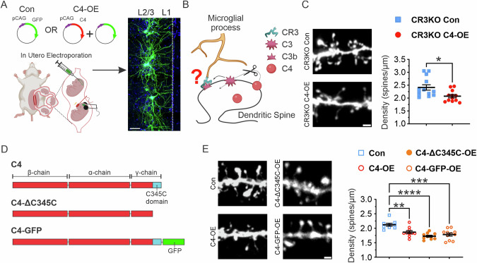
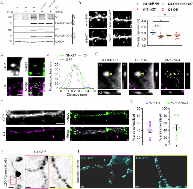
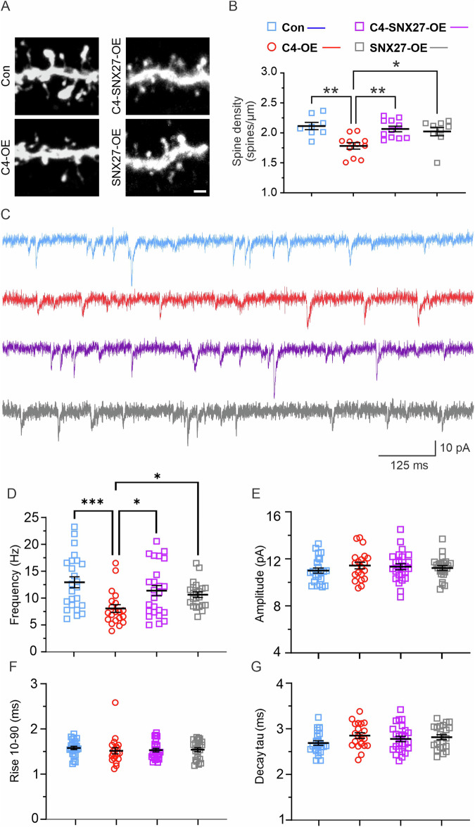
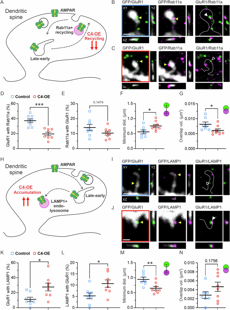
Verification
Claims the paper makes
The claims and statistics of this paper haven’t been checked yet.
Run the check — free, no account needed. It reads the open-access text and checks the sources it cites.
Claims other papers make about this one
Each is checked against this paper’s own text: does it say what the citing paper says it does?
The source backs part of the claim, not all of it — or backs it for a different population, condition or comparison.
In the citing paper
“Despite their independent cargo profiles and molecular machinery, both SNX17-Retriever and SNX27-Retromer pathways are crucial for maintaining excitatory synapse function”
From Dual endomembrane recycling pathways function in parallel to support synapse maintenance and plasticity (2025) · cites this paper as Phadke et al., 2025
Experimental — accuracy not yet measured
Self-check
Percentages, sums and reported differences re-computed from the paper’s own numbers.
The self-check needs the paper’s full text.
Citations
Cites
Microglia Sculpt Postnatal Neural Circuits in an Activity and Complement-Dependent Manner
Schafer, D. P., Lehrman, E. K., Kautzman, A. G., Koyama, R., Mardinly, A. R., Yamasaki, R., Ransohoff, R. M., Greenberg, M. E., Barres, B. A., & Stevens, B. (2012) · Neuron · doi:10.1016/j.neuron.2012.03.026 · Cited by 4,089
Complement and its role in innate and adaptive immune responses
Dunkelberger, J. R., & Song, W.-C. (2010) · Cell Research · doi:10.1038/cr.2009.139 · Cited by 1,932
Architecture of the human interactome defines protein communities and disease networks
Huttlin, E. L., Bruckner, R. J., Paulo, J. A., Cannon, J. R., Ting, L., Baltier, K., Colby, G., Gebreab, F., Gygi, M. P., Parzen, H., et al. (2017) · Nature · doi:10.1038/nature22366 · Cited by 1,437
Rare coding variants in ten genes confer substantial risk for schizophrenia
Singh, T., Poterba, T., Curtis, D., Akil, H., Al Eissa, M., Barchas, J. D., Bass, N., Bigdeli, T. B., Breen, G., Bromet, E. J., et al. (2022) · Nature · doi:10.1038/s41586-022-04556-w · Cited by 983
Dejanovic, B., Huntley, M. A., De Mazière, A., Meilandt, W. J., Wu, T., Srinivasan, K., Jiang, Z., Gandham, V., Friedman, B. A., Ngu, H., et al. (2018) · Neuron · doi:10.1016/j.neuron.2018.10.014 · Cited by 517
Dendritic spines and long-term plasticity
Segal, M. (2005) · Nature Reviews Neuroscience · doi:10.1038/nrn1649 · Cited by 489
Neural activity and the dynamics of central nervous system development
Hua, J. Y., & Smith, S. J. (2004) · Nature Neuroscience · doi:10.1038/nn1218 · Cited by 450
A Top-Down Cortical Circuit for Accurate Sensory Perception
Manita, S., Suzuki, T., Homma, C., Matsumoto, T., Odagawa, M., Yamada, K., Ota, K., Matsubara, C., Inutsuka, A., Sato, M., et al. (2015) · Neuron · doi:10.1016/j.neuron.2015.05.006 · Cited by 365
The cell biology of synapse formation
Südhof, T. C. (2021) · Journal of Cell Biology · doi:10.1083/jcb.202103052 · Cited by 358
Delayed Stabilization of Dendritic Spines in Fragile X Mice
Cruz-Martin, A., Crespo, M., & Portera-Cailliau, C. (2010) · Journal of Neuroscience · doi:10.1523/jneurosci.0577-10.2010 · Cited by 321
Research works printed but not linked: 65
- Schafer DP, Stevens B. Phagocytic glial cells: sculpting synaptic circuits in the developing nervous system. Curr Opin Neurobiol. 2013;23:1034–40. · DOI · Paper page
- Stevens B, Allen NJ, Vazquez LE, Howell GR, Christopherson KS, Nouri N, et al. The classical complement cascade mediates CNS synapse elimination. Cell. 2007;131:1164–78. · DOI · Paper page
- Sekar A, Bialas AR, De Rivera H, Davis A, Hammond TR, Kamitaki N, et al. Schizophrenia risk from complex variation of complement component 4. Nature. 2016;530:177–83. · DOI · Paper page
- Druart M, Nosten-Bertrand M, Poll S, Crux S, Nebeling F, Delhaye C, et al. Elevated expression of complement C4 in the mouse prefrontal cortex causes schizophrenia-associated phenotypes. Mol Psychiatry. 2021:1–13. https://doi.org/10.1038/s41380-021-01081-6. · DOI
- Cao W, Zheng D, Wang G, Zhang J, Ge S, Singh M, et al. Modelling biological age based on plasma peptides in Han Chinese adults. Aging. 2020;12:10676–86. · DOI
- Carpanini SM, Torvell M, Morgan BP. Therapeutic inhibition of the complement system in diseases of the central nervous system. Front Immunol. 2019;10:362. · DOI
- Reus LM, Pasaniuc B, Posthuma D, Boltz T, Ferrari R, Hernandez DG, et al. Gene expression imputation across multiple tissue types provides insight into the genetic architecture of frontotemporal dementia and its clinical subtypes. Biol Psychiatry. 2021;89:825–35. · DOI
- Zinellu A, Mangoni AA. Serum complement C3 and C4 and COVID-19 severity and mortality: a systematic review and meta-analysis with meta-regression. Front Immunol. 2021;12:696085. · DOI
- Comer AL, Jinadasa T, Sriram B, Phadke RA, Kretsge LN, Nguyen TPH, et al. Increased expression of schizophrenia-associated gene C4 leads to hypoconnectivity of prefrontal cortex and reduced social interaction. PLoS Biol. 2020;18:e3000604. · DOI
- Yilmaz M, Yalcin E, Presumey J, Aw E, Ma M, Whelan CW, et al. Overexpression of schizophrenia susceptibility factor human complement C4A promotes excessive synaptic loss and behavioral changes in mice. Nat Neurosci. 2021;24:214–24. · DOI · Paper page
- Stephan AH, Barres BA, Stevens B. The complement system: an unexpected role in synaptic pruning during development and disease. Annu Rev Neurosci. 2012;35:369–89. · DOI · Paper page
- Mao Y-T, Zhu JX, Hanamura K, Iurilli G, Datta SR, Dalva MB. Filopodia conduct target selection in cortical neurons using differences in signal kinetics of a single kinase. Neuron. 2018;98:767–82.e8. · DOI
- Lüscher C, Nicoll RA, Malenka RC, Muller D. Synaptic plasticity and dynamic modulation of the postsynaptic membrane. Nat Neurosci. 2000;3:545–50. · DOI
- Kulik YD, Watson DJ, Cao G, Kuwajima M, Harris KM. Structural plasticity of dendritic secretory compartments during LTP-induced synaptogenesis. eLife. 2019;8:e46356.
- Harris KM. Structure, development, and plasticity of dendritic spines. Curr Opin Neurobiol. 1999;9:343–8. · DOI
- Gasque P, Fontaine M, Morgan BP. Complement expression in human brain. Biosynthesis of terminal pathway components and regulators in human glial cells and cell lines. J Immunol. 1995;154:4726–33. · DOI
- Chen Y, Chu JMT, Chang RCC, Wong GTC. The complement system in the central nervous system: from neurodevelopment to neurodegeneration. Biomolecules. 2022;12:337. · DOI
- Veerhuis R, Janssen I, De Groot CJA, Van Muiswinkel FL, Hack CE, Eikelenboom P. Cytokines associated with amyloid plaques in Alzheimer’s disease brain stimulate human glial and neuronal cell cultures to secrete early complement proteins, but not C1-inhibitor. Exp Neurol. 1999;160:289–99. · DOI
- Veerhuis R, Nielsen HM, Tenner AJ. Complement in the brain. Mol Immunol. 2011;48:1592–603. · DOI
- West EE, Kemper C. Complosome—the intracellular complement system. Nat Rev Nephrol. 2023;19:426–39. · DOI
- Kioussis D, Pachnis V. Immune and nervous systems: more than just a superficial similarity? Immunity. 2009;31:705–10. · DOI · Paper page
- Coxon A, Rieu P, Barkalow FJ, Askari S, Sharpe AH, von Andrian UH, et al. A novel role for the β2 integrin CD11b/CD18 in neutrophil apoptosis: a homeostatic mechanism in inflammation. Immunity. 1996;5:653–66. · DOI · Paper page
- Kidmose RT, Laursen NS, Dobó J, Kjaer TR, Sirotkina S, Yatime L, et al. Structural basis for activation of the complement system by component C4 cleavage. Proc Natl Acad Sci USA. 2012;109:15425–30. · DOI
- Hussain NK, Diering GH, Sole J, Anggono V, Huganir RL. Sorting Nexin 27 regulates basal and activity-dependent trafficking of AMPARs. Proc Natl Acad Sci USA. 2014;111:11840–5. · DOI · Paper page
- Lauffer BEL, Melero C, Temkin P, Lei C, Hong W, Kortemme T, et al. SNX27 mediates PDZ-directed sorting from endosomes to the plasma membrane. J Cell Biol. 2010;190:565–74. · DOI · Paper page
- Loo LS, Tang N, Al-Haddawi M, Stewart Dawe G, Hong W. A role for sorting nexin 27 in AMPA receptor trafficking. Nat Commun. 2014;5:3176. · DOI · Paper page
- Hill TC, Zito K. LTP-induced long-term stabilization of individual nascent dendritic spines. J Neurosci. 2013;33:678–86. · DOI
- Huo Y, Gao Y, Zheng Q, Zhao D, Guo T, Zhang S, et al. Overexpression of human SNX27 enhances learning and memory through modulating synaptic plasticity in mice. Front Cell Dev Biol. 2020;8:595357. · DOI
- Park M, Salgado JM, Ostroff L, Helton TD, Robinson CG, Harris KM, et al. Plasticity-induced growth of dendritic spines by exocytic trafficking from recycling endosomes. Neuron. 2006;52:817–30. · DOI · Paper page
- Nishino H, Saito T, Wei R, Takano T, Tsutsumi K, Taniguchi M, et al. Research articles, cellular/molecular The LMTK1-TBC1D9B-Rab11A cascade regulates dendritic spine formation via endosome trafficking. J Neurosci. 2019;39:9491–502. · DOI
- Takahashi T, Svoboda K, Malinow R. Experience strengthening transmission by driving AMPA receptors into synapses. Science. 2003;299:1585–8. · DOI
- Esteves da Silva M, Adrian M, Schätzle P, Lipka J, Watanabe T, Cho S, et al. Positioning of AMPA receptor-containing endosomes regulates synapse architecture. Cell Rep. 2015;13:933–43. · DOI · Paper page
- Goo MS, Sancho L, Slepak N, Boassa D, Deerinck TJ, Ellisman MH, et al. Activity-dependent trafficking of lysosomes in dendrites and dendritic spines. J Cell Biol. 2017;216:2499–513. · DOI · Paper page
- Cheng X-T, Xie Y-X, Zhou B, Huang N, Farfel-Becker T, Sheng Z-H. Characterization of LAMP1-labeled nondegradative lysosomal and endocytic compartments in neurons. J Cell Biol. 2018;217:3127–39. · DOI · Paper page
- Wang X, Zhao Y, Zhang X, Badie H, Zhou Y, Mu Y, et al. Loss of sorting nexin 27 contributes to excitatory synaptic dysfunction via modulation of glutamate receptor recycling in Down syndrome. Nat Med. 2013;19:473–80. · DOI · Paper page
- Kopec CD, Li B, Wei W, Boehm J, Malinow R. Glutamate receptor exocytosis and spine enlargement during chemically induced long-term potentiation. J Neurosci. 2006;26:2000–9. · DOI · Paper page
- Chandra M, Kendall AK, Jackson LP. Toward understanding the molecular role of SNX27/retromer in human health and disease. Front Cell Dev Biol. 2021;9:642378. · DOI
- Pensalfini A, Kim S, Subbanna S, Bleiwas C, Goulbourne CN, Stavrides PH, et al. Endosomal dysfunction induced by directly overactivating Rab5 recapitulates prodromal and neurodegenerative features of Alzheimer’s disease. Cell Rep. 2020;33:108420. · DOI
- Ross CA, Poirier MA. Protein aggregation and neurodegenerative disease. Nat Med. 2004;10:S10–17. · DOI · Paper page
- Cox TM, Cachón-González MB. The cellular pathology of lysosomal diseases. J Pathol. 2012;226:241–54. · DOI
- Zaharija B, Bradshaw NJ. Aggregation of disrupted in schizophrenia 1 arises from a central region of the protein. Prog Neuropsychopharmacol Biol Psychiatry. 2024;130:110923. · DOI
- Zempel H, Mandelkow E. Lost after translation: missorting of Tau protein and consequences for Alzheimer disease. Trends Neurosci. 2014;37:721–32. · DOI
- Dejanovic B, Wu T, Tsai M-C, Graykowski D, Gandham VD, Rose CM, et al. Complement C1q-dependent excitatory and inhibitory synapse elimination by astrocytes and microglia in Alzheimer’s disease mouse models. Nat Aging. 2022;2:837–50. · DOI · Paper page
- van der Ende EL, Heller C, Sogorb-Esteve A, Swift IJ, McFall D, Peakman G, et al. Elevated CSF and plasma complement proteins in genetic frontotemporal dementia: results from the GENFI study. J Neuroinflammation. 2022;19:217. · DOI
- Xiong C, Liu J, Lin D, Zhang J, Terrando N, Wu A. Complement activation contributes to perioperative neurocognitive disorders in mice. J. Neuroinflammation. 2018;15:254. · DOI
- Bohlson SS, Tenner AJ. Complement in the brain: contributions to neuroprotection, neuronal plasticity, and neuroinflammation. Annu Rev Immunol. 2023;41:431–52. · DOI
- Badimon A, Strasburger HJ, Ayata P, Chen X, Nair A, Ikegami A, et al. Negative feedback control of neuronal activity by microglia. Nature. 2020;586:417–23. · DOI · Paper page
- Nakazawa K, Sapkota K. The origin of NMDA receptor hypofunction in schizophrenia. Pharmacol Ther. 2020;205:107426. · DOI
- Coyle JT, Tsai G, Goff D. Converging evidence of NMDA receptor hypofunction in the pathophysiology of schizophrenia. Ann N Y Acad Sci. 2003;1003:318–27. · DOI
- Shepard N, Baez-Nieto D, Iqbal S, Campbell AJ, Pan JQ, Sheng M, et al. Functional analysis of human GRIN2A mutations associated with schizophrenia and neurodevelopmental disorders reveals distinct pathological mechanism. [Preprint] 2023. Available from: https://doi.org/10.1101/2023.08.02.551645. · DOI
- Hasan A, Nitsche MA, Rein B, Schneider-Axmann T, Guse B, Gruber O, et al. Dysfunctional long-term potentiation-like plasticity in schizophrenia revealed by transcranial direct current stimulation. Behav Brain Res. 2011;224:15–22. · DOI · Paper page
- Frantseva MV, Fitzgerald PB, Chen R, Möller B, Daigle M, Daskalakis ZJ. Evidence for impaired long-term potentiation in schizophrenia and its relationship to motor skill leaning. Cereb Cortex. 2008;18:990–6. · DOI
- Valstad M, Roelfs D, Slapø NB, Timpe CMF, Rai A, Matziorinis AM, et al. Evidence for reduced long-term potentiation-like visual cortical plasticity in schizophrenia and bipolar disorder. Schizophr Bull. 2021;47:1751–60. · DOI
- D’Souza RD, Burkhalter A. A laminar organization for selective cortico-cortical communication. Front Neuroanat. 2017;11:71. · DOI · Paper page
- Cruz-Martín A, El-Danaf RN, Osakada F, Sriram B, Dhande OS, Nguyen PL, et al. A dedicated circuit links direction-selective retinal ganglion cells to the primary visual cortex. Nature. 2014;507:358–61. · DOI
- Barbas H, Pandya DN. Architecture and intrinsic connections of the prefrontal cortex in the rhesus monkey. J Comp Neurol. 1989;286:353–75. · DOI
- Abs E, Poorthuis RB, Apelblat D, Muhammad K, Pardi MB, Enke L, et al. Learning-related plasticity in dendrite-targeting layer 1 interneurons. Neuron. 2018;100:684–99.e6. · DOI
- Huttenlocher PR. Synapse elimination and plasticity in developing human cerebral cortex. Am J Ment Defic. 1984;88:488–96.
- Dalakas MC, Alexopoulos H, Spaeth PJ. Complement in neurological disorders and emerging complement-targeted therapeutics. Nat Rev Neurol. 2020;16:601–17. · DOI
- Matsuda T, Cepko CL. Electroporation and RNA interference in the rodent retina in vivo and in vitro. Proc Natl Acad Sci USA. 2004;101:16–22. · DOI
- Comer AL, Sriram B, Yen WW, Cruz-Martín A. A pipeline using bilateral in utero electroporation to interrogate genetic influences on rodent behavior. J Vis Exp. 2020:e61350. https://doi.org/10.3791/61350. · DOI
- dal Maschio M, Ghezzi D, Bony G, Alabastri A, Deidda G, Brondi M, et al. High-performance and site-directed in utero electroporation by a triple-electrode probe. Nat Commun. 2012;3:960. · DOI
- Beaudoin GMJ, Lee S-H, Singh D, Yuan Y, Ng Y-G, Reichardt LF, et al. Culturing pyramidal neurons from the early postnatal mouse hippocampus and cortex. Nat Protoc. 2012;7:1741–54. · DOI
- Cole RW, Jinadasa T, Brown CM. Measuring and interpreting point spread functions to determine confocal microscope resolution and ensure quality control. Nat Protoc. 2011;6:1929–41. · DOI
- Stevens B, Johnson MB. The complement cascade repurposed in the brain. Nat Rev Immunol. 2021;21:624–5. · DOI
Cited by
Sorted by citations
Chavis, G. D., Rivero-Ríos, P., Uygun, T., Tsukahara, T., Hayami, T., Lee, G. Y., Lee, M., Pace, C., Czesak, F. N., Hilde, K. L., et al. (2025) · Neuroscience · doi:10.1016/j.neuroscience.2025.11.009
Metadata
- Authors
- Phadke, R. A., Brack, A., Fournier, L. A., Kruzich, E., Sha, M., Picard, I., Johnson, C., Stroumbakis, D., Salgado, M., Yeung, C., Escude Velasco, B., Liu, Y. Y., & Cruz-Martín, A.
- Venue
- Molecular Psychiatry
- Published
- 2024-09-03
- Volume
- 30
- Issue
- 2
- Pages
- 796-809
- ISSN
- 1359-4184
- 1476-5578
- PMC
- PMC11746135
- Cited by
- 20
- References
- 13
- Language
- en
- Licence
- Funders
- U.S. Department of Health & Human Services | NIH | National Institute of Mental Health (1R01MH129732-01)
- BU | College of Arts and Sciences, Boston University (Brenton R. Lutz Award)
About these checks
Registry, retraction, Cites and statistics results are computed, not judged, with one exception: a printed reference that no index lists is read once by a model before it counts as a citation error. Claims are extracted by a model and kept only when their quote appears verbatim in the text; each cited source is then checked, with a model’s help, for whether it says what the claim relies on. Those verdicts can be wrong — read the claim’s quote and the cited source before relying on one.
Statistics and arithmetic are recomputed, not judged.
Confidence bands are shown only for fields calibrated on our annotated test set. None is calibrated yet, so no band is shown.
What counts as a claim ›How we measure confidence ›How we check this ›
Are you an author, or do you see a problem with this page? Request a review
How to cite
Cite CoChat for the checks on this page, and the paper for its own findings.
- This page
- CoChat. (2026). Citation check: The schizophrenia risk gene C4 induces pathological synaptic loss by impairing AMPAR trafficking. https://app.cochat.ai/papers/10.1038/s41380-024-02701-7
- The paper
- Phadke, R. A., Brack, A., Fournier, L. A., Kruzich, E., Sha, M., Picard, I., Johnson, C., Stroumbakis, D., Salgado, M., Yeung, C., Escude Velasco, B., Liu, Y. Y., & Cruz-Martín, A. (2025). The schizophrenia risk gene C4 induces pathological synaptic loss by impairing AMPAR trafficking. Molecular Psychiatry. https://doi.org/10.1038/s41380-024-02701-7
