Cited by 103Alzheimer's disease research and treatmentsFerroptosis and cancer prognosisNeurological diseases and metabolism

The DOI matches its Crossref record; 96 of the 96 references it cites were found in a registry or an index.

Selective ferroptosis vulnerability due to familial Alzheimer’s disease presenilin mutations

Open access

Abstract

Mutations in presenilin 1 and 2 ( PS1 and PS2 ) cause autosomal dominant familial Alzheimer’s disease (FAD). Ferroptosis has been implicated as a mechanism of neurodegeneration in AD since neocortical iron burden predicts Alzheimer’s disease (AD) progression. We found that loss of the presenilins dramatically sensitizes multiple cell types to ferroptosis, but not apoptosis. FAD causal mutations of presenilins similarly sensitizes cells to ferroptosis. The presenilins promote the expression of GPX4, the selenoprotein checkpoint enzyme that blocks ferroptosis by quenching the membrane propagation of lethal hydroperoxyl radicals. Presenilin γ-secretase activity cleaves Notch-1 to signal LRP8 expression, which then controls GPX4 expression by regulating the supply of selenium into the cell since LRP8 is the uptake receptor for selenoprotein P. Selenium uptake is thus disrupted by presenilin FAD mutations, suppressing GPX4 expression. Therefore, presenilin mutations may promote neurodegeneration by derepressing ferroptosis, which has implications for disease-modifying therapeutics.

Overview

The claims and statistics of this paper haven’t been checked yet.

Run the check — free, no account needed. It reads the open-access text and checks the sources it cites.

A few free checks a day · papers already verified open instantly · results are public

Integrity signals

Registry

Crossref · 10.1038/s41418-022-01003-1

Cites

96 of 96 references verified0 citation errors · 0 not checkable

Statistics

Figures

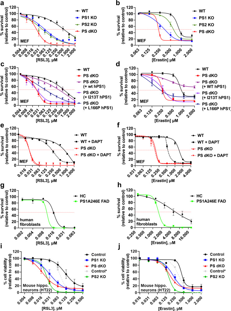
Fig. 1 Presenilin confers resistance to ferroptosis in multiple cell types and is affected by familial Alzheimer’s disease causative mutations. Cell viability analyses performed using the MTT assay to compare ferroptosis resistance in mouse embryonic fibroblasts (MEFs), primary human fibroblasts and HT22 mouse hippocampal neurons to RSL3 (a, c, e, g, i) or erastin (b, d, f, h, j) at the concentrations shown, following 24 h incubation. Note: In i and j, “control” refers to matched negative-control HT22 cells transfected with non-targeting sgRNA contemporaneously with the generation of the PS1 KO and PS dKO HT22 cells, while “control*” is the contemporaneous and matched HT22 control for the subsequently created PS2 KO HT22 cells. Hence, PS1 KO and PS dKO HT22 should be compared to “control”, while PS2 KO should be compared to “control*”. Abbreviations: wildtype (WT), presenilin 1 knockout (PS1 KO), presenilin 2 knockout (PS2 KO), presenilin double-knockout (PS dKO), wild-type human sequence presenilin 1 (wt hPS1), L166P familial Alzheimer’s disease mutant human sequence presenilin 1 (L166P hPS1), I213T familial Alzheimer’s disease mutant human sequence presenilin 1 (I213T hPS1), healthy control human fibroblast (HC), A246E familial Alzheimer’s disease mutant presenilin 1 human fibroblast (PS1A246E FAD), N-[N-(3,5-Difluorophenacetyl)-L-alanyl]-S-phenylglycine t-butyl ester (DAPT, 10 μM, γ-secretase inhibitor). Data points represent mean percentage survival relative to respective controls ± SEM, N = 12–16 from 3 to 4 independent experiments.
Fig. 1 Presenilin confers resistance to ferroptosis in multiple cell types and is affected by familial Alzheimer’s disease causative mutations. Cell viability analyses performed using the MTT assay to compare ferroptosis resistance in mouse embryonic fibroblasts (MEFs), primary human fibroblasts and HT22 mouse hippocampal neurons to RSL3 (a, c, e, g, i) or erastin (b, d, f, h, j) at the concentrations shown, following 24 h incubation. Note: In i and j, “control” refers to matched negative-control HT22 cells transfected with non-targeting sgRNA contemporaneously with the generation of the PS1 KO and PS dKO HT22 cells, while “control*” is the contemporaneous and matched HT22 control for the subsequently created PS2 KO HT22 cells. Hence, PS1 KO and PS dKO HT22 should be compared to “control”, while PS2 KO should be compared to “control*”. Abbreviations: wildtype (WT), presenilin 1 knockout (PS1 KO), presenilin 2 knockout (PS2 KO), presenilin double-knockout (PS dKO), wild-type human sequence presenilin 1 (wt hPS1), L166P familial Alzheimer’s disease mutant human sequence presenilin 1 (L166P hPS1), I213T familial Alzheimer’s disease mutant human sequence presenilin 1 (I213T hPS1), healthy control human fibroblast (HC), A246E familial Alzheimer’s disease mutant presenilin 1 human fibroblast (PS1A246E FAD), N-[N-(3,5-Difluorophenacetyl)-L-alanyl]-S-phenylglycine t-butyl ester (DAPT, 10 μM, γ-secretase inhibitor). Data points represent mean percentage survival relative to respective controls ± SEM, N = 12–16 from 3 to 4 independent experiments.CC BY · PubMed Central
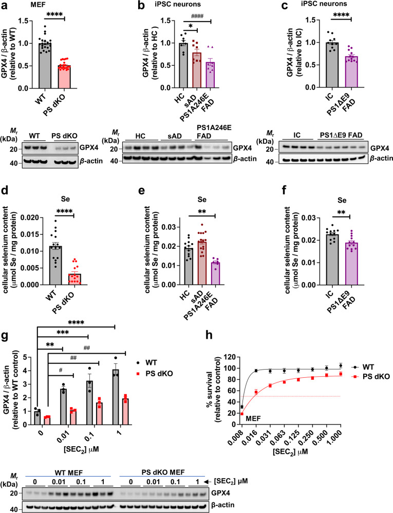
Fig. 2 Presenilin impairment suppresses selenoenzyme glutathione peroxidase-4 (GPX4), the checkpoint for ferroptosis. Densitometric quantification and representative western immunoblots of whole cell lysates probed with specific antibodies raised against GPX4 or loading control β-actin are shown for; wildtype (WT) and presenilin double-knockout (PS dKO) MEFs (a); healthy control (HC), sporadic Alzheimer’s disease (sAD) and FAD presenilin 1 mutant (PS1A246E FAD) human iPSC-derived basal cholinergic neurons (b); CRISPR-corrected isogenic control (IC) and FAD presenilin 1 mutant (PS1ΔE9 FAD) human iPSC-derived neurons (c). Total cellular selenium content in: WT, PS1 KO, PS2 KO and PS dKO MEFs (d); HC, sAD or PS1A246E FAD mutant human iPSC-derived neurons (e); and human CRISPR-corrected isogenic control (IC) and corresponding PS1ΔE9 FAD iPSC-derived neurons (f). Western blot and densitometric quantification of whole cell lysates from WT or PS dKO MEFs probed with antibodies raised against GPX4 or β-actin, following pretreatment for 6 h with 0, 0.01, 0.1 or 1 μM seleno-l-cystine (SEC2) respectively (g). Cell viability of WT and PS dKO MEFs as assessed by MTT assay following co-treatment with 0.5 μM RSL3 and a dose range of SEC2, as shown (h). Western blot data represent mean values relative to their respective mean control value (±SEM) and individual values appear as points on each graph. For the western immunoblot and selenium content in iPSC basal cholinergic neurons each data point represents three pooled wells of a 24 well plate (see Extended Data Table 1 for pluripotent stem cell line details). For the cell viability assay, data are mean values (±SEM), N = 16.
Fig. 2 Presenilin impairment suppresses selenoenzyme glutathione peroxidase-4 (GPX4), the checkpoint for ferroptosis. Densitometric quantification and representative western immunoblots of whole cell lysates probed with specific antibodies raised against GPX4 or loading control β-actin are shown for; wildtype (WT) and presenilin double-knockout (PS dKO) MEFs (a); healthy control (HC), sporadic Alzheimer’s disease (sAD) and FAD presenilin 1 mutant (PS1A246E FAD) human iPSC-derived basal cholinergic neurons (b); CRISPR-corrected isogenic control (IC) and FAD presenilin 1 mutant (PS1ΔE9 FAD) human iPSC-derived neurons (c). Total cellular selenium content in: WT, PS1 KO, PS2 KO and PS dKO MEFs (d); HC, sAD or PS1A246E FAD mutant human iPSC-derived neurons (e); and human CRISPR-corrected isogenic control (IC) and corresponding PS1ΔE9 FAD iPSC-derived neurons (f). Western blot and densitometric quantification of whole cell lysates from WT or PS dKO MEFs probed with antibodies raised against GPX4 or β-actin, following pretreatment for 6 h with 0, 0.01, 0.1 or 1 μM seleno-l-cystine (SEC2) respectively (g). Cell viability of WT and PS dKO MEFs as assessed by MTT assay following co-treatment with 0.5 μM RSL3 and a dose range of SEC2, as shown (h). Western blot data represent mean values relative to their respective mean control value (±SEM) and individual values appear as points on each graph. For the western immunoblot and selenium content in iPSC basal cholinergic neurons each data point represents three pooled wells of a 24 well plate (see Extended Data Table 1 for pluripotent stem cell line details). For the cell viability assay, data are mean values (±SEM), N = 16.CC BY · PubMed Central
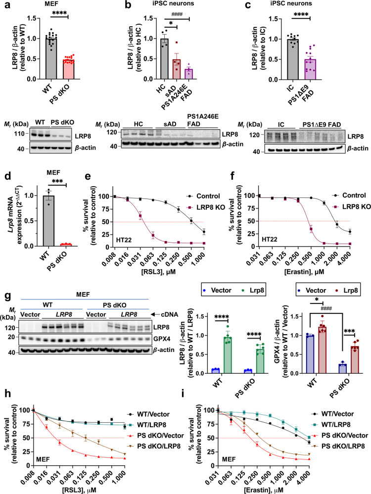
Fig. 3 LRP8 mediates resilience against ferroptosis. Densitometric quantification and representative Western immunoblots of whole cell lysates probed with specific antibodies against LRP8 or loading control β-actin^ are shown for; wildtype (WT) and presenilin double-knockout (PS dKO) MEFs (a); healthy control (HC), sporadic Alzheimer’s disease (sAD) and FAD presenilin 1 mutant (PS1A246E FAD) human iPSC-derived basal cholinergic neurons (b); isogenic control (IC) and FAD presenilin 1 mutant (PS1ΔE9 FAD) human iPSC-derived basal cholinergic neurons (c) ^Note: Fig. 2b/3b and 2c/3c share the same β-actin blots as the displayed blots derive from the same original membranes that were successively probed with Lrp8, Gpx4 and β-actin antibodies (see “Supplemental Material – Original Blots”). Lrp8 mRNA in WT and PS dKO MEFs as measured by RT-qPCR (d). MTT cell viability assay of LRP8 KO and control MEFs following 24 h exposure to a discriminating dose range of RSL3 (e) or erastin (f). Western immunoblot and quantification of LRP8 and GPX4 in WT and PS dKO MEFs that have been transiently transfected with human sequence LRP8 or Vector cDNA (g). MTT cell viability assay of WT and PS dKO MEFs that have been transiently transfected with human sequence LRP8 or Vector cDNA after 24 h exposure to a dose range of RSL3 (h) or erastin (i). Densitometric analyses of proteins relative to β-actin are shown as mean values (±SEM) and individual points represent independent wells of cultured cells except in the iPSC neurons which are pooled from three wells. For the cell viability assay, data are mean values (±SEM), N = 12.
Fig. 3 LRP8 mediates resilience against ferroptosis. Densitometric quantification and representative Western immunoblots of whole cell lysates probed with specific antibodies against LRP8 or loading control β-actin^ are shown for; wildtype (WT) and presenilin double-knockout (PS dKO) MEFs (a); healthy control (HC), sporadic Alzheimer’s disease (sAD) and FAD presenilin 1 mutant (PS1A246E FAD) human iPSC-derived basal cholinergic neurons (b); isogenic control (IC) and FAD presenilin 1 mutant (PS1ΔE9 FAD) human iPSC-derived basal cholinergic neurons (c) ^Note: Fig. 2b/3b and 2c/3c share the same β-actin blots as the displayed blots derive from the same original membranes that were successively probed with Lrp8, Gpx4 and β-actin antibodies (see “Supplemental Material – Original Blots”). Lrp8 mRNA in WT and PS dKO MEFs as measured by RT-qPCR (d). MTT cell viability assay of LRP8 KO and control MEFs following 24 h exposure to a discriminating dose range of RSL3 (e) or erastin (f). Western immunoblot and quantification of LRP8 and GPX4 in WT and PS dKO MEFs that have been transiently transfected with human sequence LRP8 or Vector cDNA (g). MTT cell viability assay of WT and PS dKO MEFs that have been transiently transfected with human sequence LRP8 or Vector cDNA after 24 h exposure to a dose range of RSL3 (h) or erastin (i). Densitometric analyses of proteins relative to β-actin are shown as mean values (±SEM) and individual points represent independent wells of cultured cells except in the iPSC neurons which are pooled from three wells. For the cell viability assay, data are mean values (±SEM), N = 12.CC BY · PubMed Central
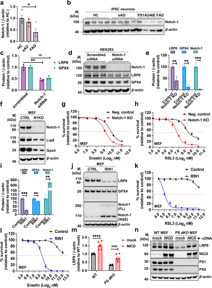
Fig. 4 Notch-1 signaling regulates sensitivity to ferroptosis. Densitometric quantification of Notch-1 in whole cell lysates from human iPSC-derived basal cholinergic neurons (a) and corresponding western blot (b). Densitometric quantification of LRP8 and GPX4 in HEK293 cells following treatment with Notch-1 specific siRNA or scrambled control siRNA (c) with the corresponding western blot shown in d. Densitometric quantification of LRP8, GPX4 and Notch-1 in Notch-1 CRISPR knockout (N1KO) and control (CTRL) MEF cell lysates (e) and a representative western blot shown in f. MTT cell viability assay of Notch-1 KO CRISPR knockout and CRISPR negative control MEFs following a 24 h incubation with a dose range of RSL3 (g) or erastin (h). Western blot of MEF cells incubated with RIN-1 or vehicle (CTRL) and probed with specific antibodies to LRP8, GPX4, Notch-1 and β-actin, as quantified in i with a representative western blot shown in j. MTT cell viability assays of MEF cells pre-incubated with RIN-1 (10 µM) or vehicle control (0.1% DMSO) for 48 h and following 24 h incubation with a dose range of RSL3 (k) or erastin (l). Quantification of LRP8 in WT and PS dKO MEFs 24 h post transfection with an NICD-containing plasmid or vector-only (mock) plasmid (m) and representative western blots demonstrating relative levels of LRP8, NICD, PS1, PS2 and β-actin (n). Densitometric analyses of proteins relative to β-actin are shown as mean values (±SEM) and individual points represent independent wells of cultured cells except in the iPSC neurons which are pooled from three wells. Abbreviations: HC = healthy control, sAD = sporadic Alzheimer’s disease, FAD PS1A246E = familial Alzheimer’s disease mutant presenilin 1 A246E, NICD = Notch intracellular domain, RIN-1 = RBPJ inhibitor-1; 2-(2-Fluorophenoxy)−4-(1-methyl-1H-pyrazol-5-yl) benzamide.
Fig. 4 Notch-1 signaling regulates sensitivity to ferroptosis. Densitometric quantification of Notch-1 in whole cell lysates from human iPSC-derived basal cholinergic neurons (a) and corresponding western blot (b). Densitometric quantification of LRP8 and GPX4 in HEK293 cells following treatment with Notch-1 specific siRNA or scrambled control siRNA (c) with the corresponding western blot shown in d. Densitometric quantification of LRP8, GPX4 and Notch-1 in Notch-1 CRISPR knockout (N1KO) and control (CTRL) MEF cell lysates (e) and a representative western blot shown in f. MTT cell viability assay of Notch-1 KO CRISPR knockout and CRISPR negative control MEFs following a 24 h incubation with a dose range of RSL3 (g) or erastin (h). Western blot of MEF cells incubated with RIN-1 or vehicle (CTRL) and probed with specific antibodies to LRP8, GPX4, Notch-1 and β-actin, as quantified in i with a representative western blot shown in j. MTT cell viability assays of MEF cells pre-incubated with RIN-1 (10 µM) or vehicle control (0.1% DMSO) for 48 h and following 24 h incubation with a dose range of RSL3 (k) or erastin (l). Quantification of LRP8 in WT and PS dKO MEFs 24 h post transfection with an NICD-containing plasmid or vector-only (mock) plasmid (m) and representative western blots demonstrating relative levels of LRP8, NICD, PS1, PS2 and β-actin (n). Densitometric analyses of proteins relative to β-actin are shown as mean values (±SEM) and individual points represent independent wells of cultured cells except in the iPSC neurons which are pooled from three wells. Abbreviations: HC = healthy control, sAD = sporadic Alzheimer’s disease, FAD PS1A246E = familial Alzheimer’s disease mutant presenilin 1 A246E, NICD = Notch intracellular domain, RIN-1 = RBPJ inhibitor-1; 2-(2-Fluorophenoxy)−4-(1-methyl-1H-pyrazol-5-yl) benzamide.CC BY · PubMed Central
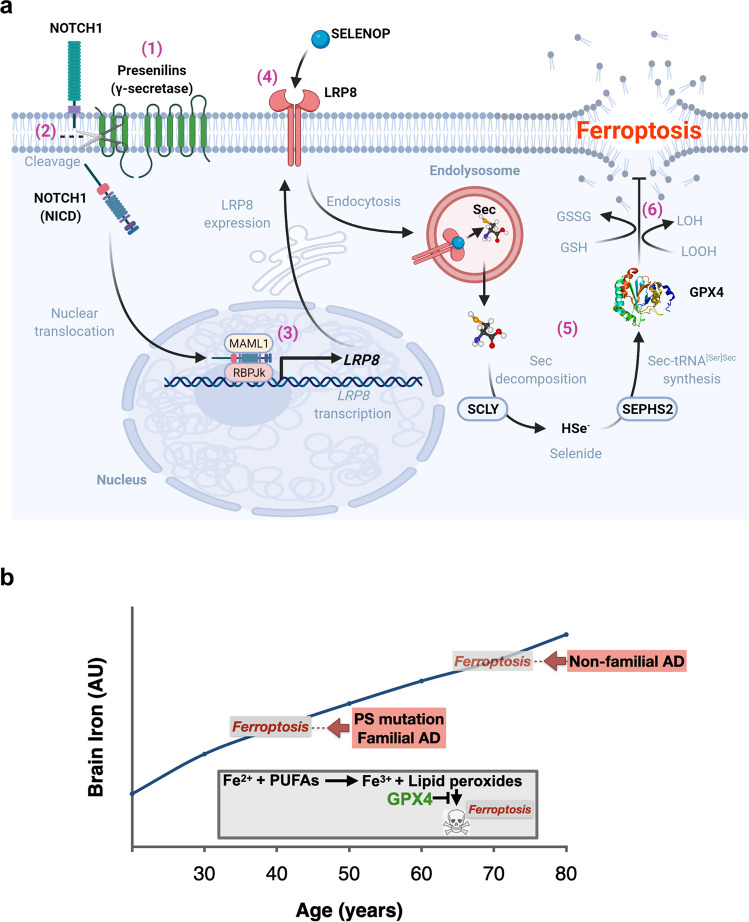
Fig. 5 A hypothesis of how loss of γ-secretase function causes ferroptotic stress. a (1) Presenilins combine with other proteins of the γ-secretase complex to (2) cleave Notch-1, releasing the intracellular domain of Notch-1 (NICD). (3) NICD translocates to the nucleus and binds the co-activator, RBPJκ, to transcribe LRP8. (4) LRP8 is the neuronal receptor for the selenium transport protein, SELENOP. (5) Following internalization, SELENOP is degraded in the lysosome, releasing selenocysteine (Sec). Sec is decomposed to selenide by Sec lyase (SCLY), which is used to synthesize Sec-tRNA[Ser]Sec in a pathway involving selenophosphate synthetase 2 (SEPHS2), ultimately incorporating Sec into de novo translated Glutathione Peroxidase 4 (GPX4). (6) GPX4, which detoxifies lethal iron-catalyzed lipid hydroperoxides (LOOH), is a critical checkpoint in the ferroptosis pathway. Diminished GPX4 activity causes ferroptosis. Created with BioRender.com. b Model for ferroptosis causing AD. Brain iron levels rise needlessly but universally with aging. The burden of brain iron increases the chance of ferroptosis occurring, held in better check by wild type PS than mutant PS. As a result, neuroferroptosis commences at an earlier age in people expressing mutant PS (familial AD) than the typical age of onset of non-familial AD in people expressing wild type PS. The insert shows the canonical ferroptosis events: cytoplasmic Fe2+ reacts with polyunsaturated fatty acids (PUFAs) to generate lipid peroxides, which, beyond a certain threshold, can disrupt the cell membrane. PS mutations suppress the expression of GPX4, which is a major checkpoint enzyme that prevent lipid peroxide accumulation from reaching lethal levels.
Fig. 5 A hypothesis of how loss of γ-secretase function causes ferroptotic stress. a (1) Presenilins combine with other proteins of the γ-secretase complex to (2) cleave Notch-1, releasing the intracellular domain of Notch-1 (NICD). (3) NICD translocates to the nucleus and binds the co-activator, RBPJκ, to transcribe LRP8. (4) LRP8 is the neuronal receptor for the selenium transport protein, SELENOP. (5) Following internalization, SELENOP is degraded in the lysosome, releasing selenocysteine (Sec). Sec is decomposed to selenide by Sec lyase (SCLY), which is used to synthesize Sec-tRNA[Ser]Sec in a pathway involving selenophosphate synthetase 2 (SEPHS2), ultimately incorporating Sec into de novo translated Glutathione Peroxidase 4 (GPX4). (6) GPX4, which detoxifies lethal iron-catalyzed lipid hydroperoxides (LOOH), is a critical checkpoint in the ferroptosis pathway. Diminished GPX4 activity causes ferroptosis. Created with BioRender.com. b Model for ferroptosis causing AD. Brain iron levels rise needlessly but universally with aging. The burden of brain iron increases the chance of ferroptosis occurring, held in better check by wild type PS than mutant PS. As a result, neuroferroptosis commences at an earlier age in people expressing mutant PS (familial AD) than the typical age of onset of non-familial AD in people expressing wild type PS. The insert shows the canonical ferroptosis events: cytoplasmic Fe2+ reacts with polyunsaturated fatty acids (PUFAs) to generate lipid peroxides, which, beyond a certain threshold, can disrupt the cell membrane. PS mutations suppress the expression of GPX4, which is a major checkpoint enzyme that prevent lipid peroxide accumulation from reaching lethal levels.CC BY · PubMed Central

Verification

Claims the paper makes

The claims and statistics of this paper haven’t been checked yet.

Run the check — free, no account needed. It reads the open-access text and checks the sources it cites.

A few free checks a day · papers already verified open instantly · results are public

Claims other papers make about this one

Each is checked against this paper’s own text: does it say what the citing paper says it does?

Self-check

Percentages, sums and reported differences re-computed from the paper’s own numbers.

The self-check needs the paper’s full text.

Citations

Cites

Show all 96 references

Metadata

Authors
Greenough, M. A., Lane, D. J. R., Balez, R., Anastacio, H. T. D., Zeng, Z., Ganio, K., McDevitt, C. A., Acevedo, K., Belaidi, A. A., Koistinaho, J., Ooi, L., Ayton, S., & Bush, A. I.
Affiliations
  • The University of Melbourne (AU)
  • University of Wollongong (AU)
  • Shenzhen KangNing Hospital (CN)
Corresponding author
  • Ayton, Scott
  • Bush, Ashley I.
Venue
Cell Death & Differentiation
Published
2022-04-21
Volume
29
Issue
11
Pages
2123-2136
ISSN
  • 1350-9047 (ISSN-L)
  • 1476-5403
OpenAlex
W4224302612
Cited by
102
References
96
Citations by year
2026: 20 · 2025: 26 · 2024: 28 · 2023: 26 · 2022: 2
FWCI
12.85
Type
journal article
Language
en
Open access
Locations
  • Cell Death and Differentiation (publishedVersion, cc-by)
  • PubMed Central (publishedVersion, cc-by)
  • PubMed (publishedVersion)
  • Research Online (University of Wollongong) (submittedVersion)
  • Adelaide Research & Scholarship (AR&S) (University of Adelaide) (submittedVersion, cc-by)
  • Figshare (submittedVersion, cc-by)
  • Työväentutkimus Vuosikirja (submittedVersion)
  • Minerva Access (University of Melbourne) (submittedVersion, cc-by)
Funders
  • Itä-Suomen Yliopisto
  • University College London
  • KU Leuven
  • National Health and Medical Research Council
Topics
  • Life Sciences › Neuroscience › Cellular and Molecular Neuroscience › Alzheimer's disease research and treatments
  • Life Sciences › Biochemistry, Genetics and Molecular Biology › Cancer Research › Ferroptosis and cancer prognosis
  • Life Sciences › Neuroscience › Neurology › Neurological diseases and metabolism
Keywords
ferroptosis, familial Alzheimer's disease, presenilin 1, presenilin 2, GPX4, Notch1, LRP8, selenoprotein P, selenium uptake, gamma-secretase, neurodegeneration, iron accumulation, apoptosis
MeSH terms
Ferroptosis, Alzheimer Disease, Humans, Mutation, Selenium, Presenilins, Presenilin-1, Amyloid Precursor Protein Secretases

About these checks

Registry, retraction, Cites and statistics results are computed, not judged, with one exception: a printed reference that no index lists is read once by a model before it counts as a citation error. Claims are extracted by a model and kept only when their quote appears verbatim in the text; each cited source is then checked, with a model’s help, for whether it says what the claim relies on. Those verdicts can be wrong — read the claim’s quote and the cited source before relying on one.

Statistics and arithmetic are recomputed, not judged.

Confidence bands are shown only for fields calibrated on our annotated test set. None is calibrated yet, so no band is shown.

What counts as a claim ›How we measure confidence ›How we check this ›

Are you an author, or do you see a problem with this page? Request a review