

The DOI matches its Crossref record; 96 of the 96 references it cites were found in a registry or an index.
Selective ferroptosis vulnerability due to familial Alzheimer’s disease presenilin mutations
Open accessAbstract
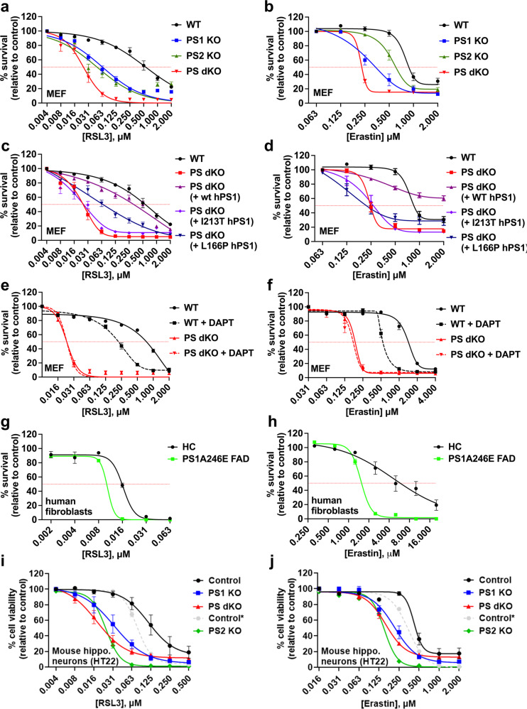
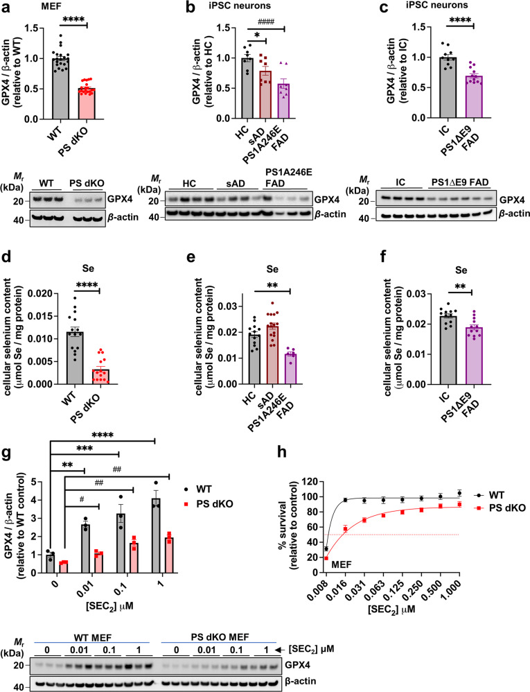
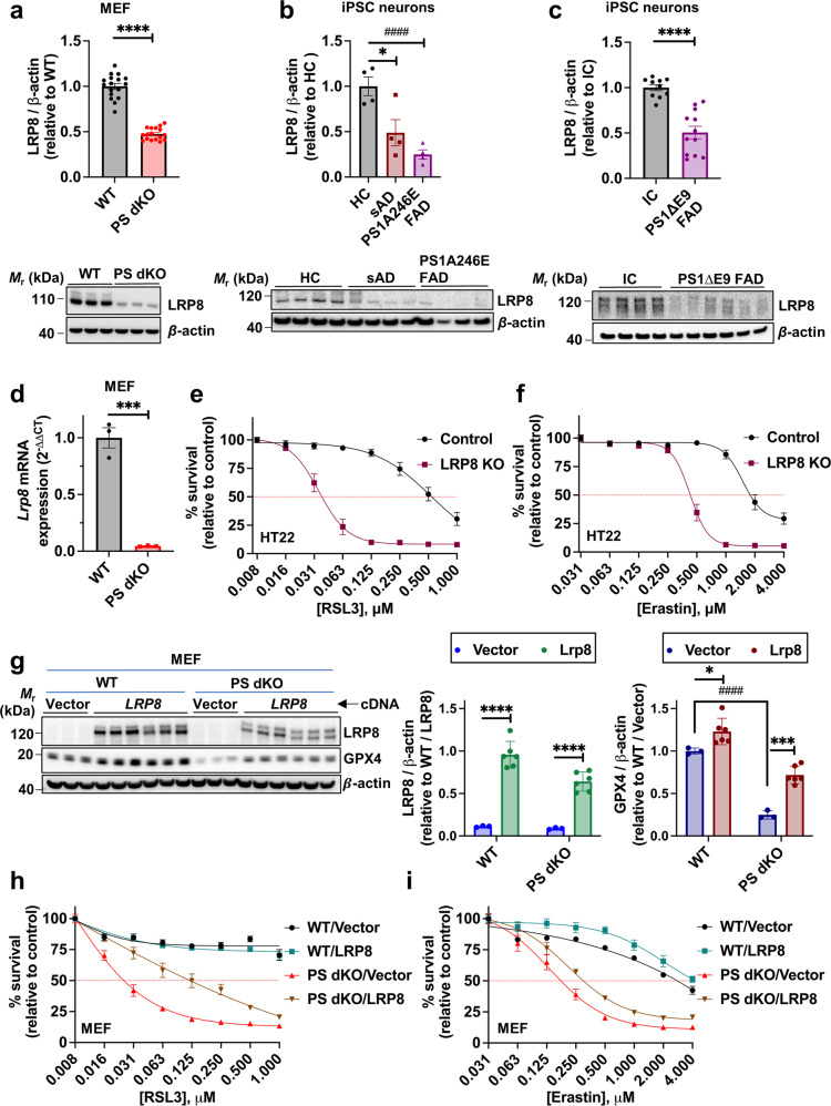
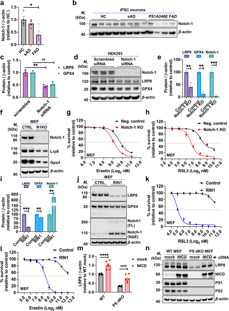
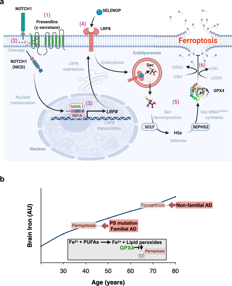
Mutations in presenilin 1 and 2 ( PS1 and PS2 ) cause autosomal dominant familial Alzheimer’s disease (FAD). Ferroptosis has been implicated as a mechanism of neurodegeneration in AD since neocortical iron burden predicts Alzheimer’s disease (AD) progression. We found that loss of the presenilins dramatically sensitizes multiple cell types to ferroptosis, but not apoptosis. FAD causal mutations of presenilins similarly sensitizes cells to ferroptosis. The presenilins promote the expression of GPX4, the selenoprotein checkpoint enzyme that blocks ferroptosis by quenching the membrane propagation of lethal hydroperoxyl radicals. Presenilin γ-secretase activity cleaves Notch-1 to signal LRP8 expression, which then controls GPX4 expression by regulating the supply of selenium into the cell since LRP8 is the uptake receptor for selenoprotein P. Selenium uptake is thus disrupted by presenilin FAD mutations, suppressing GPX4 expression. Therefore, presenilin mutations may promote neurodegeneration by derepressing ferroptosis, which has implications for disease-modifying therapeutics.
Overview
The claims and statistics of this paper haven’t been checked yet.
Run the check — free, no account needed. It reads the open-access text and checks the sources it cites.
Integrity signals
Registry
Cites
Statistics
Figures
Verification
Claims the paper makes
The claims and statistics of this paper haven’t been checked yet.
Run the check — free, no account needed. It reads the open-access text and checks the sources it cites.
Claims other papers make about this one
Each is checked against this paper’s own text: does it say what the citing paper says it does?
The source backs part of the claim, not all of it — or backs it for a different population, condition or comparison.
In the citing paper
“LRP8 has been recently shown to affect ferroptosis by regulating the level of selenoprotein GPX4 in cancer cells (54–56).”
From Secreted Apoe rewires melanoma cell state vulnerability to ferroptosis (2024) · cites this paper as [55]
Experimental — accuracy not yet measured
Self-check
Percentages, sums and reported differences re-computed from the paper’s own numbers.
The self-check needs the paper’s full text.
Citations
Cites
Ferroptosis: An Iron-Dependent Form of Nonapoptotic Cell Death
Dixon, S. J., Lemberg, K. M., Lamprecht, M. R., Skouta, R., Zaitsev, E. M., Gleason, C. E., Patel, D. N., Bauer, A. J., Cantley, A. M., Yang, W. S., et al. (2012) · Cell · doi:10.1016/j.cell.2012.03.042 · Cited by 18,691
Regulation of Ferroptotic Cancer Cell Death by GPX4
Yang, W. S., SriRamaratnam, R., Welsch, M. E., Shimada, K., Skouta, R., Viswanathan, V. S., Cheah, J. H., Clemons, P. A., Shamji, A. F., Clish, C. B., et al. (2014) · Cell · doi:10.1016/j.cell.2013.12.010 · Cited by 7,713
Ferroptosis: A Regulated Cell Death Nexus Linking Metabolism, Redox Biology, and Disease
Stockwell, B. R., Friedmann Angeli, J. P., Bayir, H., Bush, A. I., Conrad, M., Dixon, S. J., Fulda, S., Gascón, S., Hatzios, S. K., Kagan, V. E., et al. (2017) · Cell · doi:10.1016/j.cell.2017.09.021 · Cited by 7,695
Self-citationThe amyloid hypothesis of Alzheimer's disease at 25 years
Selkoe, D. J., & Hardy, J. (2016) · EMBO Molecular Medicine · doi:10.15252/emmm.201606210 · Cited by 6,164
Inactivation of the ferroptosis regulator Gpx4 triggers acute renal failure in mice
Friedmann Angeli, J. P., Schneider, M., Proneth, B., Tyurina, Y. Y., Tyurin, V. A., Hammond, V. J., Herbach, N., Aichler, M., Walch, A., Eggenhofer, E., et al. (2014) · Nature Cell Biology · doi:10.1038/ncb3064 · Cited by 3,732
A presenilin-1-dependent γ-secretase-like protease mediates release of Notch intracellular domain
De Strooper, B., Annaert, W., Cupers, P., Saftig, P., Craessaerts, K., Mumm, J. S., Schroeter, E. H., Schrijvers, V., Wolfe, M. S., Ray, W. J., et al. (1999) · Nature · doi:10.1038/19083 · Cited by 2,100
Dependency of a therapy-resistant state of cancer cells on a lipid peroxidase pathway
Viswanathan, V. S., Ryan, M. J., Dhruv, H. D., Gill, S., Eichhoff, O. M., Seashore-Ludlow, B., Kaffenberger, S. D., Eaton, J. K., Shimada, K., Aguirre, A. J., et al. (2017) · Nature · doi:10.1038/nature23007 · Cited by 2,011
The role of iron in brain ageing and neurodegenerative disorders
Ward, R. J., Zucca, F. A., Duyn, J. H., Crichton, R. R., & Zecca, L. (2014) · The Lancet Neurology · doi:10.1016/s1474-4422(14)70117-6 · Cited by 1,819
Selenium Utilization by GPX4 Is Required to Prevent Hydroperoxide-Induced Ferroptosis
Ingold, I., Berndt, C., Schmitt, S., Doll, S. G., Poschmann, G., Buday, K., Roveri, A., Peng, X., Freitas, F. P., Seibt, T., et al. (2017) · Cell · doi:10.1016/j.cell.2017.11.048 · Cited by 1,712
Ferroptosis: Mechanisms and links with diseases
Yan, H.-F., Zou, T., Tuo, Q.-Z., Xu, S., Li, H., Belaidi, A. A., & Lei, P. (2021) · Signal Transduction and Targeted Therapy · doi:10.1038/s41392-020-00428-9 · Cited by 1,471
Self-citationAnalysis of Cell Viability by the MTT AssayPreprint
Kumar, P., Nagarajan, A. G., & Uchil, P. D. (2018) · Cold Spring Harbor Protocols · doi:10.1101/pdb.prot095505 · Cited by 1,286
A Phase 3 Trial of Semagacestat for Treatment of Alzheimer's Disease
Doody, R. S., Raman, R., Farlow, M., Iwatsubo, T., Vellas, B., Joffe, S., Kieburtz, K., He, F., Sun, X., Thomas, R. G., et al. (2013) · New England Journal of Medicine · doi:10.1056/nejmoa1210951 · Cited by 1,224
Zilka, O., Shah, R., Li, B., Angeli, J. P. F., Grießer, M., Conrad, M., & Pratt, D. A. (2017) · ACS Central Science · doi:10.1021/acscentsci.7b00028 · Cited by 1,084
The chemical basis of ferroptosis
Conrad, M., & Pratt, D. A. (2019) · Nature Chemical Biology · doi:10.1038/s41589-019-0408-1 · Cited by 1,047
Global survey of cell death mechanisms reveals metabolic regulation of ferroptosis
Shimada, K., Skouta, R., Kaplan, A., Yang, W. S., Hayano, M., Dixon, S. J., Brown, L. M., Valenzuela, C. A., Wolpaw, A. J., & Stockwell, B. R. (2016) · Nature Chemical Biology · doi:10.1038/nchembio.2079 · Cited by 1,017
Skeletal and CNS Defects in Presenilin-1-Deficient Mice
Shen, J., Bronson, R. T., Chen, D. F., Xia, W., Selkoe, D. J., & Tonegawa, S. (1997) · Cell · doi:10.1016/s0092-8674(00)80244-5 · Cited by 959
Saura, C. A., Choi, S.-Y., Beglopoulos, V., Malkani, S., Zhang, D., Rao, B. S. S., Chattarji, S., Kelleher, R. J., Kandel, E. R., Duff, K., et al. (2004) · Neuron · doi:10.1016/s0896-6273(04)00182-5 · Cited by 757
Loss of ferroportin induces memory impairment by promoting ferroptosis in Alzheimer’s disease
Bao, W.-D., Pang, P., Zhou, X.-T., Hu, F., Xiong, W., Chen, K., Wang, J., Wang, F., Xie, D., Ya⁃zhuo, H. U., et al. (2021) · Cell Death and Differentiation · doi:10.1038/s41418-020-00685-9 · Cited by 682
The Notch signaling pathway: Transcriptional regulation at Notch target genes
Borggrefe, T., & Oswald, F. (2009) · Cellular and Molecular Life Sciences · doi:10.1007/s00018-009-8668-7 · Cited by 661
Chaperone-mediated autophagy is involved in the execution of ferroptosis
Wu, Z., Geng, Y., Lu, X., Shi, Y., Wu, G., Zhang, M., Shan, B., Pan, H., & Yuan, J. (2019) · Proceedings of the National Academy of Sciences · doi:10.1073/pnas.1819728116 · Cited by 582
HSPA5 Regulates Ferroptotic Cell Death in Cancer Cells
Zhu, S., Zhang, Q., Sun, X., Zeh, H. J., Lotze, M. T., Kang, R., & Tang, D. (2017) · Cancer Research · doi:10.1158/0008-5472.can-16-1979 · Cited by 576
The mechanism of γ‐Secretase dysfunction in familial Alzheimer disease
Chávez‐Gutiérrez, L., Bammens, L., Benilova, I., Vandersteen, A., Benurwar, M., Borgers, M., Lismont, S., Zhou, L., Van Cleynenbreugel, S., Esselmann, H., et al. (2012) · The EMBO Journal · doi:10.1038/emboj.2012.79 · Cited by 495
Ferroptosis occurs through an osmotic mechanism and propagates independently of cell rupture
Riegman, M., Sagie, L., Galed, C., Levin, T., Steinberg, N. J., Dixon, S. J., Wiesner, U. B., Bradbury, M. S., Niethammer, P., Zaritsky, A., et al. (2020) · Nature Cell Biology · doi:10.1038/s41556-020-0565-1 · Cited by 483
Mice lacking both presenilin genes exhibitearlyembryonic patterningdefectsPreprint
Donoviel, D. B., Hadjantonakis, A., Ikeda, M., Zheng, H., George‐Hyslop, P. S., & Bernstein, A. (1999) · Genes & Development · doi:10.1101/gad.13.21.2801 · Cited by 464
Presenilin clinical mutations can affect γ‐secretase activity by different mechanisms
Bentahir, M., Nyabi, O., Verhamme, J., Tolia, A., Horré, K., Wiltfang, J., Esselmann, H., & Strooper, B. D. (2006) · Journal of Neurochemistry · doi:10.1111/j.1471-4159.2005.03578.x · Cited by 427
M ECHANISM AND R EGULATION OF S ELENOPROTEIN S YNTHESIS
Driscoll, D. M., & Copeland, P. R. (2003) · Annual Review of Nutrition · doi:10.1146/annurev.nutr.23.011702.073318 · Cited by 409
The role of selenium peroxidases in the protection against oxidative damage of membranes
Ursini, F., & Bindoli, A. (1987) · Chemistry and Physics of Lipids · doi:10.1016/0009-3084(87)90053-3 · Cited by 391
Sun, L., Zhou, R., Yang, G., & Shi, Y. (2017) · Proceedings of the National Academy of Sciences · doi:10.1073/pnas.1618657114 · Cited by 382
Brain iron is associated with accelerated cognitive decline in people with Alzheimer pathology
Ayton, S., Wang, Y., Diouf, I., Schneider, J. A., Brockman, J. D., Morris, M. C., & Bush, A. I. (2019) · Molecular Psychiatry · doi:10.1038/s41380-019-0375-7 · Cited by 372
Self-citationSong, W., Nadeau, P., Yuan, M., Yang, X., Shen, J., & Yankner, B. A. (1999) · Proceedings of the National Academy of Sciences · doi:10.1073/pnas.96.12.6959 · Cited by 362
Ayton, S., Faux, N. G., Bush, A. I., Initiative, A. D. N., Weiner, M. W., Aisen, P. S. S., Petersen, R. C., Jack, C. R., Jagust, W., Trojanowki, J. Q., et al. (2015) · Nature Communications · doi:10.1038/ncomms7760 · Cited by 359
Self-citationBrain Glutathione Levels – A Novel Biomarker for Mild Cognitive Impairment and Alzheimer’s Disease
Mandal, P. K., Saharan, S., Tripathi, M., & Murari, G. (2015) · Biological Psychiatry · doi:10.1016/j.biopsych.2015.04.005 · Cited by 344
PSEN1 Mutant iPSC-Derived Model Reveals Severe Astrocyte Pathology in Alzheimer's Disease
Oksanen, M., Petersen, A. J., Naumenko, N., Puttonen, K., Lehtonen, Š., Gubert Olivé, M., Shakirzyanova, A., Leskelä, S., Sarajärvi, T., Viitanen, M., et al. (2017) · Stem Cell Reports · doi:10.1016/j.stemcr.2017.10.016 · Cited by 332
Kwart, D., Gregg, A., Scheckel, C., Murphy, E. A., Paquet, D., Duffield, M. D., Fak, J. J., Olsen, O., Darnell, R. B., & Tessier‐Lavigne, M. (2019) · Neuron · doi:10.1016/j.neuron.2019.07.010 · Cited by 327
Steiner, H., Duff, K., Capell, A., Romig, H., Grim, M. G., Lincoln, S., Hardy, J., Yu, X., Picciano, M., Fechteler, K., et al. (1999) · Journal of Biological Chemistry · doi:10.1074/jbc.274.40.28669 · Cited by 307
Ashraf, A., Jeandriens, J., Parkes, H. G., & So, P. (2020) · Redox Biology · doi:10.1016/j.redox.2020.101494 · Cited by 306
Cerebral quantitative susceptibility mapping predicts amyloid-β-related cognitive decline
Ayton, S., Fazlollahi, A., Bourgeat, P., Raniga, P., Ng, A., Lim, Y. Y., Diouf, I., Farquharson, S., Fripp, J., Ames, D. J., et al. (2017) · Brain · doi:10.1093/brain/awx137 · Cited by 292
Self-citationIron and Alzheimer’s Disease: An Update on Emerging Mechanisms
Lane, D. J. R., Ayton, S., & Bush, A. I. (2018) · Journal of Alzheimer s Disease · doi:10.3233/jad-179944 · Cited by 287
Self-citationBalez, R., Steiner, N., Engel, M., Muñoz, S. S., Lum, J. S., Wu, Y., Wang, D., Vallotton, P., Sachdev, P. S., O’Connor, M. D., et al. (2016) · Scientific Reports · doi:10.1038/srep31450 · Cited by 280
Self-citationMoehlmann, T., Winkler, E., Xia, X., Edbauer, D., Murrell, J. R., Capell, A., Kaether, C., Zheng, H., Ghetti, B. F., Haass, C., et al. (2002) · Proceedings of the National Academy of Sciences · doi:10.1073/pnas.112686799 · Cited by 264
Gpx4 ablation in adult mice results in a lethal phenotype accompanied by neuronal loss in brain
Yoo, S.-E., Chen, L., Na, R., Liu, Y., Rios, C. I., Remmen, H. V., Richardson, A. G., & Ran, Q. (2012) · Free Radical Biology and Medicine · doi:10.1016/j.freeradbiomed.2012.02.043 · Cited by 256
Iron Metabolism, Ferroptosis, and the Links With Alzheimer’s Disease
Yan, N., & Zhang, J. (2020) · Frontiers in Neuroscience · doi:10.3389/fnins.2019.01443 · Cited by 256
Alzheimer’s-Causing Mutations Shift Aβ Length by Destabilizing γ-Secretase-Aβn Interactions
Szaruga, M., Munteanu, B., Lismont, S., Veugelen, S., Horré, K., Mercken, M., Saido, T. C., Ryan, N. S., De Vos, T., Savvides, S. N., et al. (2017) · Cell · doi:10.1016/j.cell.2017.07.004 · Cited by 248 · cited 2×
“While the impact of PS mutations on increasing the ratio of Αβ42/40 has been highlighted as a signature of neurodegenerative toxicity [3], a systematic analysis of 138 familial PS mutations in vitro [4] revealed that the 42/40 ratio was not consistently elevated and, more tellingly, 90% of these mutations reduced acti…”
BackgroundResultWang, H., Zou, J., Zhao, B., Johannsen, E., Ashworth, T. D., Wong, H., Pear, W. S., Schug, J., Blacklow, S. C., Arnett, K. L., et al. (2011) · Proceedings of the National Academy of Sciences · doi:10.1073/pnas.1109023108 · Cited by 236
Presenilin-1 Knockin Mice Reveal Loss-of-Function Mechanism for Familial Alzheimer’s Disease
Xia, D., Watanabe, H., Wu, B., Lee, S. H., Li, Y., Tsvetkov, E., Bolshakov, V. Y., Shen, J., & Kelleher, R. J. (2015) · Neuron · doi:10.1016/j.neuron.2015.02.010 · Cited by 234
Venkateshappa, C., Harish, G., Mahadevan, A., Srinivas Bharath, M. M., & Shankar, S. K. (2012) · Neurochemical Research · doi:10.1007/s11064-012-0755-8 · Cited by 232
Liu, Y., Wang, Y., Liu, J., Kang, R., & Tang, D. (2020) · Cancer Gene Therapy · doi:10.1038/s41417-020-0182-y · Cited by 227
Target Selectivity of Vertebrate Notch Proteins
Ong, C., Cheng, H., Chang, L., Ohtsuka, T., Kageyama, R., Stormo, G. D., & Kopan, R. (2005) · Journal of Biological Chemistry · doi:10.1074/jbc.m506108200 · Cited by 218
Lichtenthaler, S. F., Haass, C., & Steiner, H. (2011) · Journal of Neurochemistry · doi:10.1111/j.1471-4159.2011.07248.x · Cited by 205
Baumeister, R., Leimer, U., Zweckbronner, I., Jakubek, C., Grünberg, J., & Haass, C. (1997) · Genes and Function · doi:10.1046/j.1365-4624.1997.00012.x · Cited by 203
The substrate repertoire of γ-secretase/presenilin
Güner, G., & Lichtenthaler, S. F. (2020) · Seminars in Cell and Developmental Biology · doi:10.1016/j.semcdb.2020.05.019 · Cited by 202
Selenium and GPX4, a vital symbiosis
Angeli, J. P. F., & Conrad, M. (2018) · Free Radical Biology and Medicine · doi:10.1016/j.freeradbiomed.2018.03.001 · Cited by 197
Walker, E. S., Martínez, M., Brunkan, A. L., & Goate, A. (2004) · Journal of Neurochemistry · doi:10.1111/j.1471-4159.2004.02858.x · Cited by 184
Woodruff, G., Young, J. E., Martinez, F. J., Buen, F., Gore, A., Kinaga, J., Li, Z., Yuan, S. H., Zhang, K., & Goldstein, L. S. B. (2013) · Cell Reports · doi:10.1016/j.celrep.2013.10.018 · Cited by 177
Zhu, Z., Li, Q. V., Lee, K., Rosen, B. P., Gonzãlez, F., Soh, C.-L., & Huangfu, D. (2016) · Cell stem cell · doi:10.1016/j.stem.2016.03.015 · Cited by 172
Ayton, S., Portbury, S. D., Kalinowski, P., Agarwal, P., Diouf, I., Schneider, J. A., Morris, M. C., & Bush, A. I. (2021) · Alzheimer s & Dementia · doi:10.1002/alz.12282 · Cited by 168
Self-citationBermejo‐Bescós, P., Martín‐Aragón, S., Benedı́, J., Susín, C., Felici, E., Gil, P., Ribera, J. M., & Villar, Á. (2008) · Free Radical Research · doi:10.1080/10715760701861373 · Cited by 167
Ferroptosis and NRF2: an emerging battlefield in the neurodegeneration of Alzheimer's disease
Lane, D. J. R., Metselaar, B., Greenough, M., Bush, A. I., & Ayton, S. (2021) · Essays in Biochemistry · doi:10.1042/ebc20210017 · Cited by 160
Self-citationQualitative changes in human γ-secretase underlie familial Alzheimer’s disease
Szaruga, M., Veugelen, S., Benurwar, M., Lismont, S., Sepulveda-Falla, D., Lleo, A., Ryan, N. S., Lashley, T., Fox, N. C., Murayama, S., et al. (2015) · Journal of Experimental Medicine · doi:10.1084/jem.20150892 · Cited by 155
Beglopoulos, V., Sun, X., Saura, C. A., Lemere, C. A., Kim, R. D., & Shen, J. (2004) · Journal of Biological Chemistry · doi:10.1074/jbc.m409544200 · Cited by 154
Berezovska, O., Xia, M., & Hyman, B. T. (1998) · Journal of Neuropathology & Experimental Neurology · doi:10.1097/00005072-199808000-00003 · Cited by 150
Burk, R. F., Hill, K. E., Motley, A. K., Winfrey, V. P., Kurokawa, S., Mitchell, S. L., & Zhang, W. (2014) · The FASEB Journal · doi:10.1096/fj.14-252874 · Cited by 149
Derry, P. J., Hegde, M. L., Jackson, G. R., Kayed, R., Tour, J. M., Tsai, A.-L., & Kent, T. A. (2019) · Progress in Neurobiology · doi:10.1016/j.pneurobio.2019.101716 · Cited by 146
Alzheimer’s disease and Notch signaling
Woo, H., Park, J. S., Gwon, A., Arumugam, T. V., & Jo, D. (2009) · Biochemical and Biophysical Research Communications · doi:10.1016/j.bbrc.2009.10.093 · Cited by 143
Zhang, Z.-H., Wu, Q., Zheng, R., Chen, C., Chen, Y., Liu, Q., Hoffmann, P. R., Ni, J., & Song, G.-L. (2017) · Journal of Neuroscience · doi:10.1523/jneurosci.3229-16.2017 · Cited by 130
Quintero-Monzon, O., Martin, M. M., Fernandez, M. A., Cappello, C. A., Krzysiak, A. J., Osenkowski, P., & Wolfe, M. S. (2011) · Biochemistry · doi:10.1021/bi2007146 · Cited by 118
Treating Alzheimer's disease by targeting iron
Nikseresht, S., Bush, A. I., & Ayton, S. (2019) · British Journal of Pharmacology · doi:10.1111/bph.14567 · Cited by 98
Self-citationMaynard, C. J., Cappai, R., Volitakis, I., Cherny, R. A., Masters, C. L., Li, Q., & Bush, A. I. (2006) · Journal of Inorganic Biochemistry · doi:10.1016/j.jinorgbio.2006.02.010 · Cited by 97
Self-citationTabuchi, K., Chen, G., Südhof, T. C., & Shen, J. (2009) · Journal of Neuroscience · doi:10.1523/jneurosci.1320-09.2009 · Cited by 96
Wines-Samuelson, M., Schulte, E. C., Smith, M. J., Aoki, C. J., Liu, X., Kelleher, R. J., & Shen, J. (2010) · PLoS ONE · doi:10.1371/journal.pone.0010195 · Cited by 93
Amtul, Z., Lewis, P. A., Piper, S. C., Crook, R., Baker, M. C., Findlay, K. A., Singleton, A., Hogg, M. C., Younkin, L. H., Younkin, S. G., et al. (2002) · Neurobiology of Disease · doi:10.1006/nbdi.2001.0473 · Cited by 89
Heilig, E. A., Xia, W., Shen, J., & Kelleher, R. J. (2010) · Journal of Biological Chemistry · doi:10.1074/jbc.m110.116962 · Cited by 88
Evidence that iron accelerates Alzheimer’s pathology: a CSF biomarker study
Ayton, S., Diouf, I., & Bush, A. I. (2017) · Journal of Neurology Neurosurgery & Psychiatry · doi:10.1136/jnnp-2017-316551 · Cited by 86
Self-citationSong, G.-L., Zhang, Z.-H., Wen, L., Chen, C., Shi, Q., Zhang, Y., Ni, J., & Liu, Q. (2014) · Journal of Alzheimer s Disease · doi:10.3233/jad-131805 · Cited by 81
Ayton, S., Faux, N. G., & Bush, A. I. (2016) · JAMA Neurology · doi:10.1001/jamaneurol.2016.4406 · Cited by 80
Self-citationCharacterization of AD-like phenotype in aged APPSwe/PS1dE9 mice
Huang, H., Nie, S., Cao, M., Marshall, C., Gao, J., Xiao, N., Hu, G., & Xiao, M. (2016) · AGE · doi:10.1007/s11357-016-9929-7 · Cited by 79
Spotlight on Ferroptosis: Iron-Dependent Cell Death in Alzheimer’s Disease
Ashraf, A., & So, P. (2020) · Frontiers in Aging Neuroscience · doi:10.3389/fnagi.2020.00196 · Cited by 76
Brai, E., Marathe, S. V., Astori, S., Fredj, N. B., Perry, E., Lamy, C. M., Scotti, A. L., & Alberi, L. A. (2015) · Frontiers in Cellular Neuroscience · doi:10.3389/fncel.2015.00447 · Cited by 74
Diouf, I., Fazlollahi, A., Bush, A. I., & Ayton, S. (2018) · Neurobiology of Disease · doi:10.1016/j.nbd.2018.12.010 · Cited by 72
Self-citationHeilig, E. A., Gutti, U., Tai, T., Shen, J., & Kelleher, R. J. (2013) · Journal of Neuroscience · doi:10.1523/jneurosci.0954-13.2013 · Cited by 70
Byrns, C. N., Pitts, M. W., Gilman, C. A., Hashimoto, A. C., & Berry, M. J. (2014) · Journal of Biological Chemistry · doi:10.1074/jbc.m113.540682 · Cited by 64
Notch1 hallmarks fibrillary depositions in sporadic Alzheimer’s disease
Brai, E., Raio, N. A., & Alberi, L. A. (2016) · Acta Neuropathologica Communications · doi:10.1186/s40478-016-0327-2 · Cited by 64
Disruption of NOTCH signaling by a small molecule inhibitor of the transcription factor RBPJ
Hurtado, C., Safarova, A. G., Smith, M. P., Chung, R., Bruyneel, A. A. N., Gomez‐Galeno, J., Oswald, F., Larson, C. J., Cashman, J. R., Ruiz‐Lozano, P., et al. (2019) · Scientific Reports · doi:10.1038/s41598-019-46948-5 · Cited by 58
der Jeugd, A. V., Parra‐Damas, A., Baeta-Corral, R., Soto-Faguás, C. M., Ahmed, T., LaFerla, F. M., Giménez‐Llort, L., D’Hooge, R., & Saura, C. A. (2018) · Scientific Reports · doi:10.1038/s41598-018-24741-0 · Cited by 50
Muñoz, S. S., Engel, M., Balez, R., Do‐Ha, D., Cabral-da-Silva, M. C., Hernández, D., Berg, T., Fifita, J. A., Grima, N., Yang, S., et al. (2020) · Cells · doi:10.3390/cells9092018 · Cited by 49
Self-citationPartial Loss of Presenilin Impairs Age-Dependent Neuronal Survival in the Cerebral Cortex
Watanabe, H., Iqbal, M., Zheng, J., Wines-Samuelson, M., & Shen, J. (2014) · Journal of Neuroscience · doi:10.1523/jneurosci.3261-14.2014 · Cited by 48
The Role of Reelin Signaling in Alzheimer’s Disease
Yu, N., Tan, M., Yu, J., Xie, A., & Tan, L. (2015) · Molecular Neurobiology · doi:10.1007/s12035-015-9459-9 · Cited by 47
Modeling Clinically Heterogeneous Presenilin Mutations with Transgenic Drosophila
Seidner, G. A., Ye, Y., Faraday, M. M., Alvord, W. G., & Fortini, M. (2006) · Current Biology · doi:10.1016/j.cub.2006.04.004 · Cited by 45
Ayton, S., & Bush, A. I. (2021) · Ageing Research Reviews · doi:10.1016/j.arr.2020.101212 · Cited by 45
Self-citationLoss of Aβ43 Production Caused by Presenilin-1 Mutations in the Knockin Mouse Brain
Xia, D., Kelleher, R. J., & Shen, J. (2016) · Neuron · doi:10.1016/j.neuron.2016.03.009 · Cited by 34
Molecular and Functional Characterization of Neurogenin-2 Induced Human Sensory Neurons
Hulme, A. E., McArthur, J. R., Maksour, S., Miellet, S., Ooi, L., Adams, D. J., Finol‐Urdaneta, R. K., & Dottori, M. (2020) · Frontiers in Cellular Neuroscience · doi:10.3389/fncel.2020.600895 · Cited by 33
Self-citationPresenilin 1 mutations influence processing and trafficking of the ApoE receptor apoER2
Wang, W., Moerman, A., Slaton, A., & Barger, S. W. (2016) · Neurobiology of Aging · doi:10.1016/j.neurobiolaging.2016.10.005 · Cited by 23
SeP, ApoER2 and megalin as necessary factors to maintain Se homeostasis in mammals
Król, M. B., Gromadzińska, J., & Wąsowicz, W. (2012) · Journal of Trace Elements in Medicine and Biology · doi:10.1016/j.jtemb.2012.03.003 · Cited by 17
Muñoz, S. S., Balez, R., Cabral-da-Silva, M. C., Berg, T., Engel, M., Bax, M., Do‐Ha, D., Stevens, C. H., Greenough, M. A., Bush, A. I., et al. (2018) · Stem Cell Research · doi:10.1016/j.scr.2018.08.006 · Cited by 17
Self-citationEngel, M., Balez, R., Muñoz, S. S., Cabral-da-Silva, M. C., Stevens, C. H., Bax, M., Do‐Ha, D., Sidhu, K., Sachdev, P. S., & Ooi, L. (2018) · Stem Cell Research · doi:10.1016/j.scr.2018.09.014 · Cited by 13
Self-citationSong, H., Boo, J. H., Kim, K. H., Kim, C., Kim, Y.-E., Ahn, J.-H., Jeon, G. S., Ryu, H., Kang, D. E., & Mook‐Jung, I. (2013) · Cell Death and Differentiation · doi:10.1038/cdd.2012.162 · Cited by 12
Cited by
Sorted by citations
The Cell Biology of Ferroptosis
Dixon, S. J., & Olzmann, J. A. (2024) · Nature Reviews Molecular Cell Biology · doi:10.1038/s41580-024-00703-5 · Cited by 1,596
Iron Homeostasis and Ferroptosis in Human Diseases: Mechanisms and Therapeutic Prospects
Ru, Q., Li, Y., Chen, L., Wu, Y., Min, J., & Wang, F. (2024) · Signal Transduction and Targeted Therapy · doi:10.1038/s41392-024-01969-z · Cited by 724
Zhou, N., Yuan, X., Du, Q., Zhang, Z., Shi, X., Bao, J., Ning, Y., & Peng, L. (2022) · Nucleic Acids Research · doi:10.1093/nar/gkac935 · Cited by 514
GPX4, ferroptosis, and diseases
Zhang, W., Liu, Y., Yan, L., Zhu, C., & Zou, Z. (2024) · Biomedicine & Pharmacotherapy · doi:10.1016/j.biopha.2024.116512 · Cited by 474
Ferroptosis in health and disease
Berndt, C., Alborzinia, H., Skafar, V., Ayton, S., Barayeu, U., Bartelt, A., Bayır, H., Bebber, C. M., Birsoy, K., Böttcher, J. P., et al. (2024) · Redox Biology · doi:10.1016/j.redox.2024.103211 · Cited by 372
Self-citation
Metadata
- Authors
- Greenough, M. A., Lane, D. J. R., Balez, R., Anastacio, H. T. D., Zeng, Z., Ganio, K., McDevitt, C. A., Acevedo, K., Belaidi, A. A., Koistinaho, J., Ooi, L., Ayton, S., & Bush, A. I.
- Affiliations
- The University of Melbourne (AU)
- University of Wollongong (AU)
- Shenzhen KangNing Hospital (CN)
- Corresponding author
- Ayton, Scott
- Bush, Ashley I.
- ORCID
- Venue
- Cell Death & Differentiation
- Published
- 2022-04-21
- Volume
- 29
- Issue
- 11
- Pages
- 2123-2136
- ISSN
- 1350-9047 (ISSN-L)
- 1476-5403
- PMC
- PMC9613996
- OpenAlex
- W4224302612
- Cited by
- 102
- References
- 96
- Citations by year
- 2026: 20 · 2025: 26 · 2024: 28 · 2023: 26 · 2022: 2
- FWCI
- 12.85
- Type
- journal article
- Language
- en
- Open access
- hybrid
- doi.org
- Locations
- Cell Death and Differentiation (publishedVersion, cc-by)
- PubMed Central (publishedVersion, cc-by)
- PubMed (publishedVersion)
- Research Online (University of Wollongong) (submittedVersion)
- Adelaide Research & Scholarship (AR&S) (University of Adelaide) (submittedVersion, cc-by)
- Figshare (submittedVersion, cc-by)
- Työväentutkimus Vuosikirja (submittedVersion)
- Minerva Access (University of Melbourne) (submittedVersion, cc-by)
- Funders
- Itä-Suomen Yliopisto
- University College London
- KU Leuven
- National Health and Medical Research Council
- Topics
- Life Sciences › Neuroscience › Cellular and Molecular Neuroscience › Alzheimer's disease research and treatments
- Life Sciences › Biochemistry, Genetics and Molecular Biology › Cancer Research › Ferroptosis and cancer prognosis
- Life Sciences › Neuroscience › Neurology › Neurological diseases and metabolism
- Keywords
- ferroptosis, familial Alzheimer's disease, presenilin 1, presenilin 2, GPX4, Notch1, LRP8, selenoprotein P, selenium uptake, gamma-secretase, neurodegeneration, iron accumulation, apoptosis
- MeSH terms
- Ferroptosis, Alzheimer Disease, Humans, Mutation, Selenium, Presenilins, Presenilin-1, Amyloid Precursor Protein Secretases
About these checks
Registry, retraction, Cites and statistics results are computed, not judged, with one exception: a printed reference that no index lists is read once by a model before it counts as a citation error. Claims are extracted by a model and kept only when their quote appears verbatim in the text; each cited source is then checked, with a model’s help, for whether it says what the claim relies on. Those verdicts can be wrong — read the claim’s quote and the cited source before relying on one.
Statistics and arithmetic are recomputed, not judged.
Confidence bands are shown only for fields calibrated on our annotated test set. None is calibrated yet, so no band is shown.
What counts as a claim ›How we measure confidence ›How we check this ›
Are you an author, or do you see a problem with this page? Request a review
