

The DOI matches its Crossref record; 37 of the 37 references it cites were found in a registry or an index; 8 of the 19 sources its claims cite were checked for support.
A PROTAC peptide induces durable β-catenin degradation and suppresses Wnt-dependent intestinal cancer
Open accessAbstract
Aberrant activation of Wnt/β-catenin signaling has been associated with the onset and progression of many types of tumors and thus β-catenin represents one attractive intracellular target for cancer therapy. Based on the Axin-derived peptide that binds to β-catenin, two stapled peptides SAHPA1 and xStAx were reported to enhance or impair Wnt/β-catenin signaling, respectively. In this study, we designed PROTACs (proteolysis targeting chimeras) by coupling SAHPA1 or xStAx with the VHL ligand to achieve efficient β-catenin degradation. The obtained xStAx-VHLL sustained β-catenin degradation and manifested strong inhibition of Wnt signaling in cancer cells and in APC−/− organoids. Furthermore, xStAx-VHLL could effectively restrain tumor formation in BALB/C nude mice, and diminish the existing tumors in APCmin/+ mice. More importantly, xStAx-VHLL could potently inhibit the survival of colorectal cancer patient-derived organoids. These findings suggest that xStAx-VHLL exhibits the ability of cancer prevention and cure, highlighting the potential of β-catenin degrader PROTACs as a new class of promising anticancer agent.
Overview
AI snapshot
Written from the paper’s full text.
TL;DR
The study designed peptide-based PROTACs by coupling the β-catenin-binding stapled peptides SAHPA1 or xStAx to a VHL ligand, yielding xStAx-VHLL as a potent β-catenin degrader. xStAx-VHLL sustained β-catenin degradation, inhibited Wnt signaling, and suppressed tumor growth in cell, organoid, xenograft, APCmin/+, and patient-derived colorectal cancer organoid models.
Key findings
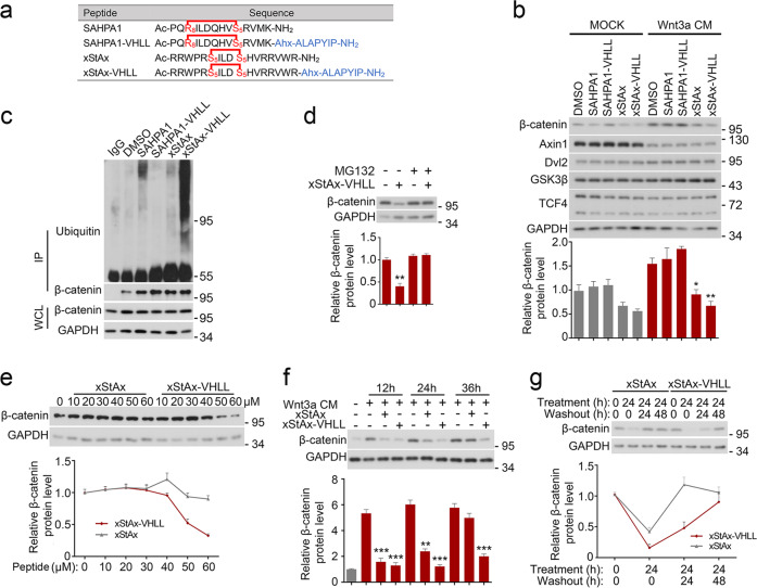
- xStAx-VHLL induced dose-dependent β-catenin degradation in HEK293T and CRC cells, promoted β-catenin ubiquitination, and its effect was blocked by the proteasome inhibitor MG132 and the VHL inhibitor VH-298.
- β-catenin remained low after 24 h of xStAx-VHLL washout, whereas β-catenin recovered after xStAx or MSAB removal; xStAx-VHLL also maintained low β-catenin up to 36 h, unlike xStAx.
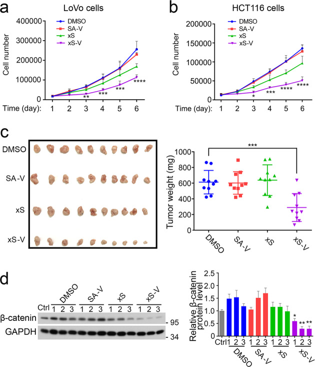
- In BALB/C nude mice injected with 3 × 10^6 LoVo cells pretreated with 50 μM peptide, xStAx-VHLL-derived tumors were significantly smaller in size and weight, with decreased β-catenin and reduced Wnt target genes Axin2, Cyclin D1, and Myc.
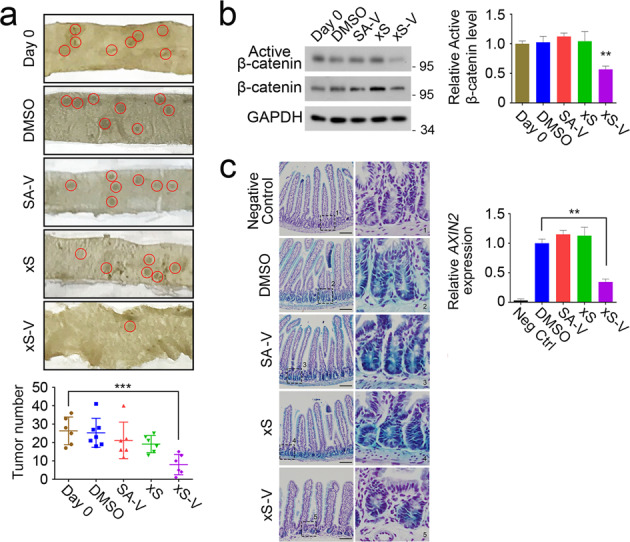
- In 11-week-old APCmin/+ mice treated intraperitoneally with 30 mg/kg every other day for 14 days, xStAx-VHLL dramatically reduced intestinal tumor number; xStAx-VHLL also inhibited survival of 11/12 CRC patient-derived organoids at 10 μM over 4 days.
Methods
- The authors synthesized SAHPA1-VHLL and xStAx-VHLL by conjugating SAHPA1 or xStAx to the VHL ligand ALAPYIP via a 6-aminocaproic acid linker, purified by HPLC, and verified by ESI-MS.
- Cell models included HEK293T and CRC lines HCT116, SW480, and LoVo; organoid models included wild-type, APC−/−, and 12 patient-derived CRC organoids; mouse models included BALB/C nude xenografts, APCmin/+ mice, and Axin2LacZ reporter mice.
- Main assays included immunoblotting, immunoprecipitation for β-catenin ubiquitination, Topflash luciferase reporter assays, qRT-PCR for Wnt target genes, cell proliferation counting, organoid survival/budding assays, tumor weight/number measurements, and β-galactosidase staining; statistics used one-way or two-way ANOVA with mean ± SD.
Limitations
- The paper states that C-11 patient-derived organoids were insensitive to xStAx-VHLL even though β-catenin decreased, possibly because other signaling pathways drive their growth and survival.
- The paper states that xStAx-VHLL is a first-generation PROTAC and requires further optimization, including non-natural residues in the VHL ligand, double-stapling, alternative E3 ligands, improved β-catenin-binding peptides such as NLS-StAx-h, and more polar PEG linkers of varying length.
- The paper notes that SAHPA1-VHLL had minimal effect on β-catenin stability and Wnt signaling, possibly due to SAHPA1’s promoting effect and lower membrane permeability compared with xStAx-VHLL; no clinical testing or pharmacokinetic data are reported in the text.
Model-written summary. Check it against the paper before relying on it.
Integrity signals
Registry
Cites
Statistics
Figures
Verification
Claims the paper makes
62 Own result · 12 Own interpretation · 8 No full text of the sources · 3 Own method · 2 Supported · 2 Partly supported · 2 No basis given · 1 Supported so far · accuracy not yet measured
Every source the paper cites for this claim was checked and supports it.
In the paper
“SAHPA1 was shown to activate Wnt/β-catenin signaling, while xStAx inhibited it13,14.”
§ A PROTAC peptide induces durable β-catenin degradation and suppresses Wnt-dependent intestinal cancer > Introduction
What the sources say
- Supportedcites [13] Cui, H. K. et al. Design of stapled alpha-helical peptides to specifically activate Wnt/beta-catenin signaling. Cell Res. 23, 581–584 (2013). source
In the Dear Editor section (page unavailable), Cui et al. report that fStAx-35R inhibited Wnt/β-catenin signaling in both HEK293T and SW480 cells. The paper uses the more specific name fStAx-35R rather than the citing sentence’s shorthand, xStAx. (LLM, conf=0.86)
- Supportedcites [14] Grossmann, T. N. et al. Inhibition of oncogenic Wnt signaling through direct targeting of beta-catenin. Proc. Natl Acad. Sci. USA 109, 17942–17947 (2012). source
The Results (Fig. 5) report that active StAx peptides inhibit Wnt3a-stimulated β-catenin reporter activity and reduce expression of Wnt/β-catenin target genes. The Results also describe their transcriptional antagonism as specific to the Wnt/β-catenin pathway. (LLM, conf=0.90)
plaindirect- Asserted: passes — The authors put it forward as true.
- Checkable: passes — It can be checked against evidence.
- Load-bearing: passes — The paper’s conclusions depend on it.
- Based: passes — The paper gives a reason why it holds.
- Supported
The sources with a verdict support this claim; the rest weren’t checked yet, have no full text we could read, or their verdicts aren’t rated yet.
In the paper
“Wnt/β-catenin signaling is essential to maintain intestinal organoid growth26,27”
§ A PROTAC peptide induces durable β-catenin degradation and suppresses Wnt-dependent intestinal cancer > Results > xStAx-VHLL impedes the growth of wild-type and APC−/− organoids
What the sources say
- Not checked — no full text of the sourcecites [26] Tuveson, D. & Clevers, H. Cancer modeling meets human organoid technology. Science 364, 952–955 (2019). source
- Supportedcites [27] Liu, Y. & Chen, Y. G. 2D-based and 3D-based intestinal stem cell cultures for personalized medicine. Cells 7, E225 (2018). source
Section 2 (3D-Based Organoid Culture System; page unavailable) describes the Wnt agonist R-spondin1 as a growth factor in intestinal organoid medium and says Wnt3a is required to generate patient-derived organoids. Section 3 (2D-Based Culture System; page unavailable) says activating Wnt/β-catenin signaling maximizes intestinal stem-cell expansion. Together, these passages support the claim’s broad point about Wnt signaling and organoid growth. (LLM, conf=0.85)
plaindirect- Asserted: passes — The authors put it forward as true.
- Checkable: passes — It can be checked against evidence.
- Load-bearing: passes — The paper’s conclusions depend on it.
- Based: passes — The paper gives a reason why it holds.
The cited sources support only part of this claim — read each source’s verdict below.
In the paper
“The PROTAC technique could aid small molecule inhibitors or peptides to exert better biological activities in vivo as PROTAC can link the ubiquitin-proteasome system to induce effective degradation of targeted proteins16.”
§ A PROTAC peptide induces durable β-catenin degradation and suppresses Wnt-dependent intestinal cancer > Discussion
What the sources say
- Partly supportedcites [16] Lai, A. C. & Crews, C. M. Induced protein degradation: an emerging drug discovery paradigm. Nat. Rev. Drug Discov. 16, 101–114 (2017). sourceExperimental — accuracy not yet measured
The Small-molecule PROTACs section (§4; page not provided) explains that PROTACs recruit an E3 ligase to degrade a target and reports mouse-xenograft evidence of better in vivo efficacy than parent inhibitors. The Peptide-based PROTACs section (§4; page not provided) describes peptide-based degradation mainly in cells and notes poor cell penetration; it does not establish better in vivo activity for peptide-based PROTACs. (LLM, conf=0.86)
hedgeddirect- Asserted: passes — The authors put it forward as true.
- Checkable: passes — It can be checked against evidence.
- Load-bearing: passes — The paper’s conclusions depend on it.
- Based: passes — The paper gives a reason why it holds.
- Partly supported
A result the paper reports from its own data — it cites no other source for it, so there is none to check. The quote shows it is in the paper.
In the paper
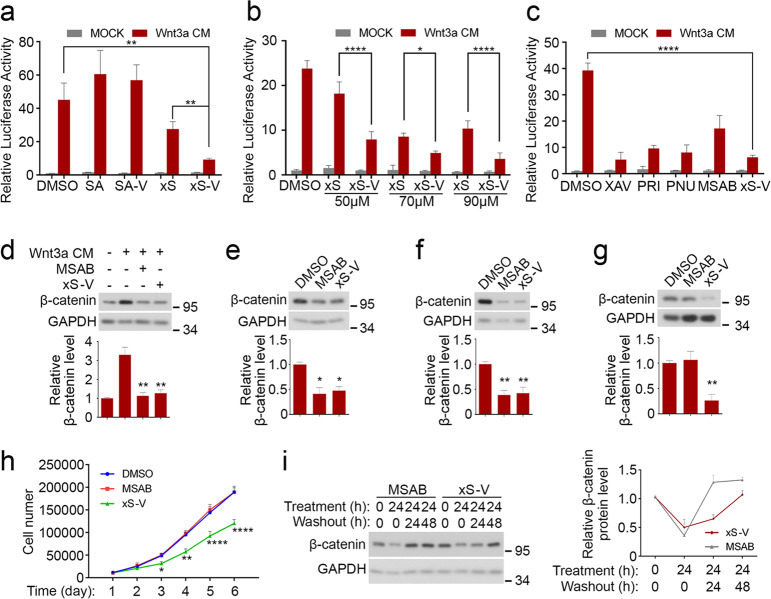
“Wnt3a induced the expression of the reporter”
§ A PROTAC peptide induces durable β-catenin degradation and suppresses Wnt-dependent intestinal cancer > Results > xStAx-VHLL inhibits Wnt/β-catenin signaling
plaindirect- Asserted: passes — The authors put it forward as true.
- Checkable: passes — It can be checked against evidence.
- Load-bearing: passes — The paper’s conclusions depend on it.
- Based: passes — The paper gives a reason why it holds.
A result the paper reports from its own data — it cites no other source for it, so there is none to check. The quote shows it is in the paper.
In the paper
“xStAx-VHLL also efficiently inhibited Wnt signaling as well as cell proliferation, intestinal organoid growth, and tumor formation.”
§ A PROTAC peptide induces durable β-catenin degradation and suppresses Wnt-dependent intestinal cancer > Discussion
boosteddirect- Asserted: passes — The authors put it forward as true.
- Checkable: passes — It can be checked against evidence.
- Load-bearing: passes — The paper’s conclusions depend on it.
- Based: passes — The paper gives a reason why it holds.
A result the paper reports from its own data — it cites no other source for it, so there is none to check. The quote shows it is in the paper.
In the paper
“xStAx-VHLL also downregulated the expression of the Wnt target genes Axin2, Cyclin D1, and Myc in APC−/− organoids”
§ A PROTAC peptide induces durable β-catenin degradation and suppresses Wnt-dependent intestinal cancer > Results > xStAx-VHLL impedes the growth of wild-type and APC−/− organoids
plaindirect- Asserted: passes — The authors put it forward as true.
- Checkable: passes — It can be checked against evidence.
- Load-bearing: passes — The paper’s conclusions depend on it.
- Based: passes — The paper gives a reason why it holds.
A result the paper reports from its own data — it cites no other source for it, so there is none to check. The quote shows it is in the paper.
In the paper
“β-catenin was remained low after 24 h of xStAx-VHLL removal”
§ A PROTAC peptide induces durable β-catenin degradation and suppresses Wnt-dependent intestinal cancer > Results > xStAx-VHLL induces dose-dependent and durable β-catenin degradation
plain- Asserted: passes — The authors put it forward as true.
- Checkable: passes — It can be checked against evidence.
- Load-bearing: passes — The paper’s conclusions depend on it.
- Based: passes — The paper gives a reason why it holds.
A result the paper reports from its own data — it cites no other source for it, so there is none to check. The quote shows it is in the paper.
In the paper
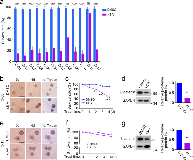
“The exon sequencing indicated the mutations of Wnt/β-catenin pathway components in all tumor tissue”
§ A PROTAC peptide induces durable β-catenin degradation and suppresses Wnt-dependent intestinal cancer > Results > xStAx-VHLL restrains the survival of CRC patient-derived organoids
hedgedn = 12- Asserted: passes — The authors put it forward as true.
- Checkable: passes — It can be checked against evidence.
- Load-bearing: passes — The paper’s conclusions depend on it.
- Based: passes — The paper gives a reason why it holds.
A result the paper reports from its own data — it cites no other source for it, so there is none to check. The quote shows it is in the paper.
In the paper
“the active of β-catenin protein levels were also decreased compared with the vehicle group”
§ A PROTAC peptide induces durable β-catenin degradation and suppresses Wnt-dependent intestinal cancer > Results > xStAx-VHLL inhibits tumor growth in APCmin/+ mouse model
plain- Asserted: passes — The authors put it forward as true.
- Checkable: passes — It can be checked against evidence.
- Load-bearing: passes — The paper’s conclusions depend on it.
- Based: passes — The paper gives a reason why it holds.
A result the paper reports from its own data — it cites no other source for it, so there is none to check. The quote shows it is in the paper.
In the paper
“the proteasome inhibitor MG132 blocked β-catenin degradation”
§ A PROTAC peptide induces durable β-catenin degradation and suppresses Wnt-dependent intestinal cancer > Results > xStAx-VHLL induces dose-dependent and durable β-catenin degradation
plaindirect- Asserted: passes — The authors put it forward as true.
- Checkable: passes — It can be checked against evidence.
- Load-bearing: passes — The paper’s conclusions depend on it.
- Based: passes — The paper gives a reason why it holds.
- It may state two separate claims as one. 27
- Probably not a substantial claim. 6
- The quote only partly states this claim. 1
The cited sources support only part of this claim — read each source’s verdict below.
In the paper
“Both SAHPA1 and xStAx are capable of penetrating the cell membrane and binding with β-catenin.”
§ A PROTAC peptide induces durable β-catenin degradation and suppresses Wnt-dependent intestinal cancer > Introduction
What the sources say
- Partly supportedcites [13] Cui, H. K. et al. Design of stapled alpha-helical peptides to specifically activate Wnt/beta-catenin signaling. Cell Res. 23, 581–584 (2013). sourceExperimental — accuracy not yet measured
In the Dear Editor section (page not available), the paper reports that SAHPA1 binds β-catenin and that FITC-labeled SAHPA1 enters the cytoplasm of HeLa cells. It does not identify a peptide called xStAx or establish both properties for it; it discusses a different peptide, fStAx-35R. (LLM, conf=0.87)
- Partly supportedcites [14] Grossmann, T. N. et al. Inhibition of oncogenic Wnt signaling through direct targeting of beta-catenin. Proc. Natl Acad. Sci. USA 109, 17942–17947 (2012). sourceExperimental — accuracy not yet measured
The Results (Fig. 3) report cellular uptake of StAx peptides and their binding to β-catenin, supporting that part of the claim. The paper does not identify a peptide called xStAx or address SAHPA1, so it does not establish the claim for both named peptides. (LLM, conf=0.84)
plaindirect- Asserted: passes — The authors put it forward as true.
- Checkable: passes — It can be checked against evidence.
- Load-bearing: passes — The paper’s conclusions depend on it.
- Based: passes — The paper gives a reason why it holds.
It may state two separate claims as one.
- Partly supported
Every source the paper cites for this claim was checked and supports it.
In the paper
“SAHPA1 was shown to activate Wnt/β-catenin signaling, while xStAx inhibited it13,14.”
§ A PROTAC peptide induces durable β-catenin degradation and suppresses Wnt-dependent intestinal cancer > Introduction
What the sources say
- Supportedcites [13] Cui, H. K. et al. Design of stapled alpha-helical peptides to specifically activate Wnt/beta-catenin signaling. Cell Res. 23, 581–584 (2013). source
The Dear Editor section reports that SAHPA1 enhanced Wnt3a-stimulated reporter activity and target-gene expression, and concludes that it activates Wnt/β-catenin signaling. The paper specifies that this activation is ligand-dependent: SAHPA1 had no obvious effect without Wnt3a. (LLM, conf=0.92)
- Supportedcites [14] Grossmann, T. N. et al. Inhibition of oncogenic Wnt signaling through direct targeting of beta-catenin. Proc. Natl Acad. Sci. USA 109, 17942–17947 (2012). source
The cited paper supports the xStAx-inhibition part of the sentence: the Results (Fig. 5 caption; page not available in our copy) report that StAx peptides inhibit β-catenin-mediated transcriptional activity and reduce Wnt/β-catenin target-gene expression. The SAHPA1 activation statement is the other half of the sentence, not the finding attributed to this StAx paper. (LLM, conf=0.87)
plaindirect- Asserted: passes — The authors put it forward as true.
- Checkable: passes — It can be checked against evidence.
- Load-bearing: fails — The paper’s conclusions may not depend on it.
- Based: passes — The paper gives a reason why it holds.
Probably not a substantial claim. The paper’s conclusions may not depend on it.
- Supported
A result the paper reports from its own data — it cites no other source for it, so there is none to check. The quote shows it is in the paper.
In the paper
“the proliferation of LoVo cells was hampered by xStAx-VHLL but not by MSAB”
§ A PROTAC peptide induces durable β-catenin degradation and suppresses Wnt-dependent intestinal cancer > Results > xStAx-VHLL inhibits Wnt/β-catenin signaling
plaindirect- Asserted: passes — The authors put it forward as true.
- Checkable: passes — It can be checked against evidence.
- Load-bearing: passes — The paper’s conclusions depend on it.
- Based: passes — The paper gives a reason why it holds.
It may state two separate claims as one.
A result the paper reports from its own data — it cites no other source for it, so there is none to check. The quote shows it is in the paper.
In the paper
“SAHPA1 slightly enhanced the Wnt3a effect while xStAx exhibited an inhibitory effect”
§ A PROTAC peptide induces durable β-catenin degradation and suppresses Wnt-dependent intestinal cancer > Results > xStAx-VHLL inhibits Wnt/β-catenin signaling
plaindirect- Asserted: passes — The authors put it forward as true.
- Checkable: passes — It can be checked against evidence.
- Load-bearing: unclear — It is unclear whether the paper’s conclusions depend on it.
- Based: passes — The paper gives a reason why it holds.
It may state two separate claims as one.
A result the paper reports from its own data — it cites no other source for it, so there is none to check. The quote shows it is in the paper.
In the paper
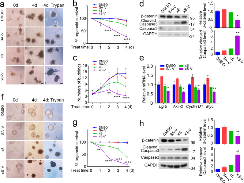
“We demonstrated that xStAx-VHLL acted as a potent PROTAC β-catenin degrader, efficiently promoting the proteasomal degradation of endogenous β-catenin in cancer cells and intestinal organoids.”
§ A PROTAC peptide induces durable β-catenin degradation and suppresses Wnt-dependent intestinal cancer > Discussion
boosteddirect- Asserted: passes — The authors put it forward as true.
- Checkable: passes — It can be checked against evidence.
- Load-bearing: passes — The paper’s conclusions depend on it.
- Based: passes — The paper gives a reason why it holds.
It may state two separate claims as one.
A result the paper reports from its own data — it cites no other source for it, so there is none to check. The quote shows it is in the paper.
In the paper
“xStAx-VHLL treatment reduced the β-catenin protein level while increased the cleaved caspase 3 level”
§ A PROTAC peptide induces durable β-catenin degradation and suppresses Wnt-dependent intestinal cancer > Results > xStAx-VHLL impedes the growth of wild-type and APC−/− organoids
plaindirect- Asserted: passes — The authors put it forward as true.
- Checkable: passes — It can be checked against evidence.
- Load-bearing: passes — The paper’s conclusions depend on it.
- Based: passes — The paper gives a reason why it holds.
It may state two separate claims as one.
A result the paper reports from its own data — it cites no other source for it, so there is none to check. The quote shows it is in the paper.
In the paper
“xStAx-VHLL induced the durable degradation of β-catenin in HEK293T cells as long as 24 h post washout, while β-catenin protein level was restored after 24 h of MSAB removal”
§ A PROTAC peptide induces durable β-catenin degradation and suppresses Wnt-dependent intestinal cancer > Results > xStAx-VHLL inhibits Wnt/β-catenin signaling
plaindirect- Asserted: passes — The authors put it forward as true.
- Checkable: passes — It can be checked against evidence.
- Load-bearing: passes — The paper’s conclusions depend on it.
- Based: passes — The paper gives a reason why it holds.
It may state two separate claims as one.
A result the paper reports from its own data — it cites no other source for it, so there is none to check. The quote shows it is in the paper.
In the paper
“C-11 organoids, which harbored the mutations of LRP6, APC, TCF7L2, KRAS, TP53, and others, were insensitive to xStAx-VHLL, even though the β-catenin protein level was decreased”
§ A PROTAC peptide induces durable β-catenin degradation and suppresses Wnt-dependent intestinal cancer > Results > xStAx-VHLL restrains the survival of CRC patient-derived organoids
plainSingle case- Asserted: passes — The authors put it forward as true.
- Checkable: passes — It can be checked against evidence.
- Load-bearing: unclear — It is unclear whether the paper’s conclusions depend on it.
- Based: passes — The paper gives a reason why it holds.
It may state two separate claims as one.
A result the paper reports from its own data — it cites no other source for it, so there is none to check. The quote shows it is in the paper.
In the paper
“β-catenin protein level in of the organoids was greatly decreased after 24 h treatment of xStAx-VHLL, whereas the cleaved caspase 3 increased”
§ A PROTAC peptide induces durable β-catenin degradation and suppresses Wnt-dependent intestinal cancer > Results > xStAx-VHLL impedes the growth of wild-type and APC−/− organoids
plaindirect- Asserted: passes — The authors put it forward as true.
- Checkable: passes — It can be checked against evidence.
- Load-bearing: passes — The paper’s conclusions depend on it.
- Based: passes — The paper gives a reason why it holds.
It may state two separate claims as one.
A result the paper reports from its own data — it cites no other source for it, so there is none to check. The quote shows it is in the paper.
In the paper
“Lgr5+ intestinal stem cells were reduced after 36 h treatment of xStAx-VHLL, and the viable cell population also decreased”
§ A PROTAC peptide induces durable β-catenin degradation and suppresses Wnt-dependent intestinal cancer > Results > xStAx-VHLL impedes the growth of wild-type and APC−/− organoids
plaindirect- Asserted: passes — The authors put it forward as true.
- Checkable: passes — It can be checked against evidence.
- Load-bearing: passes — The paper’s conclusions depend on it.
- Based: passes — The paper gives a reason why it holds.
It may state two separate claims as one.
A result the paper reports from its own data — it cites no other source for it, so there is none to check. The quote shows it is in the paper.
In the paper
“The obtained xStAx-VHLL sustained β-catenin degradation and manifested strong inhibition of Wnt signaling in cancer cells and in APC−/− organoids.”
§ A PROTAC peptide induces durable β-catenin degradation and suppresses Wnt-dependent intestinal cancer > Abstract
boosteddirect- Asserted: passes — The authors put it forward as true.
- Checkable: passes — It can be checked against evidence.
- Load-bearing: passes — The paper’s conclusions depend on it.
- Based: passes — The paper gives a reason why it holds.
It may state two separate claims as one.
A result the paper reports from its own data — it cites no other source for it, so there is none to check. The quote shows it is in the paper.
In the paper
“in LoVo cells, only xStAx-VHLL was able to reduce the β-catenin protein level, while MSAB had no effect”
§ A PROTAC peptide induces durable β-catenin degradation and suppresses Wnt-dependent intestinal cancer > Results > xStAx-VHLL inhibits Wnt/β-catenin signaling
plaindirect- Asserted: passes — The authors put it forward as true.
- Checkable: passes — It can be checked against evidence.
- Load-bearing: passes — The paper’s conclusions depend on it.
- Based: passes — The paper gives a reason why it holds.
It may state two separate claims as one.
A result the paper reports from its own data — it cites no other source for it, so there is none to check. The quote shows it is in the paper.
In the paper
“This inhibitory effect was observed not only in the systems with intact Wnt/β-catenin signaling, but also in the systems with APC-defective and constitutively active β-catenin mutations.”
§ A PROTAC peptide induces durable β-catenin degradation and suppresses Wnt-dependent intestinal cancer > Discussion
plaindirect- Asserted: passes — The authors put it forward as true.
- Checkable: passes — It can be checked against evidence.
- Load-bearing: passes — The paper’s conclusions depend on it.
- Based: passes — The paper gives a reason why it holds.
It may state two separate claims as one.
A result the paper reports from its own data — it cites no other source for it, so there is none to check. The quote shows it is in the paper.
In the paper
“the tumors originated from xStAx-VHLL pretreating cells were significantly smaller in size and weight”
§ A PROTAC peptide induces durable β-catenin degradation and suppresses Wnt-dependent intestinal cancer > Results > xStAx-VHLL restrains cell proliferation and tumor formation of colorectal cancer cancers
plaindirect- Asserted: passes — The authors put it forward as true.
- Checkable: passes — It can be checked against evidence.
- Load-bearing: passes — The paper’s conclusions depend on it.
- Based: passes — The paper gives a reason why it holds.
It may state two separate claims as one.
A result the paper reports from its own data — it cites no other source for it, so there is none to check. The quote shows it is in the paper.
In the paper
“Further, in CRC patient-derived tumor organoids, xStAx-VHLL effectively restrained the survival of tumor organoids, which highlighted its clinical potential.”
- Also stated in: § Discussion · boosted
“In this study, we have found that a potent inhibition of 11/12 patient-derived tumor organoids by xStAx-VHLL not only reveals that xStAx-VHLL is a promising anti-CRC drug candidate, but also confirms that the patient-derived tumor organoids exhibit their advances in testing the effectiveness of PROTAC peptides.”
§ A PROTAC peptide induces durable β-catenin degradation and suppresses Wnt-dependent intestinal cancer > Discussion
boosteddirectn = 12- Asserted: passes — The authors put it forward as true.
- Checkable: passes — It can be checked against evidence.
- Load-bearing: passes — The paper’s conclusions depend on it.
- Based: passes — The paper gives a reason why it holds.
It may state two separate claims as one.
- Also stated in: § Discussion · boosted
A result the paper reports from its own data — it cites no other source for it, so there is none to check. The quote shows it is in the paper.
In the paper
“SAHPA1 slightly enhanced the β-catenin level while xStAx decreased it.”
§ A PROTAC peptide induces durable β-catenin degradation and suppresses Wnt-dependent intestinal cancer > Results > xStAx-VHLL induces dose-dependent and durable β-catenin degradation
plaindirect- Asserted: passes — The authors put it forward as true.
- Checkable: passes — It can be checked against evidence.
- Load-bearing: unclear — It is unclear whether the paper’s conclusions depend on it.
- Based: passes — The paper gives a reason why it holds.
It may state two separate claims as one.
A result the paper reports from its own data — it cites no other source for it, so there is none to check. The quote shows it is in the paper.
In the paper
“xStAx-VHLL achieved a strong inhibition on APC−/− organoid survival, whereas MSAB had a minimal effect”
§ A PROTAC peptide induces durable β-catenin degradation and suppresses Wnt-dependent intestinal cancer > Results > xStAx-VHLL impedes the growth of wild-type and APC−/− organoids
boosteddirect- Asserted: passes — The authors put it forward as true.
- Checkable: passes — It can be checked against evidence.
- Load-bearing: passes — The paper’s conclusions depend on it.
- Based: passes — The paper gives a reason why it holds.
It may state two separate claims as one.
A result the paper reports from its own data — it cites no other source for it, so there is none to check. The quote shows it is in the paper.
In the paper
“Both xStAx and xStAx-VHLL started to induce β-catenin degradation as soon as 12 h post treatment.”
§ A PROTAC peptide induces durable β-catenin degradation and suppresses Wnt-dependent intestinal cancer > Results > xStAx-VHLL induces dose-dependent and durable β-catenin degradation
plaindirect- Asserted: passes — The authors put it forward as true.
- Checkable: passes — It can be checked against evidence.
- Load-bearing: unclear — It is unclear whether the paper’s conclusions depend on it.
- Based: passes — The paper gives a reason why it holds.
It may state two separate claims as one.
A result the paper reports from its own data — it cites no other source for it, so there is none to check. The quote shows it is in the paper.
In the paper
“both xStAx and xStAx-VHLL had similar membrane permeability and could efficiently enter into a cell”
§ A PROTAC peptide induces durable β-catenin degradation and suppresses Wnt-dependent intestinal cancer > Results > Design of PROTAC β-catenin degraders
plain- Asserted: passes — The authors put it forward as true.
- Checkable: passes — It can be checked against evidence.
- Load-bearing: passes — The paper’s conclusions depend on it.
- Based: passes — The paper gives a reason why it holds.
It may state two separate claims as one.
A result the paper reports from its own data — it cites no other source for it, so there is none to check. The quote shows it is in the paper.
In the paper
“The trypan blue staining at day 4 indicated the dead organoids.”
§ A PROTAC peptide induces durable β-catenin degradation and suppresses Wnt-dependent intestinal cancer > Results > xStAx-VHLL restrains the survival of CRC patient-derived organoids
hedgeddirectSingle case- Asserted: passes — The authors put it forward as true.
- Checkable: passes — It can be checked against evidence.
- Load-bearing: passes — The paper’s conclusions depend on it.
- Based: passes — The paper gives a reason why it holds.
The quote only partly states this claim.
The paper states this without a source or data behind it here, so there is nothing to check it against. The quote shows it is in the paper.
In the paper
“Although both SAHPA1 and xStAx were derived the same β-catenin-binding peptide, unlike xStAx-VHLL, SAHPA1-VHLL had minimal effect on β-catenin stability and thus no much effect on Wnt/β-catenin signaling.”
§ A PROTAC peptide induces durable β-catenin degradation and suppresses Wnt-dependent intestinal cancer > Discussion
plain- Asserted: passes — The authors put it forward as true.
- Checkable: passes — It can be checked against evidence.
- Load-bearing: unclear — It is unclear whether the paper’s conclusions depend on it.
- Based: passes — The paper gives a reason why it holds.
It may state two separate claims as one.
A result the paper reports from its own data — it cites no other source for it, so there is none to check. The quote shows it is in the paper.
In the paper
“Indeed, xStAx-VHLL was more potent than xStAx to inhibit Wnt/β-catenin signaling and attenuate cell proliferation and organoid growth.”
§ A PROTAC peptide induces durable β-catenin degradation and suppresses Wnt-dependent intestinal cancer > Discussion
plaindirect- Asserted: passes — The authors put it forward as true.
- Checkable: passes — It can be checked against evidence.
- Load-bearing: passes — The paper’s conclusions depend on it.
- Based: passes — The paper gives a reason why it holds.
It may state two separate claims as one.
In the paper
“APCmin/+ mice that harbored a point mutation in the APC gene, resulting in APC deletion and constitutive activation of Wnt signaling25”
§ A PROTAC peptide induces durable β-catenin degradation and suppresses Wnt-dependent intestinal cancer > Results > xStAx-VHLL inhibits tumor growth in APCmin/+ mouse model
What the sources say
- Not checked — no full text of the sourcecites [25] Fearon, E. R. & Vogelstein, B. A genetic model for colorectal tumorigenesis. Cell 61, 759–767 (1990). source
plaindirect- Asserted: passes — The authors put it forward as true.
- Checkable: passes — It can be checked against evidence.
- Load-bearing: passes — The paper’s conclusions depend on it.
- Based: passes — The paper gives a reason why it holds.
It may state two separate claims as one.
A conclusion the paper draws from its own results — it cites no other source for it, so there is none to check. The quote shows it is in the paper.
In the paper
“These data suggest that xStAx-VHLL impedes CRC cells proliferation and restrains tumor formation.”
§ A PROTAC peptide induces durable β-catenin degradation and suppresses Wnt-dependent intestinal cancer > Results > xStAx-VHLL restrains cell proliferation and tumor formation of colorectal cancer cancers
hedgeddirect- Asserted: passes — The authors put it forward as true.
- Checkable: passes — It can be checked against evidence.
- Load-bearing: passes — The paper’s conclusions depend on it.
- Based: passes — The paper gives a reason why it holds.
It may state two separate claims as one.
A conclusion the paper draws from its own results — it cites no other source for it, so there is none to check. The quote shows it is in the paper.
In the paper
“These findings suggest that xStAx-VHLL exhibits the ability of cancer prevention and cure”
§ A PROTAC peptide induces durable β-catenin degradation and suppresses Wnt-dependent intestinal cancer > Abstract
hedgeddirect- Asserted: passes — The authors put it forward as true.
- Checkable: passes — It can be checked against evidence.
- Load-bearing: passes — The paper’s conclusions depend on it.
- Based: passes — The paper gives a reason why it holds.
It may state two separate claims as one.
- Rests on an assumption: In the majority of CRC patients, Wnt/β-catenin signaling is aberrantly activated.
- Rests on an assumption: The 12 CRC patient-derived organoids are representative of CRC patient tumors and their responses to xStAx-VHLL predict clinical response.
What the paper did — it cites no source for this, so there is none to check. The quote shows it is in the paper.
In the paper
“the synthesized PROTACs were readily obtained with over 95% purities, as verified by ESI-MS”
§ A PROTAC peptide induces durable β-catenin degradation and suppresses Wnt-dependent intestinal cancer > Results > Design of PROTAC β-catenin degraders
plain- Asserted: passes — The authors put it forward as true.
- Checkable: passes — It can be checked against evidence.
- Load-bearing: fails — The paper’s conclusions may not depend on it.
- Based: passes — The paper gives a reason why it holds.
Probably not a substantial claim. The paper’s conclusions may not depend on it.
A claim is checked only against the cited paper’s full text, never its abstract, and none is available for its sources.
In the paper
“As Wnt/β-catenin signaling pathway is activated in various cancers, including colorectal cancer, targeting β-catenin has been regarded as an important approach to treat these diseases4,5.”
§ A PROTAC peptide induces durable β-catenin degradation and suppresses Wnt-dependent intestinal cancer > Discussion
What the sources say
- Not checked — no full text of the sourcecites [4] Nusse, R. & Clevers, H. Wnt/beta-catenin signaling, disease, and emerging therapeutic modalities. Cell 169, 985–999 (2017). source
- Not checked — no full text of the sourcecites [5] Cui, C., Zhou, X., Zhang, W., Qu, Y. & Ke, X. Is beta-catenin a druggable target for cancer therapy? Trends Biochem. Sci. 43, 623–634 (2018). source
plain- Asserted: passes — The authors put it forward as true.
- Checkable: passes — It can be checked against evidence.
- Load-bearing: passes — The paper’s conclusions depend on it.
- Based: passes — The paper gives a reason why it holds.
Probably not a substantial claim.
A conclusion the paper draws from its own results — it cites no other source for it, so there is none to check. The quote shows it is in the paper.
In the paper
“Nonetheless, as the first-generation PROTAC β-catenin degrader, xStAx-VHLL has already showed great promise in inhibiting tumor growth in vivo, and it could be employed as the leading PROTAC for further optimization in Wnt signaling-interfering cancer therapy.”
§ A PROTAC peptide induces durable β-catenin degradation and suppresses Wnt-dependent intestinal cancer > Discussion
hedgeddirect- Asserted: passes — The authors put it forward as true.
- Checkable: passes — It can be checked against evidence.
- Load-bearing: passes — The paper’s conclusions depend on it.
- Based: passes — The paper gives a reason why it holds.
Probably not a substantial claim. It may state two separate claims as one.
In the paper
“The patient-derived tumor organoids, which recapitulate the complexity and heterogeneity of tumors, have been shown to be an ideal model for drug screening and precision medicine26.”
§ A PROTAC peptide induces durable β-catenin degradation and suppresses Wnt-dependent intestinal cancer > Discussion
What the sources say
- Not checked — no full text of the sourcecites [26] Tuveson, D. & Clevers, H. Cancer modeling meets human organoid technology. Science 364, 952–955 (2019). source
boosted- Asserted: passes — The authors put it forward as true.
- Checkable: passes — It can be checked against evidence.
- Load-bearing: passes — The paper’s conclusions depend on it.
- Based: passes — The paper gives a reason why it holds.
It may state two separate claims as one.
In the paper
“Although xStAx designed by Grossmann and coworkers exhibited an impressive inhibition on Wnt/β-catenin signaling, it could be less efficient for in vivo use to treat tumors as the newly synthesized β-catenin can rapidly accumulate30.”
§ A PROTAC peptide induces durable β-catenin degradation and suppresses Wnt-dependent intestinal cancer > Discussion
What the sources say
- Not checked — no full text of the sourcecites [30] Li, V. S. et al. Wnt signaling through inhibition of beta-catenin degradation in an intact Axin1 complex. Cell 149, 1245–1256 (2012). source
hedgedconditional- Asserted: passes — The authors put it forward as true.
- Checkable: passes — It can be checked against evidence.
- Load-bearing: passes — The paper’s conclusions depend on it.
- Based: passes — The paper gives a reason why it holds.
It may state two separate claims as one.
What the paper did — it cites no source for this, so there is none to check. The quote shows it is in the paper.
In the paper
“All experiments were carried out with at least three biological replicates.”
- Also stated in: § xStAx-VHLL restrains the survival of CRC patient-derived organoids · plain
“Data from three independent experiments are displayed as the mean ± SD by two-way ANOVA (a, c) and one-way ANOVA (d, g).”
§ A PROTAC peptide induces durable β-catenin degradation and suppresses Wnt-dependent intestinal cancer > Materials and methods > Statistical analysis
plain- Asserted: passes — The authors put it forward as true.
- Checkable: passes — It can be checked against evidence.
- Load-bearing: passes — The paper’s conclusions depend on it.
- Based: passes — The paper gives a reason why it holds.
Probably not a substantial claim.
- Also stated in: § xStAx-VHLL restrains the survival of CRC patient-derived organoids · plain
What the paper did — it cites no source for this, so there is none to check. The quote shows it is in the paper.
In the paper
“All samples were obtained with informed consent, and the study was approved by the Peking University Third Hospital Medical Science Research Ethics Committee (M2018083). All relevant ethical regulations of Peking University Third Hospital Medical Science Research Ethics Committee were followed.”
§ A PROTAC peptide induces durable β-catenin degradation and suppresses Wnt-dependent intestinal cancer > Materials and methods > CRC patient tumor tissue collection and ethics statement
plain- Asserted: passes — The authors put it forward as true.
- Checkable: passes — It can be checked against evidence.
- Load-bearing: fails — The paper’s conclusions may not depend on it.
- Based: passes — The paper gives a reason why it holds.
Probably not a substantial claim. The paper’s conclusions may not depend on it. It may state two separate claims as one.
The paper relies on this without support.
In the majority of CRC patients, Wnt/β-catenin signaling is aberrantly activated.
“In the majority of CRC patients, Wnt/β-catenin signaling is aberrantly activated.”
The paper relies on this without support.
The 12 CRC patient-derived organoids are representative of CRC patient tumors and their responses to xStAx-VHLL predict clinical response.
“we were able to test the clinical potential of β-catenin targeted degrader xStAx-VHLL ex vivo”
The paper relies on this without support.
The specificity of small molecules is a general concern.
“However, the specificity of small molecules is a general concern.”
The paper relies on this without support.
Traditional drug discovery relies on cell-based or animal models, and few candidate drugs succeed in patient-based clinical studies.
“Traditional drug discovery heavily relies on cell-based or animal models, few of the candidate drugs succeed in patient-based clinical studies.”
Claims other papers make about this one
Each is checked against this paper’s own text: does it say what the citing paper says it does?
In the citing paper
“In 2020, Chen’s team reported the use of peptide-based PROTACs for TPD, introducing xStAx-VHL, a peptide-based PROTAC, to degrade β-catenin. This PROTAC directly recognizes the protein and promotes its degradation via the ubiquitin-proteasome pathway, inhibiting Wnt signaling at the cellular level and demonstrating oncogenic effects in various mouse tumor models and colon cancer patients.532”
From Recent advances in targeting the “undruggable” proteins: from drug discovery to clinical trials (2023) · cites this paper as [532]
Self-check
Percentages, sums and reported differences re-computed from the paper’s own numbers.
Each contradiction between two passages listed here was confirmed by an independent check.
Rated minor: it would not touch a result — a value no result rests on, a p-value that stays on the same side of the significance level, or a conflict that is clerical, resolved elsewhere in the paper, or outside its own findings.
In the paper
§ A PROTAC peptide induces durable β-catenin degradation and suppresses Wnt-dependent intestinal cancer > Results > xStAx-VHLL restrains the survival of CRC patient-derived organoids
“e The C-08 organoids were treated with DMSO and 10 μM xStAx-VHLL for 4 days.”
§ A PROTAC peptide induces durable β-catenin degradation and suppresses Wnt-dependent intestinal cancer > Results > xStAx-VHLL restrains the survival of CRC patient-derived organoids
“However, C-11 organoids, which harbored the mutations of LRP6, APC, TCF7L2, KRAS, TP53, and others, were insensitive to xStAx-VHLL, even though the β-catenin protein level was decreased (Fig. 6e–g).”
Rated minor: it would not touch a result — a value no result rests on, a p-value that stays on the same side of the significance level, or a conflict that is clerical, resolved elsewhere in the paper, or outside its own findings.
In the paper
§ A PROTAC peptide induces durable β-catenin degradation and suppresses Wnt-dependent intestinal cancer > Results > xStAx-VHLL inhibits Wnt/β-catenin signaling
“As shown in Fig. 2c, xStAx-VHLL (70 μM) exhibited a comparable inhibition on Wnt-induced Topflash-luciferase to other inhibitors at the working concentration (10 μM).”
§ A PROTAC peptide induces durable β-catenin degradation and suppresses Wnt-dependent intestinal cancer > Results > xStAx-VHLL inhibits Wnt/β-catenin signaling
“c HEK293T cells transfected with Topflash luciferase were treated with Wnt3a CM and 10 μM XAV939 (XAV), 20 μM PRI-724 (PRI), 100 μM PNU-74654 (PNU), 10 μM MSAB or 70 μM xStAx-VHLL (xS-V) for 24 h”
Rated minor: it would not touch a result — a value no result rests on, a p-value that stays on the same side of the significance level, or a conflict that is clerical, resolved elsewhere in the paper, or outside its own findings.
In the paper
§ A PROTAC peptide induces durable β-catenin degradation and suppresses Wnt-dependent intestinal cancer > Results > xStAx-VHLL inhibits Wnt/β-catenin signaling
“Both 10 μM MSAB and 50 μM xStAx-VHLL decreased β-catenin protein to a comparable level after 24 h treatment in HEK293T, HCT116, and SW480 cells (Fig. 2d–f).”
§ A PROTAC peptide induces durable β-catenin degradation and suppresses Wnt-dependent intestinal cancer > Results > xStAx-VHLL inhibits Wnt/β-catenin signaling
“d HEK293T cells were treated with MSAB (10 μM) or xStAx-VHLL (70 μM) and Wnt3a CM for 24 h and then harvested for immunoblotting.”
Rated minor: it would not touch a result — a value no result rests on, a p-value that stays on the same side of the significance level, or a conflict that is clerical, resolved elsewhere in the paper, or outside its own findings.
In the paper
§ A PROTAC peptide induces durable β-catenin degradation and suppresses Wnt-dependent intestinal cancer > Results > xStAx-VHLL impedes the growth of wild-type and APC−/− organoids
“f APC−/− organoids were cultured with 50 μM peptides for 4 days.”
§ A PROTAC peptide induces durable β-catenin degradation and suppresses Wnt-dependent intestinal cancer > Results > xStAx-VHLL impedes the growth of wild-type and APC−/− organoids
“g The percentage of survived APC–/– organoids was calculated each day with 10 μM peptides treatment.”
Citations
Cites
A genetic model for colorectal tumorigenesis
Fearon, E. R., & Vogelstein, B. (1990) · Cell · doi:10.1016/0092-8674(90)90186-i · Cited by 12,132
Single Lgr5 stem cells build crypt-villus structures in vitro without a mesenchymal niche
Sato, T., Vries, R. G., Snippert, H. J., van de Wetering, M., Barker, N., Stange, D. E., van Es, J. H., Abo, A., Kujala, P., Peters, P. J., et al. (2009) · Nature · doi:10.1038/nature07935 · Cited by 7,204
Wnt/β-Catenin Signaling: Components, Mechanisms, and Diseases
MacDonald, B. T., Tamai, K., & He, X. (2009) · Developmental Cell · doi:10.1016/j.devcel.2009.06.016 · Cited by 5,583
Wnt/β-catenin signaling and disease
Clevers, H., & Nusse, R. (2012) · Cell · doi:10.1016/j.cell.2012.05.012 · Cited by 5,554
Wnt/β-Catenin Signaling, Disease, and Emerging Therapeutic Modalities
Nusse, R., & Clevers, H. (2017) · Cell · doi:10.1016/j.cell.2017.05.016 · Cited by 4,342
Next-generation characterization of the Cancer Cell Line Encyclopedia
Ghandi, M., Huang, F. W., Jané-Valbuena, J., Kryukov, G. V., Lo, C. C., McDonald, E. R., Barretina, J., Gelfand, E. T., Bielski, C. M., Li, H., et al. (2019) · Nature · doi:10.1038/s41586-019-1186-3 · Cited by 3,394
Sakamoto, K. M., Kim, K. B., Kumagai, A., Mercurio, F., Crews, C. M., & Deshaies, R. J. (2001) · Proceedings of the National Academy of Sciences · doi:10.1073/pnas.141230798 · Cited by 2,609
Tankyrase inhibition stabilizes axin and antagonizes Wnt signalling
Huang, S.-M. A., Mishina, Y., Liu, S.-M., Cheung, A. K., Stegmeier, F., Michaud, G., Charlat, O., Wiellette, E., Zhang, Y., Wiessner, S., et al. (2009) · Nature · doi:10.1038/nature08356 · Cited by 2,077
Phthalimide conjugation as a strategy for in vivo target protein degradation
Winter, G. E., Buckley, D. L., Paulk, J., Roberts, J. M., Souza, A., Dhe-Paganon, S., & Bradner, J. E. (2015) · Science · doi:10.1126/science.aab1433 · Cited by 1,760
Induced protein degradation: an emerging drug discovery paradigm
Lai, A. C., & Crews, C. M. (2017) · Nature Reviews Drug Discovery · doi:10.1038/nrd.2016.211 · Cited by 1,486
An integral program for tissue renewal and regeneration: Wnt signaling and stem cell control
Clevers, H. C., Loh, K. M., & Nusse, R. (2014) · Science · doi:10.1126/science.1248012 · Cited by 1,386
Catalytic in vivo protein knockdown by small-molecule PROTACs
Bondeson, D. P., Mares, A., Smith, I. E. D., Ko, E., Campos, S., Miah, A. H., Mulholland, K. E., Routly, N., Buckley, D. L., Gustafson, J. L., et al. (2015) · Nature Chemical Biology · doi:10.1038/nchembio.1858 · Cited by 1,356
Targeting the Wnt/beta-catenin pathway in cancer: Update on effectors and inhibitors
Krishnamurthy, N., & Kurzrock, R. (2017) · Cancer Treatment Reviews · doi:10.1016/j.ctrv.2017.11.002 · Cited by 1,044
Schafmeister, C. E., Po, J., & Verdine, G. L. (2000) · Journal of the American Chemical Society · doi:10.1021/ja000563a · Cited by 1,037
Cancer modeling meets human organoid technology
Tuveson, D., & Clevers, H. (2019) · Science · doi:10.1126/science.aaw6985 · Cited by 935
Wnt Signaling through Inhibition of β-Catenin Degradation in an Intact Axin1 Complex
Li, V. S. W., Ng, S. S., Boersema, P. J., Low, T. Y., Karthaus, W. R., Gerlach, J. P., Mohammed, S., Heck, A. J. R., Maurice, M. M., Mahmoudi, T., et al. (2012) · Cell · doi:10.1016/j.cell.2012.05.002 · Cited by 889
Structure of an HIF-1α-pVHL Complex: Hydroxyproline Recognition in Signaling
Min, J.-H., Yang, H., Ivan, M., Gertler, F., Kaelin, W. G., & Pavletich, N. P. (2002) · Science · doi:10.1126/science.1073440 · Cited by 765
Structural basis for the recognition of hydroxyproline in HIF-1α by pVHL
Hon, W.-C., Wilson, M. I., Harlos, K., Claridge, T. D. W., Schofield, C. J., Pugh, C. W., Maxwell, P. H., Ratcliffe, P. J., Stuart, D. I., & Jones, E. Y. (2002) · Nature · doi:10.1038/nature00767 · Cited by 751
Wang, Y., Song, W., Wang, J., Wang, T., Xiong, X., Qi, Z., Fu, W., Yang, X., & Chen, Y.-G. (2020) · Journal of Experimental Medicine · doi:10.1084/jem.20191130 · Cited by 399
Self-citationHydrocarbon double-stapling remedies the proteolytic instability of a lengthy peptide therapeutic
Bird, G. H., Madani, N., Perry, A. F., Princiotto, A. M., Supko, J. G., He, X., Gavathiotis, E., Sodroski, J., & Walensky, L. D. (2010) · Proceedings of the National Academy of Sciences · doi:10.1073/pnas.1002713107 · Cited by 354
The unravelling of the ubiquitin system
Ciechanover, A. J. (2015) · Nature Reviews Molecular Cell Biology · doi:10.1038/nrm3982 · Cited by 314
Li, Y., Yang, J., Aguilar, A., McEachern, D., Przybranowski, S., Liu, L., Yang, C.-Y., Wang, M., Han, X., & Wang, S. (2019) · Journal of Medicinal Chemistry · doi:10.1021/acs.jmedchem.8b00909 · Cited by 312
Self-citationInhibition of oncogenic Wnt signaling through direct targeting of β-catenin
Grossmann, T. N., Yeh, J. T.-H., Bowman, B. R., Chu, Q., Moellering, R. E., & Verdine, G. L. (2012) · Proceedings of the National Academy of Sciences · doi:10.1073/pnas.1208396109 · Cited by 268
Xing, Y., Clements, W. K., Kimelman, D., & Xu, W. (2003) · Genes & Development · doi:10.1101/gad.1142603 · Cited by 237
Prospective discovery of small molecule enhancers of an E3 ligase-substrate interaction
Simonetta, K. R., Taygerly, J., Boyle, K., Basham, S. E., Padovani, C., Lou, Y., Cummins, T. J., Yung, S. L., von Soly, S. K., Kayser, F., et al. (2019) · Nature Communications · doi:10.1038/s41467-019-09358-9 · Cited by 217
Hwang, S.-Y., Deng, X., Byun, S., Lee, C., Lee, S.-J., Suh, H., Zhang, J., Kang, Q., Zhang, T., Westover, K. D., et al. (2016) · Cell Reports · doi:10.1016/j.celrep.2016.05.071 · Cited by 173
Is β-Catenin a Druggable Target for Cancer Therapy?
Cui, C., Zhou, X., Zhang, W., Qu, Y., & Ke, X. (2018) · Trends in Biochemical Sciences · doi:10.1016/j.tibs.2018.06.003 · Cited by 168
Self-citationRecent Development of Wnt Signaling Pathway Inhibitors for Cancer Therapeutics
Harb, J. F., Lin, P.-J., & Hao, J. (2019) · Current Oncology Reports · doi:10.1007/s11912-019-0763-9 · Cited by 139
Dietrich, L., Rathmer, B., Ewan, K., Bange, T., Heinrichs, S., Dale, T. C., Schade, D., & Grossmann, T. N. (2017) · Cell Chemical Biology · doi:10.1016/j.chembiol.2017.06.013 · Cited by 126
Development of Stabilized Peptide-Based PROTACs against Estrogen Receptor α
Jiang, Y., Deng, Q. W., Zhao, H., Xie, M., Chen, L., Yin, F., Qin, X., Zheng, W., Zhao, Y. J., & Li, Z. (2017) · ACS Chemical Biology · doi:10.1021/acschembio.7b00985 · Cited by 125
Development of anticancer agents targeting the Wnt/β-catenin signaling.
Zhang, X., & Hao, J. (2015) · Europe PMC (PubMed Central) · Cited by 85
Dithiocarbamate-inspired side chain stapling chemistry for peptide drug design
Li, X., Tolbert, W. D., Hu, H., Gohain, N., Zou, Y., Niu, F., He, W., Yuan, W., Su, J., Pazgier, M., et al. (2018) · Chemical Science · doi:10.1039/c8sc03275k · Cited by 73
Self-citationZhao, B. T., Qi, Z., Li, Y., Wang, C., Fu, W., & Chen, Y. (2015) · Nature Communications · doi:10.1038/ncomms8166 · Cited by 67
Self-citationDesign of stapled α-helical peptides to specifically activate Wnt/β-catenin signaling
Cui, H.-K., Zhao, B., Li, Y., Guo, Y., Hu, H., Liu, L., & Chen, Y.-G. (2013) · Cell Research · doi:10.1038/cr.2013.30 · Cited by 42
Self-citationLi, X., Zou, Y., & Hu, H. (2018) · Chinese Chemical Letters · doi:10.1016/j.cclet.2018.01.018 · Cited by 39
Self-citation2D- and 3D-Based Intestinal Stem Cell Cultures for Personalized Medicine
Liu, Y., & Chen, Y.-G. (2018) · Cells · doi:10.3390/cells7120225 · Cited by 37
Self-citationTankyrases maintain homeostasis of intestinal epithelium by preventing cell death
Ye, P., Chiang, Y. J., Qi, Z., Li, Y., Wang, S., Liu, Y., Li, X., & Chen, Y. (2018) · PLoS Genetics · doi:10.1371/journal.pgen.1007697 · Cited by 13
Self-citation
Cited by
Sorted by citations
PROTACs: past, present and future
Li, K., & Crews, C. M. (2022) · Chemical Society Reviews · doi:10.1039/d2cs00193d · Cited by 869
Recent advances in targeting the “undruggable” proteins: from drug discovery to clinical trials
Xie, X., Yu, T., Li, X., Zhang, N., Foster, L. J., Peng, C., Huang, W., & He, G. (2023) · Signal Transduction and Targeted Therapy · doi:10.1038/s41392-023-01589-z · Cited by 396
Self-citationProteolysis-targeting chimeras (PROTACs) in cancer therapy
Li, X., Pu, W., Zheng, Q., Ai, M., Chen, S., & Peng, Y. (2022) · Molecular Cancer · doi:10.1186/s12943-021-01434-3 · Cited by 389
Self-citationWnt/β-catenin-driven EMT regulation in human cancers
Xue, W., Yang, L., Chen, C., Ashrafizadeh, M., Tian, Y., & Sun, R. (2024) · Cellular and Molecular Life Sciences · doi:10.1007/s00018-023-05099-7 · Cited by 321
Stapled Helical Peptides Bearing Different Anchoring Residues
Li, X., Chen, S., Zhang, W., & Hu, H. (2020) · Chemical Reviews · doi:10.1021/acs.chemrev.0c00532 · Cited by 304
Self-citation
Metadata
- Authors
- Liao, H., Li, X., Zhao, L., Wang, Y., Wang, X., Wu, Y., Zhou, X., Fu, W., Liu, L., Hu, H.-G., & Chen, Y.-G.
- Affiliations
- Center for Life Sciences (CN)
- Second Military Medical University (CN)
- Peking University Third Hospital (CN)
- Corresponding author
- Chen, Ye-Guang
- ORCID
- Venue
- Cell Discovery
- Published
- 2020-06-09
- Volume
- 6
- Issue
- 1
- Pages
- 35-35
- ISSN
- 2056-5968 (ISSN-L)
- PMC
- PMC7280531
- OpenAlex
- W3034021518
- Cited by
- 156
- References
- 37
- Citations by year
- 2026: 21 · 2025: 31 · 2024: 39 · 2023: 31 · 2022: 20 · 2021: 10 · 2020: 4
- FWCI
- 4.79
- Type
- journal article
- Language
- en
- Open access
- gold
- doi.org
- Locations
- Cell Discovery (publishedVersion, cc-by)
- PubMed Central (publishedVersion, cc-by)
- PubMed (publishedVersion)
- Greater South Information System
- Funders
- National Natural Science Foundation of China (2017YFA0103601, 91753205, 31330049, 91849129, 2017YFA0505200)
- National Key Research and Development Program of China (2017YFA0505200, 31330049, 2017YFA0103601)
- Topics
- Life Sciences › Biochemistry, Genetics and Molecular Biology › Molecular Biology › Protein Degradation and Inhibitors
- Life Sciences › Biochemistry, Genetics and Molecular Biology › Molecular Biology › Wnt/β-catenin signaling in development and cancer
- Physical Sciences › Chemistry › Organic Chemistry › Chemical Synthesis and Analysis
- Keywords
- PROTACs, β-catenin degradation, Wnt/β-catenin signaling, stapled peptides, colorectal cancer, ApcMin/+ mice, patient-derived organoids, targeted protein degradation, anticancer agents
About these checks
Registry, retraction, Cites and statistics results are computed, not judged, with one exception: a printed reference that no index lists is read once by a model before it counts as a citation error. Claims are extracted by a model and kept only when their quote appears verbatim in the text; each cited source is then checked, with a model’s help, for whether it says what the claim relies on. Those verdicts can be wrong — read the claim’s quote and the cited source before relying on one.
A language model drew the claims from the paper on 2026-10-23, under version 3 of the claim definition (model deepseek-v4.1-flash, prompt extract-v3).
A second model labelled each claim on 2026-10-23 (model jev-1.13-20260917, definition revision gold-guideline-v0.2).
Cited sources are checked with a language model’s help (gpt-6-sol).
Statistics and arithmetic are recomputed, not judged.
Confidence bands are shown only for fields calibrated on our annotated test set. None is calibrated yet, so no band is shown.
What counts as a claim ›How we measure confidence ›How we check this ›
Are you an author, or do you see a problem with this page? Request a review
